Animal Models for Human Polycystic Ovary Syndrome (PCOS) Focused on the Use of Indirect Hormonal Perturbations: A Review of the Literature - MDPI
←
→
Page content transcription
If your browser does not render page correctly, please read the page content below
International Journal of
            Molecular Sciences

Review
Animal Models for Human Polycystic Ovary
Syndrome (PCOS) Focused on the Use of Indirect
Hormonal Perturbations: A Review of the Literature
Youngjae Ryu 1 , Sung Woo Kim 2 , Yoon Young Kim 1,2 and Seung-Yup Ku 1,2, *
 1    Biomedical Research Institute, Seoul National University Hospital, Seoul 03080, Korea;
      dragonkai@naver.com (Y.R.); yoonykim@snu.ac.kr (Y.Y.K.)
 2    Department of Obstetrics and Gynecology, College of Medicine, Seoul National University, Seoul 03080,
      Korea; byulbi81@snu.ac.kr
 *    Correspondence: jyhsyk@snu.ac.kr; Tel.: +82-2-2072-4416

 Received: 2 April 2019; Accepted: 23 May 2019; Published: 3 June 2019

 Abstract: Hormonal disturbances, such as hyperandrogenism, are considered important for
 developing polycystic ovary syndrome (PCOS) in humans. Accordingly, directly hormone-regulated
 animal models are widely used for studying PCOS, as they replicate several key PCOS features.
 However, the pathogenesis and treatment of PCOS are still unclear. In this review, we aimed to
 investigate animal PCOS models and PCOS-like phenotypes in animal experiments without direct
 hormonal interventions and determine the underlying mechanisms for a better understanding of
 PCOS. We summarized animal PCOS models that used indirect hormonal interventions and suggested
 or discussed pathogenesis of PCOS-like features in animals and PCOS-like phenotypes generated
 in other animals. We presented integrated physiological insights and shared cellular pathways
 underlying the pathogenesis of PCOS in reviewed animal models. Our review indicates that the
 hormonal and metabolic changes could be due to molecular dysregulations, such as upregulated
 PI3K-Akt and extracellular signal-regulated kinase (ERK) signalling, that potentially cause PCOS-like
 phenotypes in the animal models. This review will be helpful for considering alternative animal PCOS
 models to determine the cellular/molecular mechanisms underlying PCOS symptoms. The efforts to
 determine the specific cellular mechanisms of PCOS will contribute to novel treatments and control
 methods for this complex syndrome.

 Keywords: polycystic ovary syndrome; animal models; ovary; pathogenesis

1. Introduction
      Various animal models have been developed and studied for the human polycystic ovary syndrome
(PCOS) for more than 60 years. However, the etiology of PCOS is still unclear because of its complex
manifestation as a syndrome and limitations of translational studies using animals.
      Although a huge gap exists between humans and laboratory animals, which are mainly rodents,
with respect to their reproductive physiology (e.g., differences in ovulation number and patterns,
released hormone profile, sensitivity to hormones, behavioural styles, and anatomy of the organs),
studies using animal models are essential to explore the pathophysiology of PCOS in vivo.
      According to Rotterdam’s criteria for PCOS diagnosis [1], the key features of PCOS that should
be manifested in the animal models are hyperandrogenism, too many antral follicles and abnormal
estrous cycle (amenorrhea or oligomenorrhea in case of non-human primates). The most suitable
animal PCOS model would be naturally occurring PCOS-like animals. However, laboratory rodents,
unlike humans, do not develop PCOS-like phenotypes naturally, although the persistence of ovarian
follicles with a prolonged estrous period has been reported in natural housed rats [2]. To date, natural

Int. J. Mol. Sci. 2019, 20, 2720; doi:10.3390/ijms20112720                          www.mdpi.com/journal/ijms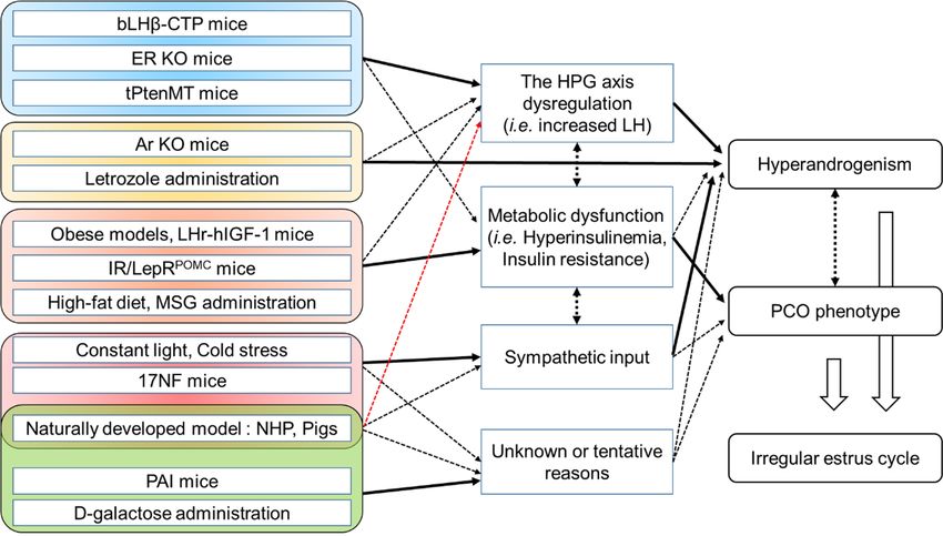
Int. J. Mol. Sci. 2019, 20, 2720                                                                     2 of 27

PCOS-like features have not been reported in rodents. Natural development of polycystic ovary (PCO)
and related hormone disturbances with clinical symptoms, has been observed in animals such as cow,
pig, and dog; however, these animals did not generally show hyperandrogenism. Naturally occurring
hyperandrogenism has been reported in non-human primates. Abbott et al. (2017) reported PCOS-like
traits in naturally hyperandrogenic female rhesus monkeys (Macaca mulatta) [3]. However, the use and
screening of non-human primates for PCOS studies have many limitations.
       Therefore, to study PCOS with laboratory animals, various artificial methods have been used;
prenatally or postnatally hormone-treated (i.e., excess androgen or estrogen) animals are extensively used.
These animal models can manifest the key features of PCOS, such as hyperandrogenism, those with
excess antral follicles, prolonged or irregular estrous cycle, and metabolic symptoms. Moreover, prenatal
perturbation using androgens has demonstrated transgenerational development of PCOS phenotypes in
the offspring. However, to define the specific pathogenesis of PCOS and explore control/treatment methods,
it is undoubtedly valuable to establish other methods along with the hormonal intervention models.
       Thus, herein we focused on highlighting up-to-date animal PCOS models that use indirect hormonal
interventions and summarizing suggested/discussed mechanisms of the pathogenesis of the developed
PCOS-like phenotypes in those models. Furthermore, we have briefly summarized naturally developed
PCO and relevant mechanisms in other animals to search for shared pathophysiology with respect to
polycystic ovarian morphology, hormonal alteration, and accompanying metabolic syndromes. We have
also discussed shared cellular/molecular mechanisms underlying the pathogenesis of PCOS in animals on
the basis of a literature review and suggested further directions for the use of animal PCOS models.

2. Methods
      We searched the literature up to June 2018 by using PubMed (https://www.ncbi.nlm.nih.
 gov/pubmed/) and Google Scholar (https://scholar.google.com/). The papers were searched
 using the following terms: ‘PCOS’, ‘polycystic ovary syndrome’, ‘animal model’, ‘mouse’, ‘rat’,
‘primate’, ‘transgenic’, ‘chemical’, ‘ovarian dysfunction’, ‘testosterone’, ‘androgen’, and ‘pathogenesis’.
We excluded studies with androgen-, estrogen-, or other directly hormone-treated animal PCOS
 models in which either prenatal or postnatal administration was performed. In addition, we excluded
 studies in which evaluation of the ovarian morphology and serum androgen levels was not performed.
 However, we included the papers when following or similar studies using same experimental methods
 backed those evaluations up.
      Furthermore, to search for new aspects of pathogenesis of PCOS in other species, we used keywords
 such as ‘cystic ovarian disease’, ‘COD’, ‘polycystic ovary’, ‘animal’, and ‘veterinary’. Among the
papers, we selected representative literature to discuss the relevant mechanisms.
      Human PCOS patients with more than 12 cystic follicles per ovary or ovarian volume over 10 mL,
 diagnosed using ultrasonography, are confirmed to have ‘excess dominant follicles’ [4]. In this review,
we use the term ‘PCO’ or for animals on the basis of histological section images and sentences in the
 reviewed literature that describe those with excess numbers of antral follicles. In addition, we used
‘hyperandrogenism’ as the status presented by significantly elevated serum testosterone concentration
when compared with the control group. All the animals described in this review are female, except
when especially noted.

2.1. Animal Polycystic Ovary Syndrome (PCOS) Models Induced by Direct Hormonal Interventions
    Because one of a clinical hallmark of PCOS patients is hyperandrogenism, to induce PCOS-like
phenotypes in laboratory animals, direct androgen treatment methods have been widely used with
agents such as testosterone propionate (TP), dihydrotestosterone (DHT), or dehydroepiandrosterone.
Other hormonal agents such as estradiol valerate (EV), glucocorticoid, human chorionic gonadotropin
(hCG), and anti-Müllerian hormone (AMH) have also been used. These hormones induce PCOS-like
phenotypes by not only postnatal administration but also prenatal administration in rodents, sheep,
and non-human primates.
Int. J. Mol. Sci. 2019, 20, 2720                                                                    3 of 27

     Many well-written reviews have already summarized these types of animal PCOS models [5–14].
In this review, we aimed to introduce mainly indirect hormonal methods for developing PCOS-like
phenotypes in animals; therefore, we have provided a short description of hormone-induced animal
PCOS models.
     On the basis of the results obtained using androgen receptor- Knockout (KO) mice, excess
androgen is sufficient to induce an irregular estrous cycle, excess dominant follicles and other metabolic
symptoms in mice. Although further studies are needed to check whether the effect of androgen
is mainly on the neuronal regulation or ovaries, it is clear that exposure to high androgen levels is
sufficient to induce PCOS-like manifestations in rodents.
     Animals such as sheep and monkeys exposed to androgens prenatally have also developed
hallmarks of PCOS phenotypes. Luteinizing hormone (LH) pulse frequency is also elevated in prenatally
androgen exposed sheep and monkeys [15]. In other animals, such as sheep, and rhesus monkey,
prenatal androgen exposure induced hyperandrogenism and oligo-anovulation in the offspring [9].
Androgen exposure during the gestation stage re-programs the hypothalamic-pituitary-gonadal (HPG)
axis through epigenetic changes or neuronal rearrangements [16,17]. Recently, mice treated with
Anti-Müllerian hormone (AMH) prenatally showed acyclic estrous and elevated serum testosterone and
LH levels [18]. The authors indicated that excess AMH exposure during the late period of pregnancy
(embryonic day 16.5 to 18.5) leads to hyperactivation of the gonadotropin-releasing hormone (GnRH)
neurons in the offspring, resulting in fluctuations in LH release from the pituitary gland. As a result,
this model induced a masculinization of the exposed female fetus and a PCOS-like reproductive and
neuroendocrine phenotype in adulthood.

Limitations and Difficulties of Hormone-Treated Animal Models
      Androgen-treated models yield good reproduction results and manifestation of the relevant PCOS
symptoms in animals. However, direct hormonal interventions may not be suitable for finding a basal
and common pathway for its pathogenesis. However, the experiments demonstrated that androgen
exposure can alter endocrine homeostasis and lead to the development of ovarian dysfunction as well
as its accompanying metabolic symptoms. There are many mechanisms driving increased androgen
production in PCOS include increased LH stimulation resulting from abnormal LH secretory dynamics
and increased LH bioactivity, and hyperinsulinemia due to insulin resistance. Considering that PCOS
is a hyperandrogenic disorder [19,20], hyperandrogenism is one of the most important features to be
elaborately manifested in the animal model. In addition, considering both human males and females
may not be exposed to such excessive hormonal disturbance in their lives.
      LH stimulation and alteration of GnRH neuron activation may be the fundamental reasons
for hyperandrogenism in PCOS patients [21]. However, increased insulin levels (hyperinsulinemia)
and insulin resistance have been suggested as the key triggers for the development of PCOS [22,23].
Other factors such as alterations in AMH and Follicle-stimulating hormone (FSH) levels may induce
the occurrence of PCOS [24]. Indeed, all these factors are related to PCOS pathogenesis on the basis of
both clinical data and animal experiments [25,26]. However, the shared molecular pathway/mechanism
and causal relationship regarding the pathogenesis of PCOS are still unclear. Therefore, animal PCOS
models that do not use direct hormonal intervention may be useful.

2.2. Animal Models with PCOS or Similar Symptoms Induced by Indirect Hormonal Perturbations

2.2.1. Genetically Engineered or Genetic Animal Models Including PCOS-Like Symptoms
     To understand the role of specific genes and their pathways related to the development of PCOS,
various genetically modified rodents have been studied. We reviewed literature focused on elevated
serum testosterone levels and the polycystic ovarian appearance in the animals. In addition, we noted
suggested/discussed mechanisms responsible for the developed phenotypes (Table 1).
Int. J. Mol. Sci. 2019, 20, 2720 4 of 27
Table 1. Transgenic and genetically modified animal models including PCOS-like symptom.
Time point of
Estrous Androgen
Types Author Year Species Strain Ovarian Cyst Metabolic Features LH Level Examination Notes
Cycle Level
(Post-Natal)
Enhanced sympathetic input;
Yes NGF affects trkA receptor and
Transgenic [27] 2009 Mouse 17NF Prolonged ↑ (+PMSG) N/R ↓ 28 days
(+hCG) p75NTR ; apoptosis signaling
in the ovarian follicles
Obesity, glucose
Increased ovarian
Transgenic [28] 2014 Mouse 17NF Prolonged N/R ↑ intolerance, N/A 10, 20 weeks
sympathetic input
hyperinsulinemia
Yes Absence of estrogen and
Transgenic [29] 2010 Mouse Ar KO Acyclic ↑ N/R ↑ 19 weeks
(hemorrhagic) elevated LH concentration
1995, The HPG axis dysregulation;
Transgenic [30,31] Mouse LH β-CTP Prolonged Yes ↑ N/R ↑ 14 - 42 days
1997 Inadequate negative feedback
Yes ERα KO in theca cells
Theca-specific
Transgenic [32] 2009 Mouse Irregular (hemorrhagic, + ↑ N/R ↓ 2 - 6 months predispose the ovary to
(CYP17) ERα KO
PMSG or hCG) develop cysts
Obesity, Leptin receptor malfunction;
6 weeks, 12
Genetic [33] 2009 Rat JCR:LA-cp strain Irregular Yes ↑ dyslipidemia, N/R increased insulin and leptin
weeks
insulin resistance level
# = papers supporting similar study; N.S. = non-significance; N/R = not reported; ↑ = significantly increased/up-regulated; ↓ = significantly decreased/down-regulated; Ar = aromatase;
AT2R = angiotensin II type 2 receptor; ER = estrogen receptor; hCG = human chorionic gonadotropin; HPG axis = Hypothalamic-pituitary-gonadal axis; IGF = insulin-like growth factor;
LH = luteinizing hormone; NGF = Nerve growth factor; PMSG = pregnant mare serum gonadotropin.Int. J. Mol. Sci. 2019, 20, 2720 5 of 27
PCOS-Like Phenotypes Caused by Metabolic Dysfunctions
In the numerous genetically manipulated or spontaneously mutated rodent models, the rodent
strains or transgenic mice that show dysregulations in lipid or glucose metabolism commonly manifest
ovarian cysts and metabolic dysfunctions. The New Zealand obese mice and Zucker (fa/fa) rat, which are
both spontaneous genetic obesity rodent models, commonly present hyperinsulinemia, ovarian cysts,
and poor fertility. However, these strains did not show elevated (or even significantly decreased) serum
testosterone or LH levels [33–35]. Because leptin receptor mutations and reduced leptin signalling
(leptin resistance) were observed in these rodents [36,37], it is assumed that leptin-mediated LH
release was compromised and, therefore, synthesis of androgen by LH stimulation was not accelerated.
These findings suggest that hyperinsulinemia and cystic ovarian morphology induced by metabolic
abnormalities do not cause hyperandrogenemia, at least, in rodents. Nevertheless, species difference
should be taken into account. It is well established that increased circulating insulin levels cause
or contribute to hyperandrogenism in women with PCOS, as opposite to rodents, in at least two
important ways, by stimulating increased ovarian androgen production, and by inhibiting hepatic
Sex hormone-binding globulin (SHBG) production [38]. Honnma et al. indicated that downregulated
ovarian adiponectin levels may related to ovarian dysfunction, although serum adiponectin level was
significantly higher in the Zucker (fa/fa) rats than in the control group. However, a recent study showed
that overexpression of adiponectin in mice did not ameliorate PCO morphology as well as the estrous
cycle induced by DHT administration [39]. Kajihara et al. reported increased Forkhead box O (FOXO)-1
expression in the ovary tissue of Zucker (fa/fa) rats, especially in the granulosa cells. FOXOs regulate
cell proliferation, response to oxidative stress, and downstream targets of the phosphatidylinositol
3-kinase (PI3K)–Akt pathway [40].
On the other hand, JCR:LA-cp rats that also had a defect in the leptin receptor (LepR) and
concurrent hyperleptinemia showed a spontaneous increase in serum testosterone levels, irregular
estrous cycle, cystic follicles, hyperinsulinemia, obesity, and dyslipidemia [41]. Although increased
insulin level is considered a major regulatory factor of ovarian androgen genesis [42], the discrepancies
among the obese rodent models (New Zealand obese mouse, Zucker rat, and JCR:LA-cp rat) may
not elicit simple connections between insulin level and hyperandrogenism. Significantly increased
leptin levels were detected in both the New Zealand obese mice and Zucker (fa/fa) rats [36,43].
Therefore, it is possible that the frequency of LH stimulation or expression of ovarian enzymes related
to steroidogenesis may differ between those models. A previous study has reported that the increased
uptake of non-esterified fatty acid in the ovary of JCR:LA-cp rats is correlated to the serum testosterone
level [44]. These results suggest that intracellular ovarian lipotoxicity may be related to androgen
production. The degree of fatty acid-mediated oxidation stress to the ovary may differ among the
obese rodent models.
Mice lacking the insulin receptor (IR) and LepR in hypothalamic pro-opiomelanocortin neurons
(POMC; IR/LepRPOMC ) manifested prolonged estrous cycle, hyperinsulinemia, hyperleptinemia,
increased body fat mass, and elevated serum LH and testosterone levels [45,46]. However, the mice
did not develop typical PCO morphology. In addition, adipose dysfunction, macrophage infiltration,
and elevated cytokine levels (including IL-1β and IL-6) in adipose tissues such as perigonadal fat were
observed in this model [29]. On the basis of these results, elevated LH stimulation and hyperinsulinemia
may be related to hyperandrogenism; however, the increased insulin and serum testosterone levels did
not result in the development of prominent follicular cysts in the IR/LepRPOMC mice.
Another transgenic obese mouse, Mito-ob, that overexpressed prohibitin in adipocytes showed
the formation of ovarian cysts without any significant changes in the serum levels of insulin, leptin,
LH, and testosterone [47]. Instead, increased body fat mass and serum estradiol level were observed in
the Mito-ob mice. Thus, increased estradiol levels or overgrowth of the periovarian adipose tissue may
be the reasons for the cystic ovarian morphology in this model.
Mice overexpressing the human insulin-like growth factor-I (IGF-I) controlled by the LH receptor
(LHr) expression (LHr-hIGF-I) showed ovarian cysts and elevated serum testosterone levels. The LHInt. J. Mol. Sci. 2019, 20, 2720 6 of 27
level was decreased, and the estradiol level was increased, but not significantly, in this model.
Upregulated IGF-I production resulted in elevated serum testosterone levels in all LHr-hIGF-I
female mice; however, only 40% manifested PCO morphology, even though the histomorphological
characteristics of the cysts differed from those of human PCOS patients. Therefore, the authors
suggested that increased IGF-I expression may be a direct cause of hyperandrogenism in this model.
Protein kinase B-β (PKBβ/Akt2) KO mice manifested severe ovarian cysts (not the typical PCO
morphology), hyperinsulinemia, and hyperandrogenemia at 90 weeks of age [48]. The serum LH level
did not differ significantly when compared with the wild-type group. In addition, marked extracellular
signal-regulated kinase (ERK) as well as cAMP response element-binding protein expressions and
fat (lipid) accumulation aside to the ovary were observed. The 20-week-old PKBβ KO mice did not
show increased serum insulin or testosterone levels. Restuccia et al. (2012) indicated that upregulated
ERK signalling due to the loss of PKBβ and lipid accumulation may induce hyperandrogenism and
ovarian cysts. However, Akt2 KO mice showed hyperinsulinemia and insulin intolerance at 8 weeks of
age [49]; therefore, further studies are needed to clarify the mechanisms underlying the PCOS-like
appearance by using this model.
Lan et al. (2017) reported that Ptenfl/fl –Cyp17iCre (tPtenMT) mice in which Pten was selectively
deleted in theca cells presented a prolonged estrous cycle, formation of numerous antral follicles,
and elevated serum testosterone levels [50]. The theca cells of tPtenMT showed increased Akt
phosphorylation and upregulated expressions of Lhcgr, Star, Cyp11a1, and Cyp17a1. In addition to the
upregulation of Lhcgr expression, which means a more elevated response to LH, the authors reported
that the phosphorylation of FOXO-1 was increased in the ovaries of the tPtenMT mice and augmented
by LH administration. Moreover, Lhcgr may be a direct downstream target gene of FOXO-1 in the theca
cells. This is an important finding: Dysregulation of the PI3K-Akt pathway and related signalling,
consistently implied by the other aforementioned genetic models, can change the responsiveness to
LH, resulting in hyperandrogenism.
PCOS-Like Phenotypes Induced by Changes in the Endocrine System
Specific deletion or overexpression of endocrine system-related genes results in the occurrence
of PCOS-like features in animals. Multiple cystic follicles with haemorrhagic cysts and elevated LH
levels were reported in estrogen receptor-α (ERα) KO mice by Couse et al. [51]. The ERα KO mice
showed significantly increased testosterone and estradiol levels. However, ERβ KO mice did not show
the same elevated levels [52]. Expression of Lhb in the pituitary gland and Cyp17 and Cyp19 in the
ovary was upregulated in the ERα KO mice. Administration of the GnRH antagonist suppressed
the symptoms in these mice. In addition, Lee et al. (2009) demonstrated that theca cell-specific
ERα KO (thEsr1 KO), mice using 17β-hydroxylase a1 (Cyp17-iCre), developed PCOS-like phenotypes
such as irregular estrous cycle, cystic ovaries (induced by pregnant mare’s serum gonadotropin and
hCG injection), and increased serum testosterone levels. However, the LH level was significantly
decreased, or even undetectable, in the mice [32]. Moreover, ERβ KO mice with the bLHβ–CTP
transgene (estrogen receptor-β KO with increased LH level; bLHβ–CTP mice will be described
below) presented suppressed development of cystic follicles and reduced testosterone levels when
compared with wild-type bLHβ–CTP transgenic mice [53]. It is noteworthy that a loss of ERα function
leads to dysregulation of LH release and an increase in CYP17 expression in the ovary, resulting in
hyperandrogenism. It can be postulated that the development of PCOS-like phenotypes is related to
stimulation via ERβ, which is observed mainly in granulosa cells, and impaired functions of ERα,
which is expressed in the theca or interstitial cells of the ovary as well as the hypothalamus.
Aromatase KO (Ar KO) mice developed haemorrhagic ovarian cysts, increased serum LH levels,
and extensively elevated testosterone levels [54]. This model is consistent with the letrozole-treated
model (described later). Interestingly, haemorrhagic ovarian cysts were not prominent in the
letrozole-treated rodent model, unlike in ERα KO as well as Ar KO mice [55]. The Ar KO
mice showed increased body weight, fat accumulation, and impaired lipid metabolism, such asInt. J. Mol. Sci. 2019, 20, 2720 7 of 27
hypercholesterolemia [56]. Estrogen replacement inversed the described symptoms in the Ar KO
mice. The increase in LH release caused by estrogen depletion stimulates androgen synthesis in the
theca cells, and elevated androgen levels mediate metabolic symptoms in the animals. On the basis
of the results obtained using the ER KO and Ar KO mice, the formation of hyperemic ovarian cysts
(haemorrhagic cysts observed in these models) may be due to the absence of estrogenic functions and
dysregulation of LH stimulation in the ovary.
Risma et al. developed transgenic mice that chronically expressed the LH hormone by using the
chimeric bovine LHβ gene ligated into the coding region of the carboxyl terminal peptide (CTP) of
the hCG β subunit [30]. The bLHβ-CTP mice presented significantly elevated serum testosterone and
estradiol levels. In addition, PCO morphology with haemorrhagic or fluid-filled cysts and anovulation
was observed. The elevated serum testosterone and LH levels were observed before puberty in
this model [31]. Kero et al. (2003) reported that bLHβ-CTP mice showed increased body weight
with fat mass accumulation, hyperinsulinemia, hyperleptinemia, and significantly elevated serum
corticosterone levels at 3 to 5 months of age [57]. In addition, they observed reduced thermogenesis in
the brown adipose tissue in this model. Ovariectomized bLHβ-CTP mice did not show a significant
gain in body weight when compared with the control group. Although the data time points could
not be matched in the papers, these results demonstrate that increased steroidogenesis induced by
chronically elevated LH levels results in the development of PCO morphology at an early stage (from
3 weeks of age) with accompanying metabolic symptoms.
Transgenic mice that overexpressed inhibin α-subunit also developed severe, but not typical
PCO, ovarian cysts, and elevated serum testosterone levels [58]. The serum estradiol level was
decreased, and the rate of cyst formation was gradually increased with senescence. Because inhibin
stimulates LH-induced androgen synthesis in ovarian theca cells [59], hyperandrogenemia in this
model is reasonable. However, based on the relatively long observation period (more than 12 months),
the characteristics of the ovarian cysts (few and very large follicular cysts in the ovary) may not match
other typical androgen-induced PCO morphologies reported in other studies.
Other Transgenic Rodent Animal Models
The plasminogen activator inhibitor-1 (PAI-1) expressing mouse, another transgenic animal
model with PCO morphology and hyperandrogenemia, was reported [60]. Although the authors
did not examine other factors related to PCOS, a hypertrophied theca cell layer of the ovary and
highly expressed PAI-1 in the granulosa cells were described. Furthermore, considering that the
PAI-1 overexpressed mouse showed hyperinsulinemia and PAI-1 is closely related to metabolic
syndromes, the effects of metabolic alteration may not exclude elevated testosterone levels as well as
PCO morphology [61,62].
Mice overexpressing the nerve growth factor (NGF) selectively with the promoter 17α-hydroxylase/
C17–20 lyase (17NF mice) manifested irregular estrous cycle, hyperinsulinemia, increased fat mass,
glucose intolerance, and elevated estradiol and testosterone levels [27,28]. Although the arrest of
antral follicle growth and granulosa cell atresia were observed, PCO morphology was not observed in
this model. NGF overexpression was to target organs that express 17α-hydroxylase, which is mainly
the ovary; however, increased circulating NGF levels may affect systematic sympathetic stimulation.
Willson et al. (2014) suggested that the direct effect of NGF or hyperinsulinemia may be the reason for
hyperandrogenism in this model. However, given that excess sympathetic activity is considered a
reason for obesity as well as insulin resistance [63], a clear relationship among the manifestation of
PCOS-like symptoms in this model seems to be elusive.
2.3. Diet- or Environmentally or Chemically Induced Animal PCOS Models
To induce PCOS-like phenotypes in laboratory animals, not only genetic but also various other
methods have been used (Table 2). We classified the methods into three groups: changing diet
(excessive calorie intake), environmental stress, and administration of chemicals.Int. J. Mol. Sci. 2019, 20, 2720 8 of 27
Table 2. Diet- or environmentally or chemically induced animal models including PCOS-like symptom.
Estrous Ovarian Androgen Metabolic Ages before the Intervention/Observation
Types Author Year Species Methods LH Level Notes
Cycle Cyst Level Features Intervention Period
Increased AMH level; Formation of
Chemical [64] 2012 Mouse D-galactose (S.C.) Irregular Yes ↑ N/R N/R 7–8 weeks 6–7 weeks
ROS and AGEs products
Altered hypothalamic SCN regulation;
Environmental [65] 2014 Rat Constant light N/R Yes ↑ N/R N/R 6 weeks 16 weeks
Melatonin absence
Constant light & Melatonin absence; Gonadotropin
Environmental [66] 2004 Rat Prolonged Yes N/R N/R N/R 3-4 months 8 month
pinealectomy release dysregulation
Increased noradrenergic activity
Environmental [67] 2008 Rat Cold stress circumstance Irregular Yes ↑ N/R N.S. 7–8 weeks 3 h/day, 8 weeks
response to cold stress
P1 to 10 treated; 4–5
Chemical [68] 2010 Rat Bisphenol A (S.C.) N/R Yes ↑ N/R N/R - months (observation GnRH pulse disruption
period)
Chemical [69] 2003 Rat Letrozole (P.O.) Acyclic Yes ↑ N/R ↑ 6 weeks 3 weeks Elevated testosterone and LH level
Insulin resistance, High androgen and low estrogen by
Chemical [70] 2013 Rat Letrozole (S.C./pellet) Acyclic Yes ↑ ↑ 3 weeks 5 weeks, 10 weeks
hyperinsulinemia inhibited aromatase activity
Obesity, fat
Monosodium-L-glutamate P 2 to 10 treated; P 75 Increased AMH level on the ovarian
Chemical [71] 2016 Rat Irregular Yes N.S. accumulation, N.S. -
(S.C.) (observation period) follicles; hyperinsulinemia
hyperinsulinemia
Obesity, increased
Cynomolgus glucose level, During >56 months Endometrial hyperplasia with
Natural [72] 2008 Naturally occurred Prolonged Yes ↑# N/R
monkey hyperinsulinemia, (observation period) hyperinsulinemia
hyperleptinemia
Suggested environmental, epigenetic,
Rhesus > 5 years
Natural [3] 2017 Naturally occurred N.S. N/R ↑ N.S. ↑ prenatally programmed
monkey (observation period)
hyperandrogenism suggested
# = papers supporting similar study; * = a review paper; N.S. = non-significance; N/R = not reported; ↑ = significantly increased/up-regulated; ↑# = increased, compared to references; ↓ =
significantly decreased/down-regulated; AGEs = advanced glycation end products; AMH = Anti-Müllerian hormone; E = embryonic; GnRH = Gonadotropin-releasing hormone; Hsp =
Heat shock protein; P = postnatal; P.O. = oral administration; ROS = reactive oxygen species; S.C. = subcutaneous injection; SCN = suprachiasmatic nucleus.Int. J. Mol. Sci. 2019, 20, 2720 9 of 27
2.3.1. Diet-Mediated PCOS-Like Phenotypes in Animals
On the basis of the relationship between endocrinal alteration and metabolic controls, animal
PCOS models using diet are consistent with obese models. A long-term (14 weeks) high-fat and
high-sugar diet (HDHS; high fat chow with 60% of the calories derived from fat and 32% sucrose
solution as daily water) significantly elevated the serum testosterone and insulin levels in rats [73].
In addition, irregular estrous cycle, increased fat mass, and cystic ovaries were observed. However,
the serum LH level was significantly decreased in this model, which is mostly consistent with previous
results obtained using genetic obese animal PCOS models. In addition, the serum estradiol and AMH
levels were not different when compared with the control group (normal diet). Therefore, the impaired
LH surge may result in the development of cystic ovaries in this model. In a similar study from the
same group, rats that received the HDHS diet for 11 weeks manifested hyperinsulinemia but not
hyperandrogenemia [74]. In addition, the authors analysed changes in gene expressions caused by
the HDHS diet in the ovary; the expressions of ovarian genes related to the primary follicle stage
(e.g., epiregulin, Ereg), estrogen metabolism (e.g., Cbr1 or Ste2), and insulin receptors (Insrr) were
mostly shifted. Lai et al. (2014) used a 60% high-fat diet in mice and showed that 20 days of the high-fat
diet induced increased serum testosterone levels and an irregular estrous cycle, but no follicular cysts
were observed [75]. In this study, the expression ratio of p-Akt/Akt did not change in both liver and
skeletal muscle tissue when compared with the control group; however, increased fatty acid uptake
and elevated oxidation were observed. In summary, excess calorie intake causes hyperandrogenism,
metabolic dysfunctions and PCO features in the end. Bishop et al. (2018) reported that phenotypic
alterations in ovarian and uterine structure/function and PCO features were induced by exogenous
testosterone in young adult rhesus monkeys fed with a western-style diet [76]. It is possible that
prolonged upregulated testosterone levels due to high fat (or high fatty acid) cause both PCO and
gonadotropin disruptions.
In another study, a soy-based diet was used; rats reared on the soy-based diet for 28 weeks
presented PCO phenotype, prolonged estrous cycle, and glucose intolerance but did not show elevated
testosterone levels [77]. Soy is a phytoestrogen, and its estrogenic effects may cause PCO morphology.
However, this type of PCO did not cause hyperandrogenism. Serum LH and insulin levels were not
measured in this study.
2.3.2. Environmental Changes Induce PCOS-Like Phenotypes in Animals
Since the 1960s, studies on the effects of continuous light exposure on the ovaries of animals
have been reported in line with anatomical or surgical interventions to the HPG axis. These studies
have been reviewed by Singh [78,79]. Rats exposed to continuous light for several weeks showed
PCO morphology and prolonged estrous cycle; the serum LH level was not changed in this method.
In addition, a previous study showed that the serum testosterone level was elevated after 16 weeks of
continuous light exposure in rats, whereas the body weight was significantly decreased [65]. Given that
continuous light abolishes melatonin rhythm, inhibition of the effects of melatonin may induce the
symptoms [80]. For instance, prolonged estrous cycle and cystic ovaries were observed in rats that
underwent pinealectomy [66]. In hamsters, melatonin is known to depress Kiss1 expression in the
hypothalamus [81]. Therefore, alteration to the HPG axis due to melatonin deprivation may be a reason
for developing PCOS-like phenotypes in hamsters. Furthermore, stress-mediated sympathetic nervous
activity during constant light exposure could be the cause of PCOS-like features in this model [82].
Although metabolic markers were not analysed in this PCOS model, constant light is known to cause
insulin intolerance in rodents and disrupt serum corticosterone levels [83,84]. In summary, several
important factors imply that the constant light exposure method induces PCOS in rodents. However,
this model has multifactorial effects on animals; thus, it may be difficult to postulate specific causes.
With respect to stressful conditions and sympathetic activity, Bernuci et al. (2008) reported
that 8 weeks of chronic cold stress (at 4 ◦ C for 3 h/day) induced PCO morphology and prolonged
estrous in rats [67]. Both serum estradiol and testosterone levels were significantly increased. The LHInt. J. Mol. Sci. 2019, 20, 2720 10 of 27
and FSH levels were not statistically different when compared with the control group. In addition,
3 weeks of cold stress upregulated the expression of heat-shock protein 90 (Hsp 90) in the ovary
and serum corticosterone levels [85]. Moreover, intraovarian NGF and NE levels were significantly
upregulated by cold stress [86]. In combination with the aforementioned 17NF mice, the direct effect
of increased sympathetic nervous activity on the ovary is strongly related to the development of
PCOS-like phenotypes in rodents. However, non-shivering (adaptive) thermogenesis will occur under
cold conditions, which would be also presented in case of excessive calorie intake models, i.e., high-fat
diet. Non-shivering thermogenesis is regulated by the thyroid hormone and NE under cold conditions.
In this process, fatty acid oxidation occurs actively [87]. Thus, chronic metabolic burdens should not
be neglected.
2.3.3. Chemically Induced PCOS-Like Phenotypes in Animals
Chemicals have been used to establish animal PCOS models. Daily subcutaneous injections of
d-galactose for 6–7 weeks in mice induced irregular estrous cycle, hyperandrogenemia, and increased
serum AMH levels [64]. The PCO morphology was manifested in about 30% of the treated population.
Chronic d-galactose administration causes decreased antioxidant activity and increased advanced
glycation end products (AGE), which means excessive formation of reactive oxygen species (ROS)
in vivo [88,89]. Thus, increased oxidative stress may have a role in altering the functions of the
HPG axis.
Neonatal rodents injected with monosodium l-glutamate (MSG) presented unique characteristics
in the later adult period. Formation of PCO morphology, irregular estrous cycle, and hyperinsulinemia
were observed in the treated mice without elevation of serum testosterone, estradiol, or LH levels [71].
Periovarian fat accumulation and adipocyte size were significantly increased. The degree of AMH
staining in the granulosa cells was higher in the MSG-treated mice than in the control group. However,
this model may not be similar to PCOS in humans because of normal testosterone levels and severely
decreased weight of the ovary and uterus. MSG damages several brain regions, including the arcuate
nuclei of the hypothalamus; this results in a significant decrease in the release of the growth hormone
and obesity [90,91]. Consequently, alteration of the HPG axis by MSG administration cannot be
excluded, e.g., necrosis of Kiss1 neurons in the hypothalamus. However, the MSG-treated model did
not show significantly changed levels of the sex-steroid and gonadotropin hormones, which indicates
that the regulatory functions of the hypothalamus seem to remain intact. This model suggests that cystic
ovaries with antretic follicles cannot generate hyperandrogenism without LH stimulation. In addition,
based on the results, development of PCO morphology may be implicated in metabolic dysfunctions.
Neonatal rats exposed to the well-known endocrine disruptor chemical bisphenol A (BPA) showed
PCOS-like phenotypes. Fernandez et al. (2010) reported that consecutive 10-day injections of BPA
from postnatal day 1 resulted in the development of the PCO morphology after 4 to 5 months.
Serum testosterone and estradiol levels were elevated, with irregular GnRH pulsatility [68]. Recently,
another report by the same group indicated that neonatal exposure to BPA alters serum levels of the
thyroid-stimulating hormone [92]. In addition, perinatal exposure of rats to BPA results in irregular
estrous cycle and reduced serum LH levels [93].
Another endocrine disruptor chemical, tributyltin chloride (TBT), induced irregular estrous
cycle and increased testosterone levels by consecutive oral administration to rats. Although the
TBT-treated rats did not show PCO morphology, increased fibrosis and apoptosis were observed in
the ovary [94]. The TBT-treated rats also presented accompanying metabolic dysfunctions such as
increased body weight and elevated serum insulin and leptin levels. The serum estradiol and LH levels
were decreased in this model. The authors suggested that the effect of TBT to Kiss1 neuron actions
associated with hyperleptinemia may alter the HPG axis. Leptin deficiency is generally accompanied
by downregulation of Kiss1 expression in the hypothalamus [95,96]; intriguingly, the TBT-treated rats
showed downregulated Kiss action with increased leptin concentration.Int. J. Mol. Sci. 2019, 20, 2720 11 of 27
Letrozole, a P450 aromatase inhibitor, has been widely used for inducing PCOS-like phenotypes
in rodents. Kafali et al. (2003) first reported that the administration of letrozole results in the formation
of ovarian cysts, acyclic estrous cycle, and elevated serum testosterone and LH levels [69]; the serum
estradiol level was significantly decreased. Moreover, prolonged letrozole treatment induced metabolic
dysfunctions such as hyperinsulinemia, and insulin intolerance [70]. Moreover, genetic expression was
significantly changed in the pituitary gland (increased Lhb and Gnrhr) and hypothalamus (increased
Kissr1) of letrozole-treated mice. Macrophage infiltration and elevated mRNA levels of IL-6, TNF-α,
and MCP-1 were observed in the adipose tissue of letrozole-treated mice [97]. Although some hormonal
profiles (e.g., serum estradiol or FSH levels) show variations in different papers, letrozole manifests good
reproducibility for PCOS-like features in rodents. This model provides concurrent hyperandrogenism
and hypoestrogenism by aromatase inhibition, which is different from other androgen-treated animal
PCOS models. However, recovery from the symptoms is observed after the cessation of intervention.
In addition, given that no mutations have been reported in P450 aromatase in human PCOS patients
and, interestingly, letrozole has been tried for treating anovulation in PCOS patients, searching for the
pathogenesis of PCOS by using aromatase inhibitors seems to be somewhat difficult [98–100].
Important studies on spontaneously occurring PCOS-like features in non-human primates have
been performed. We have added these reports in this section because the pathogenic causes have not
yet been determined and environmental factors may not be excluded. Arifin et al. (2008) reported
that a cynomolgus monkey (Macaca fascicularis) spontaneously developed PCOS-like features [72].
The animal showed a prolonged menstrual cycle, hyperinsulinemia, hyperleptinemia, and elevated
androstenedione levels. Moreover, PCO morphology and increased testosterone levels were detected
post-mortem; the serum estradiol level was decreased. Abbott et al. (2017) also reported naturally
developed PCOS-like traits in rhesus monkeys (Macaca mulatta). When they grouped a pool of
reared non-human primates by circulating testosterone to high or normal levels, hyperandrogenic
monkeys showed statistically elevated serum LH, AMH, estradiol, and cortisol levels when compared
with normal testosterone monkeys [3]. The serum testosterone level was correlated with estradiol,
LH, and FSH levels, but not serum insulin level, in the naturally hyperandrogenic rhesus monkeys.
The endometrial thickness, which was significantly increased in the hyperandrogenic primates,
was correlated with the insulin level. In this study, irregular menstrual cycle was not detected, and PCO
morphology was not successfully measured in the targeted group. It is expected that further studies
with these naturally occurring hyperandrogenic primates will provide us with a better understanding
of the pathogenesis of PCOS with respect to genetic or epigenetic aspects.
Because animals are used for studying the pathogenesis of PCOS, naturally occurring polycystic
ovaries in animals may provide some insights for the translation of animal data (Table 3). Naturally
developed cystic ovaries and hormonal changes in other animal species are not reminiscent of human
PCOS; however, some shared mechanisms may exist.Int. J. Mol. Sci. 2019, 20, 2720 12 of 27
Table 3. Alterations of ovarian phenotype in the other animals.
Estrous Ovarian Androgen Intervention/Observation
Types Author Year Species Methods Metabolic Features LH Level Notes
Cycle Cyst Level Period
Hypothalamic-pituitary dysfunction;
Low insulin and IGF-1 Premature and
Naturally low insulin and negative energy
Natural [101]* 2006 Cow Irregular Yes N/R concentration, Ketosis, aberrant LH -
occurred balance associated metabolic/hormone
liver dysfunction pulse
changes
Low IGF-1 leads
Naturally
Natural [102] 2002 Cow Irregular Yes N/R Free fatty acid ↑ N/R - Hypothalamus-pituitary axis alteration;
occurred
ketosis and free fatty acid
Naturally IGF-II and IGFBP alteration in the
Natural [103] 2010 Cow Irregular Yes ↓ N/R N/R -
occurred ovary tissue
Naturally
Natural [104] 2014 Dog Irregular Yes N/R N/R N/R - -
occurred
Naturally
Natural [105] 2010 Pig N/A Yes ↑ N/R N/R - -
occurred
Water Naturally Increased reactive oxygen species and
Natural [106] 2015 Irregular Yes N/R N/R N/R -
buffalo occurred decrease antioxidant capacity
* = a review paper; N.S. = non-significance; N/R = not reported; ↑ = significantly increased/up-regulated; ↓ = significantly decreased/down-regulated.Int. J. Mol. Sci. 2019, 20, 2720 13 of 27
In dairy cattle, the cystic ovarian disease (COD) is a very common reproductive disease, and its
prevalence has been reported to be 5% to 30%. COD is a major reason for infertility in cows, and the
ovarian cysts are similar to atretic follicles. Vanholder et al. (2006) have reviewed its pathogenesis in
cows [101]. Briefly, COD in cows is considered to be induced by the absence or a reduced LH surge at
the pre-ovulatory stage. Although the pathophysiology of COD is still unclear, given that most of the
cysts are detected at the post-partum stage when energy disturbance is severe, the reduction in the level
of insulin or IGFs is strongly related to the development of ovarian cysts. The period of negative energy
balance at the post-partum period results in low insulin and IGF levels [102,103]. Reducing these
hormone levels is not sufficient to produce enough estradiol in the bovine ovary, which leads to the
failure of the negative feedback of estradiol to the hypothalamus. Consequently, gonadotropin release
is altered. Cystic cows at this stage show more circulating fatty acids than normal cows. Moreover,
metabolic symptoms such as hepatic lipidosis and ketosis were observed in the cystic cows [102].
Recently, altered gene and protein expressions of insulin related factors such as PI3K and IRS-1 were
suggested to be attributable for the cystic follicles in the cattle [107]. Thus, metabolic hormones such as
insulin and IGF-1 are important regulators of ovarian follicle development as well as cystic formations
in dairy cows [108]. Similarly, spontaneous COD was observed in water buffalos, which showed
elevated ROS and, conversely, decreased anti-oxidant capacity in the serum and follicular fluid [106].
Therefore, ROS damage to the granulosa cells and imbalance of oxidants/anti-oxidants may be related
to the pathogenesis of COD in this animal.
Numerous spontaneously developed polycystic ovaries have been found in pigs. Generally,
these ovaries are not accompanied by apparent symptoms, except for persistent estrous and infertility.
Although hormone outcomes such as progesterone levels differ depending on the types of cyst follicles
and number of developed cysts (polycystic or oligocystic), PCO formation in sows seems to be due to
ACTH stimulation and lack of LH release [109]. Thus, COD in pigs is partly related to stress conditions.
With respect to androgens, the serum testosterone level was higher in polycystic sows than in normal
sows. In addition, the presence of ovarian cysts was correlated to estradiol and testosterone levels in
the cystic ovaries [105].
Spontaneous ovarian cysts have also been observed in dogs. Hormonally active ovarian cysts
in bitches are the source of hyperestrogenism that presented as alopecia. In addition to the cystic
follicles, persistent estrous cycle and cystic endometrial hyperplasia are often manifested in cystic
dogs [110]. The estradiol and progesterone levels are correlated to the serum and cystic fluid [104].
Although studies on the pathogenesis of ovarian cysts in dogs are limited, because many of the bitches
undergo ovarian hysterectomy in the early ages, it has been proposed that a not high enough LH
peak or reduced responsive receptors in the ovary may be related to the pathological development of
ovarian cysts.
2.4. Evidence from and Implications of the Animal PCOS Models
Although the animal PCOS models and the data from animal studies have several limitations
(e.g., hormone and metabolic profiles not evaluated rigorously, no unified standard measures to
define the quality and quantity of excess number of dominant follicles, and various time points
of the experiments and observation of the samples), the experimental outcomes and suggested or
demonstrated biological mechanisms are invaluable for understanding the pathogenesis of PCOS-like
manifestations in the animals.
2.4.1. Physiological Insights from the Animal Models with PCOS-Like Symptom
Ovary specific estrogen receptor knockout mice can have elevated testosterone but not pituitary LH
levels since negative feedback loops controlling this system are regulated by estradiol and progesterone
(Figure 1).
With respect to the regulation of LH release in the animal PCOS models reviewed here, it is
conceivable that estrogen or leptin, which is released by white adipose tissue, regulates the activityInt. J. Mol. Sci. 2019, 20, 2720 14 of 27
Kiss1
of Int. neuron
J. Mol. Sci. 2019,in
20,the PEER REVIEW [111,112]. The Kiss1 neuron activates GnRH neurons,
hypothalamus
x FOR 15 of and,
27
consequently, LH release from the pituitary gland is increased. In addition, the abrogated inhibitory
effect
effect of melatonin
of melatonin on LH onrelease
LH release or its frequency
or its frequency may be amay
reasonbefor
a hyperandrogenism
reason for hyperandrogenism
manifestation.
manifestation.
Although serum Although
LH level wasserumnotLH level was not
significantly significantly
altered altered inlight
in the constant the constant
exposurelight exposure
model and the
model and
inhibitory the inhibitory
action of melatoninaction
onofLH
melatonin on LH
release was release wasonly
recognized recognized only at the
at the neonatal neonatal
stage, stage,
it is possible
it melatonin
that is possible deprivation
that melatonin deprivation
could enhancecould enhance LHbystimulation
LH stimulation activating by activating
Kiss1 neuronsKiss1 neurons
[81,113].
[81,113].
Figure
Figure1. A1.schematic representation
A schematic of the summarized
representation pathophysiology
of the summarized to explain to
pathophysiology theexplain
manifestation
the
of manifestation
elevated testosterone levels and polycystic ovary (PCO) morphology. Solid lines indicate
of elevated testosterone levels and polycystic ovary (PCO) morphology. Solid lines direct
effects, and direct
indicate dottedeffects,
lines, relatively weak
and dotted or indirect
lines, relativelyeffects.
weak Two-headed arrowsTwo-headed
or indirect effects. present cross-actions.
arrows
present cross-actions.
Analysis of the expression of AMH, a member of the transforming growth factor-β family, in the
d-galactose and MSG-treated
Analysis PCOS
of the expression modelsa member
of AMH, has beenof performed. In these
the transforming models,
growth serumfamily,
factor-β AMHin level
the or
expression
D-galactosein and
the ovarian tissuePCOS
MSG-treated was models
statistically upregulated
has been performed. when compared
In these models,with serumtheAMH
non-treated
level
group. AMH is expressed in the granulosa cells of females, and its expression
or expression in the ovarian tissue was statistically upregulated when compared with the non-treatedis correlated to follicular
growth
group.andAMHnumber [114]. In in
is expressed PCOS females, significantly
the granulosa elevated
cells of females, and serum AMH levels
its expression are observed.
is correlated to
Moreover,
follicular PCO
growth patients with hyperandrogenism
and number [114]. In PCOS females, present higher elevated
significantly AMH levels serumthanAMH PCO patients
levels are
without hyperandrogenism
observed. [115]. The
Moreover, PCO patients withupregulation
hyperandrogenism of AMH levelshigher
present in females
AMHislevelssuggested
than PCO as the
patients without hyperandrogenism [115]. The upregulation of AMH levels
reason for HPG axis alterations and anovulation [116]. However, further studies on the role of AMH in in females is suggested
as the
PCOS arereason for HPG
required. In anaxis alterations
in vitro and anovulation
experiment, ERβ activation[116].by
However,
estradiolfurther studies
suppressed AMHon the role of
expression,
AMH in
whereas ERαPCOS are required.
activation In an in
stimulated thevitro experiment,
expression [117].ERβ activation
Thus, by estradiol
discrepancies suppressed
in the results ofAMH the ER
expression, whereas ERα activation stimulated the expression [117]. Thus, discrepancies
KO models have been detected, which indicate that a loss of function of ERα induces the development in the results
of the ER KO
of PCOS-like modelsand
features have beenofdetected,
a loss functionwhich
of ERβ indicate thattoa the
is related lossattenuation
of function of of ERα
PCOS induces the
parameters
development
in rodents. of PCOS-like features and a loss of function of ERβ is related to the attenuation of PCOS
parameters in rodents.
In the case of the animals that showed enhanced sympathetic activity or received cold stress,
In the case of the animals that showed enhanced sympathetic activity or received cold stress,
both types of models presented hyperandrogenism despite decreased or unchanged LH release.
both types of models presented hyperandrogenism despite decreased or unchanged LH release.
Intriguingly, mice overexpressing NGF did not develop ovarian cysts, whereas cystic ovaries were
Intriguingly, mice overexpressing NGF did not develop ovarian cysts, whereas cystic ovaries were
commonly observed in cold-stressed rats. Thus, hyperandrogenism in these models may be mediated by
commonly observed in cold-stressed rats. Thus, hyperandrogenism in these models may be mediated
direct nervous input (or stress-mediated inputs) to the ovary, e.g., β2 -adrenergic receptor activation [118].
by direct nervous input (or stress-mediated inputs) to the ovary, e.g., β2-adrenergic receptor
In addition,
activationit[118].
is unclear whether
In addition, it iselevated
unclear endogenous
whether elevated androgen synthesis
endogenous is solelysynthesis
androgen responsible for the
is solely
development of polycystic ovaries in these studies.
responsible for the development of polycystic ovaries in these studies.
With
Withrespect
respecttotospontaneously
spontaneously developed
developed PCO PCOininotherother animals,
animals, pigspigs
thatthat present
present moremorethan than
20
20 ovarian
ovarian cysts
cystsandandelevated
elevatedcirculating
circulatingtestosterone
testosterone levels
levels can
can bebe used
used asas a good
a good animal
animal model
model forfor
PCOS. Although a lack of or immature LH surge is considered the reason for the development ofInt. J. Mol. Sci. 2019, 20, 2720 15 of 27
PCOS. Although a lack of or immature LH surge is considered the reason for the development of cystic
Int. J. Mol. Sci. 2019, 20, x FOR PEER REVIEW 16 of 27
ovaries in animals, including pigs and cows, formation in pigs is partly related to stress-induced (e.g.,
social or heat stress) dysfunction of the ovary. Increased sympathetic innervation and catecholamines
cystic ovaries in animals, including pigs and cows, formation in pigs is partly related to stress-
were observed in sows with dexamethasone-induced cystic ovaries [119]. A study by Abbott et al.
induced (e.g., social or heat stress) dysfunction of the ovary. Increased sympathetic innervation and
(2017) showed that
catecholamines werefemale rhesus
observed monkeys
in sows with high testosteronecystic
with dexamethasone-induced levelsovaries
(about 16%Aof
[119]. the by
study total
observed
Abbott et monkeys). Thus, animals
al. (2017) showed vulnerable
that female to stresswith
rhesus monkeys mayhigh
be prone to developing
testosterone a PCOS-like
levels (about 16% of
phenotypes and infertility.
the total observed monkeys). Thus, animals vulnerable to stress may be prone to developing a PCOS-
like phenotypes and infertility.
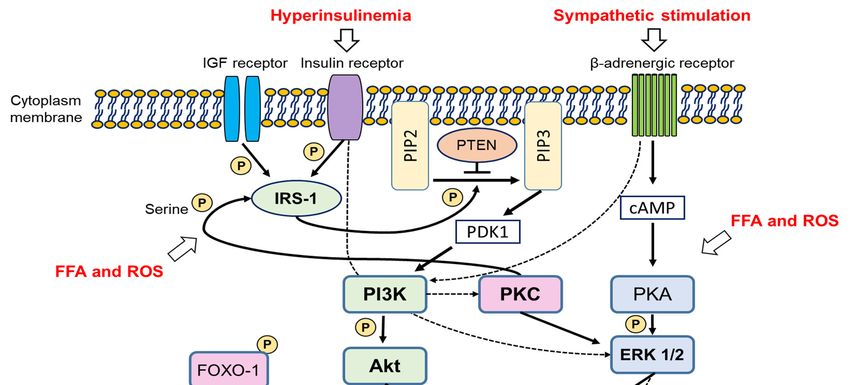
2.4.2. Molecular Views of the Pathogenesis of PCOS in the Animal PCOS Models
2.4.2.
We Molecular
found some Views of thepathways
common Pathogenesis of PCOS
related in the Animal
to PI3K-Akt PCOS
signalling Models
(Figure 2). PI3K-Akt signalling
is a crucial intracellular signalling for cellular metabolism,
We found some common pathways related to PI3K-Akt signalling (Figure proliferation, and survival2). and glucose
PI3K-Akt
homeostasis.
signalling isThus, thisintracellular
a crucial pathway is emphasized
signalling forin major metabolism,
cellular disorders, such as cancer,and
proliferation, muscular
survivalatrophy,
and
and diabetes
glucose [120–122].Thus,
homeostasis. In addition, ovarian
this pathway follicular development
is emphasized and recruitment
in major disorders, such as cancer, aremuscular
related to
thisatrophy,
conventional pathway
and diabetes [123]. Recently,
[120–122]. In addition, the ovarian
importance of this
follicular signalling in
development andtherecruitment
pathogenesis are of
related
PCOS wastoreviewed
this conventional pathway
[124]. Briefly, [123]. Recently,
the PI3K-Akt pathway theis importance
activated byofinsulin,
this signalling
IGF-1, andin other
the
pathogenesis
growth factors. ofWhen
PCOSinsulin
was reviewed
or IGF-1[124]. Briefly,
binds thereceptor,
to its PI3K-Aktinsulin
pathway is activated
receptor by insulin,
substrate-1 IGF- is
(IRS-1)
1, and other growth
phosphorylated. factors. WhenIRS-1
Phosphorylated insulin or IGF-1
recruits binds
PI3K, to its receptor,
which activatesinsulin
Akt, andreceptor substrate-1
the activated Akt
(IRS-1) is phosphorylated. Phosphorylated IRS-1 recruits PI3K, which activates Akt,
regulates the activation of several downstream proteins, including FOXO proteins, by phosphorylation. and the activated
TheAkt regulates the
phosphatase and activation of several
tensin homolog (PTEN) downstream
antagonizes proteins, includingand
Akt activation FOXO proteins, by of
phosphorylation
phosphorylation.
FOXO proteins. Among Thethe
phosphatase and tensin
FOXO proteins, homolog
upregulated FOXO1(PTEN) antagonizes
expression, which Akt activation
is mainly and
expressed
phosphorylation of FOXO proteins. Among the FOXO proteins, upregulated FOXO1
in the granulosa cells of the ovary, in cystic follicles and oxidative stress-induced apoptosis in the expression,
which is mainly expressed in the granulosa cells of the ovary, in cystic follicles and oxidative stress-
granulosa cells or rodents are observed [125,126].
induced apoptosis in the granulosa cells or rodents are observed [125,126].
Figure 2. Suggested and integrated molecular signalling possibly related to PCOS development.
Solid lines indicate direct effects, and dotted lines, indirect effects. PIP2 = phosphatidylinositol-
4,5-bisphosphate, = phosphatidylinositol-3,4,5-trisphosphate.
PIP3and
Figure 2. Suggested integrated molecular signalling possibly related to PCOS development. Solid
lines indicate direct effects, and dotted lines, indirect effects. PIP2 = phosphatidylinositol-4,5-
It is reasonablePIP3
bisphosphate, to postulate that over-activation of the PI3K-Akt pathway is the most potent
= phosphatidylinositol-3,4,5-trisphosphate.
reason for the development of PCOS-like phenotypes in the IGF-1 overexpressing mice and tPtenMT
mice. A It is reasonable
previous to postulate
study reportedthat
thatover-activation of the PI3K-Akt
alteration of PI3K-Akt pathway
signalling is the
in the mostespecially
ovary, potent
thereason
theca for theoccurred
cell, development of PCOS-like
in high-fat phenotypes
diet mice [127]; in the IGF-1
animal PCOSoverexpressing
models using mice
theand tPtenMT
high-fat diet
mice. A previous study reported that alteration of PI3K-Akt signalling in the ovary, especially the
theca cell, occurred in high-fat diet mice [127]; animal PCOS models using the high-fat diet methodYou can also read