Oncogenic and Tumor-Suppressive Functions of NOTCH Signaling in Glioma - MDPI
←
→
Page content transcription
If your browser does not render page correctly, please read the page content below
cells
Review
Oncogenic and Tumor-Suppressive Functions of
NOTCH Signaling in Glioma
Elena Parmigiani , Verdon Taylor and Claudio Giachino *
 Department of Biomedicine, University of Basel, Mattenstrasse 28, 4058 Basel, Switzerland;
 elena.parmigiani@unibas.ch (E.P.); verdon.taylor@unibas.ch (V.T.)
 * Correspondence: claudio.giachino@unibas.ch

 Received: 22 September 2020; Accepted: 14 October 2020; Published: 15 October 2020

 Abstract: Although the role of NOTCH signaling has been extensively studied in health and disease,
 many questions still remain unresolved. Being crucial for tissue homeostasis, NOTCH signaling is
 also implicated in multiple cancers by either promoting or suppressing tumor development. In this
 review we illustrate the context-dependent role of NOTCH signaling during tumorigenesis with a
 particular focus on gliomas, the most frequent and aggressive brain tumors in adults. For a long time,
 NOTCH has been considered an oncogene in glioma mainly by virtue of its neural stem cell-promoting
 activity. However, the recent identification of NOTCH-inactivating mutations in some glioma patients
 has challenged this notion, prompting a re-examination of the function of NOTCH in brain tumor
 subtypes. We discuss recent findings that might help to reconcile the controversial role of NOTCH
 signaling in this disease, and pose outstanding questions that still remain to be addressed.

 Keywords: NOTCH signaling; glioma; glioblastoma; brain tumor; oncogene; tumor suppressor;
 neural stem cells; CSL; RBPJ; ASCL1

1. Context-Dependent Roles of NOTCH Signaling in Cancer
     The NOTCH signaling pathway transduces short-range signals between adjacent cells and
therefore is highly dependent on niche architecture. In mammals, signal activation is induced by
direct interaction of one of the four NOTCH receptors (NOTCH1–4) with one of five canonical ligands
(Jagged1, Jagged2, Delta-like 1 (DLL1), DLL3, DLL4), followed by two sequential proteolytic cleavages
of the receptor by ADAM metalloproteases and the γ-secretase complex. Ligand-induced proteolysis
of NOTCH receptors liberates the NOTCH intracellular domain (NICD). The canonical signal links the
NICD to the nuclear CSL (CBF-1, Suppressor of Hairless, Lag-1; RBPJ in vertebrates) transcriptional
regulator [1], which is part of a repressor complex that includes NCoR/SMRT and histone deacetylases,
and is bound to the promoters of target genes. Upon NICD binding, the composition of the RBPJ
complex changes and co-activators including mastermind-like protein 1 (MAML-1) and histone
acetyltransferases (p300, CBP) are recruited to initiate gene transcription [2]. NOTCH controls the
expression of a wide range of target genes on the basis of the tissue and cell type that reflects its
multiple functions. The basic helix-loop-helix (bHLH) transcription factors of the HES/HEY family
were the first NOTCH target genes to be described [3]. They mainly function as transcriptional
repressors in heteromeric complexes with Transducin-Like Enhancer-of-split (TLE) proteins, and their
expression is in some cases finely tuned by autoregulatory loops that result in oscillatory expression
crucial to regulate, for example, stem cell maintenance and cell fate decision during embryonic
development of the nervous system [4]. An additional level of regulation, at least in the brain,
is exerted by a class of dominant negative regulators of bHLH proteins, the inhibitor of DNA-binding
(ID) proteins. IDs can form heterodimers with bHLH factors and, in most cases, counteract their
activity [5–8]. NICD/RBPJ can regulate the activity of gene promoters and play a fundamental role

Cells 2020, 9, 2304; doi:10.3390/cells9102304                                       www.mdpi.com/journal/cells
Cells 2020, 9, 2304 2 of 18
at distant long-range enhancers [9,10], as has been described in T cell acute lymphoblastic leukemia
(T-ALL) [11,12].
NOTCH signaling is highly conserved from invertebrates to humans and is involved in the
regulation of a variety of cellular processes throughout life, including cell proliferation, stem cell
maintenance, cell fate decisions, and differentiation. Accordingly, deregulation of NOTCH signaling
occurs in several human diseases, including cancer [1]. Due to its pleiotropic functions, NOTCH
signaling is implicated in many aspects of tumor development where it can act either as an oncogene
or a tumor suppressor, as extensively reviewed by others [13,14]. The balance between one or the other
role is determined by many factors, such as the cell type, the stage of tumor development, or the target
genes involved [13,14]. Genetic alterations in the genes of different NOTCH pathway components
that can lead to either enhanced or decreased NOTCH signaling have been identified in multiple
cancers [13–15]. Mutations that directly occur in NOTCH receptor genes can alter different portions of
the protein [15]. NOTCH activating mutations often occur in the negative regulatory region (NRR),
which regulates receptor cleavage, or in the PEST domain, which is involved in NICD degradation,
thus leading to ligand-independent or prolonged receptor activation [16–20]. In contrast, NOTCH
inactivating mutations predominantly occur in the epidermal growth factor (EGF)-like repeats of the
extracellular portion of the receptors. These mutations, including point mutations, focal deletions,
and nonsense and missense mutations, are predicted to prevent ligand binding or generate a truncated
protein and are often associated with downregulated expression of NOTCH target genes [21–26].
Historically, NOTCH signaling in cancer has been extensively studied in the context of T-ALL,
where NOTCH acts as an oncogene and NOTCH-activating mutations are present in more than 50%
of patients [17]. Different laboratories have identified translocations or mutation clusters within the
NOTCH1 receptor gene that significantly favor tumor progression by causing a ligand-independent
constitutive activation of the pathway [27]. Evidence that NOTCH can also promote tumor growth
in solid tumors comes, for example, from breast cancer. It has been demonstrated that integration of
the mouse mammary tumor virus (MMTV) causes rearrangement and activation of a particular locus
containing the Notch4 sequence and this ultimately results in cancer development [28,29]. Recurrent gene
rearrangements in NOTCH1 and NOTCH2, inducing NOTCH pathway activation, have also been
identified in human estrogen receptor (ER)-negative and triple-negative breast cancer samples and
cell lines [19]. In T-ALL and breast cancers, evidence points to c-Myc as an important effector of the
tumor-promoting function of NOTCH [17,19,30]. However, NOTCH could also cooperate with known
“cancer drivers” including breast cancer gene 1 (BRCA1), whose loss-of-function is responsible for the
majority of breast cancers [31]. Miao and colleagues (2020) have recently suggested that NOTCH1
activation can regulate the cell cycle and attenuate BRCA1 deficiency-induced G2/M blockade and
genomic instability, thus providing tumor cells with a survival advantage [31]. Moreover, it has
been proposed that NOTCH signaling could promote epithelial-to-mesenchymal transition (EMT),
enhancing tumor cell aggressiveness and metastatic potential [31,32].
The NOTCH pathway can also be tumor-suppressive in other contexts. The first evidence for
a tumor suppressor role of NOTCH came from studies in mouse and human keratinocytes [33,34].
Later, NOTCH-inactivating mutations were identified in head and neck squamous cell carcinoma
(HNSCC) [21,22] and small cell lung cancer (SCLC) [35–37]. In 2011, two independent whole-genome
sequencing studies reported NOTCH-inactivating mutations in two different cohorts of patients
with HNSCC [21,22]. These findings were further supported by a more recent and comprehensive
genomic characterization of HNSCC that found NOTCH mutations in approximately 20% of the
patients [38]. In SCLC, a highly aggressive and therapy-resistant lung cancer characterized by the
expression of neuroendocrine (NE) markers, a recent in vivo clonal analysis demonstrated that a
rare population of pulmonary NE stem cells could be induced to reactivate and differentiate upon
injury and that this process is regulated by NOTCH. However, when NOTCH signaling is inhibited,
the differentiation program is blocked and NE cells remain in a highly self-renewing state that is prone
to transformation [39]. NOTCH-inactivating mutations correlating with a poorer patient prognosisCells 2020, 9, 2304 3 of 18
have also been found in approximately 40% of patients with bladder cancer. Intriguingly, half of
those patients did not carry other concomitant mutations in well-known oncogenic drivers including
FGFR3 or RAS, suggesting a prominent role for NOTCH signaling in tumor initiation [23]. Moreover,
complete or partial loss of chromosome 9, where the NOTCH1 gene is located (9q34.3), is a common
chromosomal aberration in bladder carcinoma [40]. In these tumors, NOTCH acts as a suppressor of
cell proliferation by upregulating multiple members of the dual-specific phosphatase (DUSP) family,
which inhibit Extracellular signal-Regulated Kinase 1/2 (ERK1/2) phosphorylation. As a consequence,
NOTCH mutant tumor cells show increased ERK1/2 phosphorylation that can be reverted by NOTCH
activation [23].
Intriguingly, there is evidence that NOTCH signaling can play both tumor-promoting and
tumor-suppressive roles, even within the same organ. For instance, although the growth of HNSCC is
largely driven by NOTCH inactivation [41], occasional Notch gain-of-function mutations have been
reported in oral squamous cell carcinoma (OSCC) [42]. In the hematopoietic system, while an oncogenic
role of NOTCH has been described in both acute (T-ALL) and chronic (CLL) forms of lymphocytic
leukemia [17,18], a tumor-suppressive role has been proposed in chronic myelomonocytic leukemia
(CMML) [43] and also suggested in acute myeloid leukemia (AML) [44]. Such dualism has been linked to
the function of NOTCH in the regulation of cell fate choices during immune cell development. Multiple
in vitro and in vivo studies have demonstrated that NOTCH favors T cell over B cell commitment
and myeloid differentiation [45–47]. Consequently, NOTCH gain-of-function mutations lead to a
rapid and abnormal expansion of T cells at the expense of other cell lineages, whereas NOTCH
inactivation, particularly in the stromal compartment, causes an increase in myeloid progenitors and
granulocyte/macrophage descendants, resulting in myeloid hyperplasia and myeloproliferative-like
disease [43,48–51]. Hence, depending on the cell type, NOTCH signaling can play opposite roles in
the development of hematological malignancies. A dual role for NOTCH signaling is also evident in
some solid tumors, including lung cancer. The most prevalent form of lung tumors is non-small-cell
lung cancer (NSCLC), a heterogeneous group of neoplasms that includes lung adenocarcinoma and
squamous cell lung carcinoma in which NOTCH signaling activity has been suggested to promote and
suppress tumor growth, respectively [14,37,52–54]. However, perhaps the most emblematic example
of the fascinating complexity of how the NOTCH pathway can orchestrate tumor development is
given by SCLC, an infrequent but very aggressive subtype of lung cancer. Lim and colleagues (2017)
proposed that NOTCH signaling can simultaneously be oncogenic and tumor-suppressive in different
cell subpopulations of an individual tumor, although intratumoral heterogeneity generated by NOTCH
activity promotes overall SCLC growth [55]. The authors described the presence of two “symbiotic” cell
types: slowly-proliferating and chemoresistant non-NE cells and actively dividing NE cells. NOTCH
activation triggers a non-NE fate switch that slows tumor growth but also gives rise to non-NE cells
that sustain NE cell expansion by providing trophic support. Therefore, while NOTCH activation
delays initial tumor progression, it can provide a survival advantage after chemotherapy and fuel
rapid tumor relapse [55].
2. NOTCH Signaling in Neural Stem/Progenitor Cells and Glioma Formation
Gliomas account for approximately 30% of all brain tumors and around 80% of malignant brain
tumors [56]. They can be generically divided into diffuse low- and intermediate-grade II–III gliomas
(low-grade gliomas, LGGs) and the most aggressive grade IV gliomas or glioblastoma multiforme
(GBM). Although patients with LGGs have a more favorable prognosis, the diffuse infiltrative nature
of LGGs makes a complete neurosurgical resection almost impossible, often resulting in recurrence
and progression towards higher-grade gliomas [24]. Indeed, around 20% of GBMs are secondary
tumors arising from preexisting LGGs [26]. Recent advances in omics techniques have allowed for
the integration of clinical and histopathological data with a deep characterization of the genetic,
epigenetic, expression, and metabolic profiles of gliomas [57]. The scenario that has emerged is that of
a heterogeneous group of glioma subtypes, each characterized by different survival rates and responsesCells 2020, 9, 2304 4 of 18
to treatments. The importance of these findings has encouraged the World Health Organization to
include molecular parameters together with the classical histological markers in the diagnosis of
gliomas [58]. Given the broad heterogeneity within gliomas and the highly context-dependent roles of
NOTCH signaling in different cancers even within the same tumor type, it is perhaps not surprising
that the functions of NOTCH in glioma still remain controversial.
NOTCH activity is fundamental to maintain neural stem cell (NSC) and progenitor identity and
to regulate cell fate decisions in the developing and adult brain [4]. Since uncontrolled proliferation
and impaired differentiation of NSCs and glial progenitors may lead to glioma formation [59,60],
understanding how these processes are regulated would be important to target cancer-initiating
cells. After formation of the central nervous system is completed, NSCs persist within two restricted
regions of the mammalian brain, the subgranular zone of the hippocampal dentate gyrus (DG) and the
subventricular zone (SVZ) of the lateral ventricles [61,62]. Most adult NSCs are quiescent and only a
proportion of them divide at any given time to self-renew and generate intermediate progenitors (IPs).
IPs, in contrast, actively proliferate and differentiate mainly into neuroblasts, although glial cells can
also be generated in the adult NSC niches [63–65]. The entire process is finely tuned by a variety of
different mechanisms that ensure neuron generation and, at the same time, maintenance of a stem cell
pool. Within the neurogenic lineage, the canonical RBPJ-mediated NOTCH pathway and the NOTCH
target gene Hes5 are restricted to NSCs [66–69]. While there is evidence for adult stem and progenitor
heterogeneity, NOTCH-dependence seems to be a common feature among NSC populations [62].
Indeed, both quiescent and actively proliferating NSCs express Hes5, indicating NOTCH pathway
activation [70–72], and simultaneous Notch1 and Notch2 or Rbpj gene deletion in mice causes NSC
proliferation, differentiation, and exhaustion [67,68,73]. Interestingly, however, and in contrast to
global NOTCH pathway inhibition, blocking the function of individual NOTCH receptors differentially
affects active versus quiescent NSC populations, which seem to preferentially rely on NOTCH1 and
NOTCH2/3, respectively [73–77]. Recent data from our lab suggest that NOTCH2 can promote NSC
quiescence by inducing the expression of ID4 [77]. ID4 and NOTCH signaling synergize to inhibit
excessive accumulation of the proneural factor ASCL1 [77,78], which stimulates NSC proliferation
and differentiation [79,80]. While NOTCH target genes of the HES/HEY family repress ASCL1 gene
transcription [78], ID4 also facilitates the degradation of ASCL1 protein [81].
NOTCH signaling also regulates both the astrocyte and oligodendrocyte lineages.
During embryonic development, NOTCH favors the gliogenic switch of radial glial cells by driving the
expression of glial cell markers GFAP [82], BLBP [83], and NFIA [84]. The induction of an astrogliogenic
program also requires NOTCH signaling after ischemia in the adult brain [85]. NOTCH signaling
remains active in differentiated astrocytes but is modulated in response to injury. In striatal astrocytes,
NOTCH signaling is reduced after stroke, resulting in ectopic glial proliferation and production of
new neurons in non-neurogenic regions [86]. Reports indicate that NOTCH signaling also modulates
the fate of oligodendrocyte progenitor cells (OPCs), another important cell of origin of glioma [60].
Jagged1-induced canonical NOTCH signaling restricts oligodendrocyte maturation and maintains
OPCs during brain development and remyelination [87–89]. However, F3/Contactin can act as a
non-canonical NOTCH ligand to promote OPC differentiation in a developmental stage specific
manner [90].
Altogether, it is clear that NOTCH signaling plays a central role during homeostasis and
injury response in NSCs and glial progenitors, both of which are potential cells-of-origin of brain
tumors [60,91–94]. In line with this, NOTCH ligands, receptors, and downstream targets are expressed
in several types of brain tumor [95–100] and varying levels of NOTCH activity can contribute to
intra-tumor heterogeneity by promoting stem cell character in subpopulations of glioma cells [101,102].
Clearly, NOTCH signaling potentially regulates multiple steps of gliomagenesis, including tumor
initiation, progression, and recurrence. Yet, a consensus on the role of NOTCH in glioma development
is still missing.Cells 2020, 9, 2304 5 of 18
3. NOTCH as an Oncogene in Glioma
Evidence indicates that NOTCH signaling can promote glioma aggressiveness in some contexts
(Figure 1). The presence of self-renewing glioma stem cells (GSCs) that have increased DNA repair
capacity and expression of ATP-binding cassette (ABC) multidrug transporters, and that differentiate
into less-tumorigenic cancer cells that form the tumor bulk, is one phenomenon that can confer
therapy resistance in glioma [101,103–106]. Reminiscent of its role in healthy NSCs, NOTCH can
facilitate stem cell character in brain tumors and has therefore been considered a promising target
for the development of more effective glioma therapies. Data from both human tumors and murine
glioma models suggest that NOTCH signaling is preferentially active in subpopulations of glioma
cells [101,102,107]. In agreement with this, in vitro and xenotransplantation studies with glioma cell
lines have indicated that CD133-positive GSCs are particularly sensitive to γ-secretase inhibitors (GSI)
or NOTCH1/2 knockdown compared to CD133-negative glioma cells [108,109]. Blocking NOTCH
signaling or RBPJ reduced clonogenic potential in tumor-sphere assays and engraftment capacity in
glioma xenograft models [108–110]. Conversely NICD overexpression, although not sufficient alone to
induce brain tumors in the mouse brain [111,112], could increase cell survival due to radio-resistance
and side population phenotype in glioma cells [101,108,109]. Combinatorial treatment of GSIs and
radiation was more effective at inhibiting self-renewal than radiation alone [109,113], a synergistic effect
that was partially mediated by NOTCH enhancing Akt and Stat3 phosphorylation [108,109]. NOTCH
signaling could also promote a malignant phenotype in human glioma cell lines and xenograft models
by repressing the expression of the promyelocytic leukemia protein (PML) tumor suppressor [114]
and inducing the oncogenic long non-coding RNA TUG1 [115]. Reports have shown that knocking
down the NOTCH ligands Jagged1 or Dll1 by RNA interference reduced survival and growth of
tumor cells in multiple glioma cell lines [99,116] and high Jagged1 expression correlates with poor
prognosis of glioma patients [116]. Interestingly, the extracellular matrix glycoprotein Tenascin-C
and Jagged1 can reinforce each other’s expression, which could establish a feedback loop promoting
tumor growth [116–118]. However, it is important to note that high levels of Jagged1 can also inhibit
canonical NOTCH signaling in glioma cells, potentially through the activity of the Jagged1 intracellular
domain [119].
In the adult SVZ neurogenic niche, NOTCH signaling plays a central role in maintaining
the quiescent NSC pool [68,73], which is resistant to antimitotic treatment and can regenerate
more actively proliferating progenitor cells [120,121]. Similarly, a relatively quiescent subset of
chemotherapy-resistant glioma cells can propagate tumor growth after temozolomide administration in
mouse models of GBM [104]. In line with this finding, a recent report demonstrated that pharmacological
inhibition of receptor tyrosine kinases (RTK) prompts the emergence of slow-cycling and drug-tolerant
persister cells in a subset of PDGFRA-amplified human glioma cell lines, and that an increase in
NOTCH signaling activity allows transition to the persister state [122]. Although resistance to RTK
inhibition could also develop independent of NOTCH in some cell clones [123], these data indicate
that NOTCH can contribute to cell plasticity and drug tolerance in glioma by virtue of its stem
cell-promoting activity. In this context, NOTCH inhibition could be exploited to release high expression
of the proneural transcription factor ASCL1 in a subset of GSCs and induce their terminal neuronal
differentiation [124,125].
Adult SVZ NSCs reside in a specialized vascular niche [126,127] that can foster stem cell
character and repress differentiation through endothelial-derived factors that positively modulate
NOTCH-dependent transcription [66,128]. In analogy to the SVZ niche, it has been proposed that
GSCs reside in the proximity of blood vessels and are exposed to factors produced by endothelial
cells [129]. Among these, nitric oxide has been shown to activate NOTCH signaling and promote
stem-like character in PDGF-driven glioma [101]. Interestingly, NOTCH signaling is also augmented in
hypoxic tumor regions by Vasorin-mediated stabilization of NICD, and hypoxia-induced expression of
Vasorin promotes glioma aggressiveness [130]. Finally, there is evidence for dormant GSC populations
residing at the invasive front of the tumor [131], where Jagged1 expressed by nerve fibers could facilitateCells 2020, 9, 2304 6 of 18
NOTCH1+ CD133+ glioma cell invasion of white matter tracts through a SOX9-SOX2-NOTCH1 feedback
loop Cells
[132].2020, 9, x FOR PEER REVIEW 6 of 18
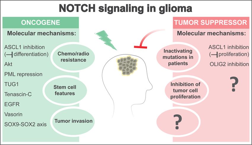
Figure Figure 1. The
1. The NOTCHNOTCH pathwaycan
pathway canact
act either
either as
as an
anoncogene
oncogeneoror as as
a tumor suppressor
a tumor in glioma,
suppressor in glioma,
depending on the context. On the one hand, NOTCH signaling activity in subpopulations of glioma
depending on the context. On the one hand, NOTCH signaling activity in subpopulations of glioma
cells can enhance stem cell features, promote resistance to radio- and chemo-therapies, and favor
cells can enhance stem cell features, promote resistance to radio- and chemo-therapies, and favor
tumor development by activating oncogenic pathways (e.g., PI3K/Akt) or inhibiting tumor
tumor development by activating oncogenic pathways (e.g., PI3K/Akt) or inhibiting tumor suppressors
suppressors (e.g., PML). NOTCH can also regulate long non-coding RNAs such as TUG1 to maintain
(e.g., stemness
PML). NOTCH can also
and suppress regulate long
differentiation. non-coding
Moreover, NOTCHRNAs such
signaling asestablish
can TUG1 to maintain
positive stemness
feedback
and suppress
loops withdifferentiation.
Tenascin-C, EGFR, Moreover, NOTCHaxis.
and a SOX9-SOX2 signaling
Finally,can
NOTCHestablish positive interactions
can modulate feedback loops
with between
Tenascin-C, EGFR,
glioma stemandcellsa SOX9-SOX2
(GSCs) and theiraxis.niche
Finally, NOTCHlocations
in different can modulate
within interactions
the tumor mass,between
glioma stem cells
including (GSCs)regions
in hypoxic and their niche Vasorin)
(through in different
and locations withinfront
at the invasive the (through
tumor mass, including in
SOX9-SOX2),
hypoxicthereby promoting
regions (through stem cell features
Vasorin) and and invasive
at the potential.
invasive On the other
front (through hand, NOTCH-inactivating
SOX9-SOX2), thereby promoting
stem cell features and invasive potential. On the other hand, NOTCH-inactivatingidentified
mutations and low expression levels of canonical NOTCH target genes have been mutations in and
patients with glioma subtypes, and genetic NOTCH inhibition accelerates glioma
low expression levels of canonical NOTCH target genes have been identified in patients with glioma formation in glioma
mouse models, pointing to a tumor-suppressive role of NOTCH in glioma similar to in epithelial
subtypes, and genetic NOTCH inhibition accelerates glioma formation in glioma mouse models,
cancers. Although the molecular mechanisms driving a tumor-suppressive role of NOTCH signaling
pointing to a tumor-suppressive role of NOTCH in glioma similar to in epithelial cancers. Although the
in glioma are still largely unknown, NOTCH can inhibit tumor cell proliferation and glioma growth
molecular mechanisms driving a tumor-suppressive role of NOTCH signaling in glioma are still
by suppressing ASCL1 and OLIG2 expression. Interestingly, NOTCH-mediated suppression of
largely unknown,
ASCL1 NOTCH
can result can
in either inhibit tumor
oncogenic cell proliferation
or tumor-suppressive and
effects byglioma growth
inhibiting by suppressing
differentiation or
ASCL1 and OLIG2 expression. Interestingly,
proliferation of glioma cells, respectively. NOTCH-mediated suppression of ASCL1 can result in
either oncogenic or tumor-suppressive effects by inhibiting differentiation or proliferation of glioma
4. NOTCH
cells, as a Tumor Suppressor in Glioma
respectively.
Surprisingly,
4. NOTCH as a Tumor recent data suggest
Suppressor that NOTCH signal inhibition may be an important molecular
in Glioma
event in the formation of some forms of glioma (Figure 1). Genome-wide analyses in patients with
Surprisingly,
LGG identifiedrecent datainsuggest
mutations NOTCHthat NOTCH
signaling signal inhibition
components may be
in a significant an important
proportion molecular
of isocitrate
eventdehydrogenase
in the formation
(IDH) mutant tumors [24–26,133]. These mutations are particularly frequent in the with
of some forms of glioma (Figure 1). Genome-wide analyses in patients
LGGgenes
identified mutations
encoding the NOTCH1in NOTCH signaling
and NOTCH2 components
receptors but less in a significant
common proportion
in NOTCH3/4, of isocitrate
and occur at
similar positions
dehydrogenase (IDH) to those
mutant demonstrated experimentally
tumors [24–26,133]. to inactivate
These mutations NOTCH1 function infrequent
are particularly epithelialin the
genescancers [24–26,133].
encoding the NOTCH1In addition, less frequent
and NOTCH2 mutations
receptors butinless
thecommon
RBPJ gene inwere detected and
NOTCH3/4, in LGG
occur at
samples and were mutually exclusive with NOTCH1 mutations [134]. Accordingly, the expression of
similar positions to those demonstrated experimentally to inactivate NOTCH1 function in epithelial
some NOTCH target genes is downregulated in NOTCH/RBPJ mutant gliomas, indicating pathway
cancers [24–26,133]. In addition, less frequent mutations in the RBPJ gene were detected in LGG
inactivation [24,26,134]. Expression of the proliferation marker MKI67 was found to be augmented in
samples and were mutually exclusive with NOTCH1 mutations [134]. Accordingly, the expression of
NOTCH1/RBPJ mutant brain tumors with low HES/HEY expression levels [134], and NOTCH
somemutations
NOTCH weretargeta genes is downregulated
high-risk NOTCH/RBPJ
factor associatedinwith shorter patientmutant gliomas,
survival [135], indicating
suggesting pathway
that
inactivation [24,26,134]. Expression of the proliferation marker MKI67 was
NOTCH signaling inhibition contributes to increased glioma aggressiveness. Consistentfound to be augmented
with this, in
NOTCH1/RBPJ mutant brain tumors with low HES/HEY expression levels [134], and NOTCH mutationsCells 2020, 9, 2304 7 of 18
were a high-risk factor associated with shorter patient survival [135], suggesting that NOTCH signaling
inhibition contributes to increased glioma aggressiveness. Consistent with this, frequent NOTCH
pathway alterations were also detected when comparing progressed tumors with their corresponding
lower-grade counterparts [26]. Although occasional NOTCH1/2 mutations could also be detected in IDH
mutant astrocytomas with TP53 inactivation [25], genetic inactivation of NOTCH pathway components
mainly occurs in oligodendrogliomas with co-deletion of chromosome arms 1p and 19q [24,25]. This is
intriguing, as the genes for NOTCH2, MIB2, and the NOTCH targets HES2-5 and HEYL are all
located on chromosome arm 1p [136,137], lending support to the hypothesis that NOTCH signaling
inhibition contributes to the initiation of tumors with oligodendroglial characteristics. Interestingly,
genetic inhibition of NOTCH can promote OPC proliferation and expansion in mouse models [138],
and excessive activation of quiescent OPC populations leads to malignant transformation [139].
In contrast to LGG, NOTCH mutations are very rare in GBM. However, it is worth noting that
levels of NOTCH signaling activation substantially vary among GBM samples and subtypes [98,107],
and hemizygous or homozygous deletions of the 1p36 locus, which could affect the MIB2 and HES2-5
genes, occur in a proportion of GBMs [140,141]. Although a number of genes have been proposed
as 1p36 candidate tumor suppressors, it is unclear if NOTCH inhibition can contribute to cancer
development after 1p36 loss.
Indirect evidence that NOTCH signaling could have tumor suppressor activity in glioma
comes from studies on the proneural transcription factor ASCL1, whose expression is normally
repressed by canonical NOTCH targets of the HES/HEY family in NSCs and GSCs [78,107,124,142].
Lineage tracing experiments in mice have shown that ASCL1+ neural progenitors can be cells of origin of
GBM [59,91] and upregulation of ASCL1 and inhibition of NOTCH signaling characterize astrocytoma
progression [143]. ASCL1 expression is maintained in both xenografts from human proneural
GBM samples and GBM mouse models, and ASCL1 can induce cell cycle genes and oncogenes,
thereby promoting glioma cell proliferation in some contexts [122,144–146]. Accordingly, ASCL1
knockdown or conditional gene knockout prolong survival of glioma-bearing mice [145,146]. In contrast,
high expression of a curated NOTCH-signaling gene set, including HES/HEY transcriptional repressors,
correlates with less proliferative glioma cell subpopulations with putative GSC character [122].
These data are in line with the role of NOTCH signaling in reducing ASCL1 expression and preserving
quiescence in NSCs [68,78,79,86,107].
More direct evidence that NOTCH signaling could restrict glioma formation in some contexts
comes from studies taking advantage of genetic conditional gene deletion or overexpression approaches
in vivo. NOTCH2 overexpression can inhibit glioma formation in mouse glioma models [107] and
HEY2 overexpression can reduce the proliferation of murine and human glioma cells [147]. Conversely,
ID2-mediated repression of HEY1 promotes NSC transformation, possibly by releasing the expression
of OLIG2 [148], an oligodendroglial lineage determinant involved in glioma cell proliferation [149].
Simultaneous genetic deletion of Notch1 and Notch2 or Rbpj accelerates the growth of PDGF-driven
GBMs in mice [107]. Moreover, inactivation of the Rbpj gene together with the tumor suppressor gene
Trp53 induces aggressive, de novo forebrain tumors with primitive neuroectodermal features [107].
These findings are in line with an unbiased in vivo CRISPR screen that recently identified NOTCH1
among potential tumor suppressors in GBM [150] and with the observation that high expression of
specific NOTCH target genes positively correlates with a better prognosis in defined subsets of LGGs
and GBMs in humans [107].
5. Open Questions and Perspectives
NOTCH receptors and their downstream targets are potential candidates for specific drug targeting,
and various strategies to modulate NOTCH for cancer therapy are being actively pursued [151].
However, understanding the molecular basis of NOTCH oncogenic and tumor-suppressive functions
is fundamental in order to develop effective strategies to therapeutically modulate NOTCH in glioma.
Although clinical trials with GSIs in patients with glioma have been reported, positive effects have onlyCells 2020, 9, 2304 8 of 18
been seen in one study [151,152]. Given the broad spectrum of gamma-secretase target proteins and the
dose-limiting toxicities of GSIs, it would be interesting to test the effects of small molecules that directly
target the NOTCH transcriptional activation complex in glioma [153–155]. Considering the extensive
intertumoral heterogeneity in glioma, it is possible that NOTCH inhibition would be beneficial only in
a proportion of molecularly selected patients in a personalized therapy. For instance, studies in vitro
suggest that PTEN and TP53 status may affect sensitivity to GSIs in GBM [156,157]. NOTCH and EGF
receptor pathways can potentiate each other in glioma cells [158–160] and, therefore, the oncogenic
function of NOTCH signaling could be more apparent in primary GBMs of the classical subtype [161]
than in GBMs with OPC-like proneural features [162] or IDH mutant LGGs [24,25]. The precise role of
NOTCH in different forms of glioma requires further studies.
To some extent, the strength of NOTCH signaling might also explain the reported discrepancies in
the oncogenic versus tumor-suppressive functions of NOTCH in the brain. NOTCH signaling is required
to maintain a low and oscillatory ASCL1 expression in order to promote NSC self-renewal [6,78].
However, sustained NOTCH signaling activation induces proliferative quiescence of NSCs [78].
Conversely, NOTCH inhibition induces activation of latent parenchymal progenitors [86] and NSC
hyperproliferation before resulting in neuronal differentiation and NSC depletion [78]. Reminiscent of
this, NOTCH inhibition could induce terminal differentiation of glioma cells in some contexts [124] but
facilitate their proliferation in others [107]. NOTCH inhibition can contribute to the initiation of epithelial
cancers by favoring the expansion of NOTCH mutant clones at the expense of wild-type cells [163].
Since analogous cell competition mechanisms play important roles during cancer progression [164],
it would be interesting to determine if NOTCH-regulated competitive interactions occur between
adjacent neural progenitors and if NOTCH mutations can contribute to glioma formation in this context.
Reversible NOTCH signal activation likely plays a role in regulating glioma cell behavior during
tumor development, but NOTCH-independent clones may arise under prolonged drug selection
pressure [123]. Hence, it will also be important to address if the role of NOTCH in glioma varies on the
basis of the stage of disease progression, as suggested for SCLC [55].
Finally, indication for a tumor-suppressive activity of the NOTCH pathway in glioma
predominantly comes from an in vivo immunocompetent setting, and a recent CRISPR screen identified
frequent co-mutation of the NOTCH1 receptor and B2m, an essential component of the MHC-I antigen
presentation complex [150]. Interestingly, findings indicate that the immune response to cancer in the
brain is shaped by the cancer type [165,166]. Whether NOTCH activity in tumor cells can regulate
interactions with the glioma microenvironment and immune evasion remains unexplored.
6. Concluding Remarks
NOTCH signaling can act as an oncogene or a tumor suppressor, depending on the context [1].
Although it is clear that NOTCH plays central roles in glioma, its precise function has remained
puzzling. The data reviewed here support the hypothesis of a dual role of the NOTCH pathway
as an oncogene and a tumor suppressor in glioma (Figure 1), similar to what has been suggested
in some other malignancies. Human gliomas comprise multiple disease subtypes that differ at the
genetic, epigenetic, and transcriptional levels, and this intertumoral heterogeneity could be one
critical factor underlying the observed discrepancies in NOTCH function. In addition, differences
in the outcome of NOTCH modulation likely relate to the stage of disease progression, crosstalk
with other signaling pathways, and intratumoral (stem) cell heterogeneity. While diverse molecular
bases of NOTCH oncogenic function in glioma have been addressed in previous studies, mechanistic
data on the NOTCH tumor-suppressive activity in brain tumor subtypes are still lacking. It is also
unclear if NOTCH-regulated cell competition mechanisms are in place during glioma initiation and
progression. Finally, whether NOTCH activity in glioma cells regulates the crosstalk with the tumor
microenvironment and immune cells in particular remains unexplored. Clearly, the multiple tasks of
NOTCH signaling in glioma deserve further scrutiny.Cells 2020, 9, 2304 9 of 18
Author Contributions: All authors contributed to manuscript conceptualization and preparation. All authors
have read and agreed to the published version of the manuscript.
Funding: Swiss Cancer League (KLS-4518-08-2018), Stiftung zur Krebsbekämpfung, Huggenberger-Bischoff
Stiftung zur Krebsforschung, Stiftung für krebskranke Kinder Regio Basiliensis, and Forschungsfonds
Nachwuchsforschende of the University of Basel.
Acknowledgments: We thank the members of the Taylor lab for critical reading of the manuscript and for
helpful discussions.
Conflicts of Interest: The authors declare no conflict of interest.
References
1. Siebel, C.; Lendahl, U. Notch Signaling in Development, Tissue Homeostasis, and Disease. Physiol. Rev. 2017,
97, 1235–1294. [CrossRef] [PubMed]
2. Kopan, R.; Ilagan, M.X. The canonical Notch signaling pathway: Unfolding the activation mechanism. Cell
2009, 137, 216–233. [CrossRef] [PubMed]
3. Fischer, A.; Gessler, M. Delta-Notch–and then? Protein interactions and proposed modes of repression by
Hes and Hey bHLH factors. Nucleic. Acids. Res. 2007, 35, 4583–4596. [CrossRef] [PubMed]
4. Pierfelice, T.; Alberi, L.; Gaiano, N. Notch in the vertebrate nervous system: An old dog with new tricks.
Neuron 2011, 69, 840–855. [CrossRef] [PubMed]
5. Boareto, M.; Iber, D.; Taylor, V. Differential interactions between Notch and ID factors control neurogenesis
by modulating Hes factor autoregulation. Development 2017, 144, 3465–3474. [CrossRef]
6. Imayoshi, I.; Isomura, A.; Harima, Y.; Kawaguchi, K.; Kori, H.; Miyachi, H.; Fujiwara, T.; Ishidate, F.;
Kageyama, R. Oscillatory control of factors determining multipotency and fate in mouse neural progenitors.
Science 2013, 342, 1203–1208. [CrossRef]
7. Bai, G.; Sheng, N.; Xie, Z.; Bian, W.; Yokota, Y.; Benezra, R.; Kageyama, R.; Guillemot, F.; Jing, N. Id sustains
Hes1 expression to inhibit precocious neurogenesis by releasing negative autoregulation of Hes1. Dev. Cell
2007, 13, 283–297. [CrossRef]
8. Nam, H.S.; Benezra, R. High levels of Id1 expression define B1 type adult neural stem cells. Cell Stem. Cell
2009, 5, 515–526. [CrossRef]
9. Castel, D.; Mourikis, P.; Bartels, S.J.; Brinkman, A.B.; Tajbakhsh, S.; Stunnenberg, H.G. Dynamic binding of
RBPJ is determined by Notch signaling status. Genes Dev. 2013, 27, 1059–1071. [CrossRef]
10. Wang, H.; Zou, J.; Zhao, B.; Johannsen, E.; Ashworth, T.; Wong, H.; Pear, W.S.; Schug, J.; Blacklow, S.C.;
Arnett, K.L.; et al. Genome-wide analysis reveals conserved and divergent features of Notch1/RBPJ binding
in human and murine T-lymphoblastic leukemia cells. Proc. Natl. Acad. Sci. USA 2011, 108, 14908–14913.
[CrossRef]
11. Herranz, D.; Ambesi-Impiombato, A.; Palomero, T.; Schnell, S.A.; Belver, L.; Wendorff, A.A.; Xu, L.;
Castillo-Martin, M.; Llobet-Navas, D.; Cordon-Cardo, C.; et al. A NOTCH1-driven MYC enhancer promotes
T cell development, transformation and acute lymphoblastic leukemia. Nat. Med. 2014, 20, 1130–1137.
[CrossRef] [PubMed]
12. Yashiro-Ohtani, Y.; Wang, H.; Zang, C.; Arnett, K.L.; Bailis, W.; Ho, Y.; Knoechel, B.; Lanauze, C.; Louis, L.;
Forsyth, K.S.; et al. Long-range enhancer activity determines Myc sensitivity to Notch inhibitors in T cell
leukemia. Proc. Natl. Acad. Sci. USA 2014, 111, E4946–E4953. [CrossRef] [PubMed]
13. Nowell, C.S.; Radtke, F. Notch as a tumour suppressor. Nat. Rev. Cancer 2017, 17, 145–159. [CrossRef]
[PubMed]
14. Ntziachristos, P.; Lim, J.S.; Sage, J.; Aifantis, I. From fly wings to targeted cancer therapies: A centennial for
notch signaling. Cancer Cell 2014, 25, 318–334. [CrossRef] [PubMed]
15. Egloff, A.M.; Grandis, J.R. Molecular pathways: Context-dependent approaches to Notch targeting as cancer
therapy. Clin. Cancer Res. 2012, 18, 5188–5195. [CrossRef]
16. Ellisen, L.W.; Bird, J.; West, D.C.; Soreng, A.L.; Reynolds, T.C.; Smith, S.D.; Sklar, J. TAN-1, the human homolog
of the Drosophila notch gene, is broken by chromosomal translocations in T lymphoblastic neoplasms. Cell
1991, 66, 649–661. [CrossRef]Cells 2020, 9, 2304 10 of 18
17. Weng, A.P.; Ferrando, A.A.; Lee, W.; Morris, J.P.T.; Silverman, L.B.; Sanchez-Irizarry, C.; Blacklow, S.C.;
Look, A.T.; Aster, J.C. Activating mutations of NOTCH1 in human T cell acute lymphoblastic leukemia.
Science 2004, 306, 269–271. [CrossRef]
18. Puente, X.S.; Pinyol, M.; Quesada, V.; Conde, L.; Ordonez, G.R.; Villamor, N.; Escaramis, G.; Jares, P.; Bea, S.;
Gonzalez-Diaz, M.; et al. Whole-genome sequencing identifies recurrent mutations in chronic lymphocytic
leukaemia. Nature 2011, 475, 101–105. [CrossRef]
19. Robinson, D.R.; Kalyana-Sundaram, S.; Wu, Y.M.; Shankar, S.; Cao, X.; Ateeq, B.; Asangani, I.A.; Iyer, M.;
Maher, C.A.; Grasso, C.S.; et al. Functionally recurrent rearrangements of the MAST kinase and Notch gene
families in breast cancer. Nat. Med. 2011, 17, 1646–1651. [CrossRef] [PubMed]
20. Wang, K.; Zhang, Q.; Li, D.; Ching, K.; Zhang, C.; Zheng, X.; Ozeck, M.; Shi, S.; Li, X.; Wang, H.; et al.
PEST domain mutations in Notch receptors comprise an oncogenic driver segment in triple-negative breast
cancer sensitive to a gamma-secretase inhibitor. Clin. Cancer Res. 2015, 21, 1487–1496. [CrossRef] [PubMed]
21. Agrawal, N.; Frederick, M.J.; Pickering, C.R.; Bettegowda, C.; Chang, K.; Li, R.J.; Fakhry, C.; Xie, T.X.;
Zhang, J.; Wang, J.; et al. Exome sequencing of head and neck squamous cell carcinoma reveals inactivating
mutations in NOTCH1. Science 2011, 333, 1154–1157. [CrossRef] [PubMed]
22. Stransky, N.; Egloff, A.M.; Tward, A.D.; Kostic, A.D.; Cibulskis, K.; Sivachenko, A.; Kryukov, G.V.;
Lawrence, M.S.; Sougnez, C.; McKenna, A.; et al. The mutational landscape of head and neck squamous cell
carcinoma. Science 2011, 333, 1157–1160. [CrossRef] [PubMed]
23. Rampias, T.; Vgenopoulou, P.; Avgeris, M.; Polyzos, A.; Stravodimos, K.; Valavanis, C.; Scorilas, A.; Klinakis, A.
A new tumor suppressor role for the Notch pathway in bladder cancer. Nat. Med. 2014, 20, 1199–1205.
[CrossRef] [PubMed]
24. Brat, D.J.; Verhaak, R.G.; Aldape, K.D.; Yung, W.K.; Salama, S.R.; Cooper, L.A.; Rheinbay, E.; Miller, C.R.;
Vitucci, M.; Morozova, O.; et al. Comprehensive, Integrative Genomic Analysis of Diffuse Lower-Grade
Gliomas. N. Engl. J. Med. 2015, 372, 2481–2498. [CrossRef] [PubMed]
25. Suzuki, H.; Aoki, K.; Chiba, K.; Sato, Y.; Shiozawa, Y.; Shiraishi, Y.; Shimamura, T.; Niida, A.; Motomura, K.;
Ohka, F.; et al. Mutational landscape and clonal architecture in grade II and III gliomas. Nat. Genet. 2015,
47, 458–468. [CrossRef] [PubMed]
26. Bai, H.; Harmanci, A.S.; Erson-Omay, E.Z.; Li, J.; Coskun, S.; Simon, M.; Krischek, B.; Ozduman, K.; Omay, S.B.;
Sorensen, E.A.; et al. Integrated genomic characterization of IDH1-mutant glioma malignant progression.
Nat. Genet. 2016, 48, 59–66. [CrossRef]
27. Radtke, F.; Fasnacht, N.; Macdonald, H.R. Notch signaling in the immune system. Immunity 2010, 32, 14–27.
[CrossRef] [PubMed]
28. Jhappan, C.; Gallahan, D.; Stahle, C.; Chu, E.; Smith, G.H.; Merlino, G.; Callahan, R. Expression of an activated
Notch-related int-3 transgene interferes with cell differentiation and induces neoplastic transformation in
mammary and salivary glands. Genes Dev. 1992, 6, 345–355. [CrossRef]
29. Robbins, J.; Blondel, B.J.; Gallahan, D.; Callahan, R. Mouse mammary tumor gene int-3: A member of the
notch gene family transforms mammary epithelial cells. J. Virol. 1992, 66, 2594–2599. [CrossRef]
30. Klinakis, A.; Szabolcs, M.; Politi, K.; Kiaris, H.; Artavanis-Tsakonas, S.; Efstratiadis, A. Myc is a Notch1
transcriptional target and a requisite for Notch1-induced mammary tumorigenesis in mice. Proc. Natl. Acad.
Sci. USA 2006, 103, 9262–9267. [CrossRef]
31. Miao, K.; Lei, J.H.; Valecha, M.V.; Zhang, A.; Xu, J.; Wang, L.; Lyu, X.; Chen, S.; Miao, Z.; Zhang, X.; et al.
NOTCH1 activation compensates BRCA1 deficiency and promotes triple-negative breast cancer formation.
Nat. Commun. 2020, 11, 3256. [CrossRef] [PubMed]
32. Wang, Z.; Li, Y.; Kong, D.; Sarkar, F.H. The role of Notch signaling pathway in epithelial-mesenchymal
transition (EMT) during development and tumor aggressiveness. Curr. Drug Targets 2010, 11, 745–751.
[CrossRef]
33. Lefort, K.; Mandinova, A.; Ostano, P.; Kolev, V.; Calpini, V.; Kolfschoten, I.; Devgan, V.; Lieb, J.; Raffoul, W.;
Hohl, D.; et al. Notch1 is a p53 target gene involved in human keratinocyte tumor suppression through
negative regulation of ROCK1/2 and MRCKalpha kinases. Genes Dev. 2007, 21, 562–577. [CrossRef]
34. Nicolas, M.; Wolfer, A.; Raj, K.; Kummer, J.A.; Mill, P.; van Noort, M.; Hui, C.C.; Clevers, H.; Dotto, G.P.;
Radtke, F. Notch1 functions as a tumor suppressor in mouse skin. Nat. Genet. 2003, 33, 416–421. [CrossRef]Cells 2020, 9, 2304 11 of 18
35. George, J.; Lim, J.S.; Jang, S.J.; Cun, Y.; Ozretic, L.; Kong, G.; Leenders, F.; Lu, X.; Fernandez-Cuesta, L.;
Bosco, G.; et al. Comprehensive genomic profiles of small cell lung cancer. Nature 2015, 524, 47–53. [CrossRef]
[PubMed]
36. Sriuranpong, V.; Borges, M.W.; Ravi, R.K.; Arnold, D.R.; Nelkin, B.D.; Baylin, S.B.; Ball, D.W. Notch signaling
induces cell cycle arrest in small cell lung cancer cells. Cancer Res. 2001, 61, 3200–3205. [PubMed]
37. Wang, N.J.; Sanborn, Z.; Arnett, K.L.; Bayston, L.J.; Liao, W.; Proby, C.M.; Leigh, I.M.; Collisson, E.A.;
Gordon, P.B.; Jakkula, L.; et al. Loss-of-function mutations in Notch receptors in cutaneous and lung
squamous cell carcinoma. Proc. Natl. Acad. Sci. USA 2011, 108, 17761–17766. [CrossRef]
38. Cancer Genome Atlas Network. Comprehensive genomic characterization of head and neck squamous cell
carcinomas. Nature 2015, 517, 576–582. [CrossRef]
39. Ouadah, Y.; Rojas, E.R.; Riordan, D.P.; Capostagno, S.; Kuo, C.S.; Krasnow, M.A. Rare Pulmonary
Neuroendocrine Cells Are Stem Cells Regulated by Rb, p53, and Notch. Cell 2019, 179, 403–416.e23.
[CrossRef]
40. Kimura, F.; Florl, A.R.; Seifert, H.H.; Louhelainen, J.; Maas, S.; Knowles, M.A.; Schulz, W.A. Destabilization
of chromosome 9 in transitional cell carcinoma of the urinary bladder. Br. J. Cancer 2001, 85, 1887–1893.
[CrossRef]
41. Loganathan, S.K.; Schleicher, K.; Malik, A.; Quevedo, R.; Langille, E.; Teng, K.; Oh, R.H.; Rathod, B.; Tsai, R.;
Samavarchi-Tehrani, P.; et al. Rare driver mutations in head and neck squamous cell carcinomas converge on
NOTCH signaling. Science 2020, 367, 1264–1269. [CrossRef] [PubMed]
42. Song, X.; Xia, R.; Li, J.; Long, Z.; Ren, H.; Chen, W.; Mao, L. Common and complex Notch1 mutations in
Chinese oral squamous cell carcinoma. Clin. Cancer Res. 2014, 20, 701–710. [CrossRef] [PubMed]
43. Klinakis, A.; Lobry, C.; Abdel-Wahab, O.; Oh, P.; Haeno, H.; Buonamici, S.; van De Walle, I.; Cathelin, S.;
Trimarchi, T.; Araldi, E.; et al. A novel tumour-suppressor function for the Notch pathway in myeloid
leukaemia. Nature 2011, 473, 230–233. [CrossRef] [PubMed]
44. Lobry, C.; Ntziachristos, P.; Ndiaye-Lobry, D.; Oh, P.; Cimmino, L.; Zhu, N.; Araldi, E.; Hu, W.; Freund, J.;
Abdel-Wahab, O.; et al. Notch pathway activation targets AML-initiating cell homeostasis and differentiation.
J. Exp. Med. 2013, 210, 301–319. [CrossRef] [PubMed]
45. Bell, J.J.; Bhandoola, A. The earliest thymic progenitors for T cells possess myeloid lineage potential. Nature
2008, 452, 764–767. [CrossRef]
46. Feyerabend, T.B.; Terszowski, G.; Tietz, A.; Blum, C.; Luche, H.; Gossler, A.; Gale, N.W.; Radtke, F.;
Fehling, H.J.; Rodewald, H.R. Deletion of Notch1 converts pro-T cells to dendritic cells and promotes thymic
B cells by cell-extrinsic and cell-intrinsic mechanisms. Immunity 2009, 30, 67–79. [CrossRef]
47. Wada, H.; Masuda, K.; Satoh, R.; Kakugawa, K.; Ikawa, T.; Katsura, Y.; Kawamoto, H. Adult T-cell progenitors
retain myeloid potential. Nature 2008, 452, 768–772. [CrossRef]
48. Kim, Y.W.; Koo, B.K.; Jeong, H.W.; Yoon, M.J.; Song, R.; Shin, J.; Jeong, D.C.; Kim, S.H.; Kong, Y.Y. Defective
Notch activation in microenvironment leads to myeloproliferative disease. Blood 2008, 112, 4628–4638.
[CrossRef]
49. Wang, L.; Zhang, H.; Rodriguez, S.; Cao, L.; Parish, J.; Mumaw, C.; Zollman, A.; Kamoka, M.M.; Mu, J.;
Chen, D.Z.; et al. Notch-dependent repression of miR-155 in the bone marrow niche regulates hematopoiesis
in an NF-kappaB-dependent manner. Cell Stem. Cell 2014, 15, 51–65. [CrossRef]
50. Yao, D.; Huang, Y.; Huang, X.; Wang, W.; Yan, Q.; Wei, L.; Xin, W.; Gerson, S.; Stanley, P.; Lowe, J.B.; et al.
Protein O-fucosyltransferase 1 (Pofut1) regulates lymphoid and myeloid homeostasis through modulation of
Notch receptor ligand interactions. Blood 2011, 117, 5652–5662. [CrossRef]
51. Yoda, M.; Kimura, T.; Tohmonda, T.; Uchikawa, S.; Koba, T.; Takito, J.; Morioka, H.; Matsumoto, M.;
Link, D.C.; Chiba, K.; et al. Dual functions of cell-autonomous and non-cell-autonomous ADAM10 activity
in granulopoiesis. Blood 2011, 118, 6939–6942. [CrossRef] [PubMed]
52. Licciulli, S.; Avila, J.L.; Hanlon, L.; Troutman, S.; Cesaroni, M.; Kota, S.; Keith, B.; Simon, M.C.; Puré, E.;
Radtke, F.; et al. Notch1 is required for Kras-induced lung adenocarcinoma and controls tumor cell survival
via p53. Cancer Res. 2013, 73, 5974–5984. [CrossRef] [PubMed]
53. Westhoff, B.; Colaluca, I.N.; D’Ario, G.; Donzelli, M.; Tosoni, D.; Volorio, S.; Pelosi, G.; Spaggiari, L.;
Mazzarol, G.; Viale, G.; et al. Alterations of the Notch pathway in lung cancer. Proc. Natl. Acad. Sci. USA
2009, 106, 22293–22298. [CrossRef] [PubMed]Cells 2020, 9, 2304 12 of 18
54. Zheng, Y.; de la Cruz, C.C.; Sayles, L.C.; Alleyne-Chin, C.; Vaka, D.; Knaak, T.D.; Bigos, M.; Xu, Y.; Hoang, C.D.;
Shrager, J.B.; et al. A rare population of CD24(+)ITGB4(+)Notch(hi) cells drives tumor propagation in
NSCLC and requires Notch3 for self-renewal. Cancer Cell 2013, 24, 59–74. [CrossRef] [PubMed]
55. Lim, J.S.; Ibaseta, A.; Fischer, M.M.; Cancilla, B.; O’Young, G.; Cristea, S.; Luca, V.C.; Yang, D.; Jahchan, N.S.;
Hamard, C.; et al. Intratumoural heterogeneity generated by Notch signalling promotes small-cell lung
cancer. Nature 2017, 545, 360–364. [CrossRef] [PubMed]
56. Ostrom, Q.T.; Bauchet, L.; Davis, F.G.; Deltour, I.; Fisher, J.L.; Langer, C.E.; Pekmezci, M.; Schwartzbaum, J.A.;
Turner, M.C.; Walsh, K.M.; et al. The epidemiology of glioma in adults: A “state of the science” review.
Neuro. Oncol. 2014, 16, 896–913. [CrossRef]
57. Reifenberger, G.; Wirsching, H.G.; Knobbe-Thomsen, C.B.; Weller, M. Advances in the molecular genetics of
gliomas—Implications for classification and therapy. Nat. Rev. Clin. Oncol. 2017, 14, 434–452. [CrossRef]
[PubMed]
58. Louis, D.N.; Perry, A.; Reifenberger, G.; von Deimling, A.; Figarella-Branger, D.; Cavenee, W.K.; Ohgaki, H.;
Wiestler, O.D.; Kleihues, P.; Ellison, D.W. The 2016 World Health Organization Classification of Tumors of
the Central Nervous System: A summary. Acta Neuropathol. 2016, 131, 803–820. [CrossRef]
59. Alcantara Llaguno, S.; Sun, D.; Pedraza, A.M.; Vera, E.; Wang, Z.; Burns, D.K.; Parada, L.F. Cell-of-origin
susceptibility to glioblastoma formation declines with neural lineage restriction. Nat. Neurosci. 2019,
22, 545–555. [CrossRef]
60. Liu, C.; Sage, J.C.; Miller, M.R.; Verhaak, R.G.; Hippenmeyer, S.; Vogel, H.; Foreman, O.; Bronson, R.T.;
Nishiyama, A.; Luo, L.; et al. Mosaic analysis with double markers reveals tumor cell of origin in glioma.
Cell 2011, 146, 209–221. [CrossRef]
61. Bond, A.M.; Ming, G.L.; Song, H. Adult Mammalian Neural Stem Cells and Neurogenesis: Five Decades
Later. Cell Stem. Cell 2015, 17, 385–395. [CrossRef] [PubMed]
62. Giachino, C.; Taylor, V. Notching up neural stem cell homogeneity in homeostasis and disease. Front. Neurosci.
2014, 8, 32. [CrossRef] [PubMed]
63. Gage, F.H.; Temple, S. Neural stem cells: Generating and regenerating the brain. Neuron 2013, 80, 588–601.
[CrossRef] [PubMed]
64. Gotz, M.; Nakafuku, M.; Petrik, D. Neurogenesis in the Developing and Adult Brain-Similarities and Key
Differences. Cold Spring Harb. Perspect Biol. 2016, 8. [CrossRef]
65. Navarro Negredo, P.; Yeo, R.W.; Brunet, A. Aging and Rejuvenation of Neural Stem Cells and Their Niches.
Cell Stem. Cell 2020, 27, 202–223. [CrossRef]
66. Andreu-Agullo, C.; Morante-Redolat, J.M.; Delgado, A.C.; Farinas, I. Vascular niche factor PEDF modulates
Notch-dependent stemness in the adult subependymal zone. Nat. Neurosci. 2009, 12, 1514–1523. [CrossRef]
67. Ehm, O.; Goritz, C.; Covic, M.; Schaffner, I.; Schwarz, T.J.; Karaca, E.; Kempkes, B.; Kremmer, E.; Pfrieger, F.W.;
Espinosa, L.; et al. RBPJkappa-dependent signaling is essential for long-term maintenance of neural stem
cells in the adult hippocampus. J. Neurosci. 2010, 30, 13794–13807. [CrossRef]
68. Imayoshi, I.; Sakamoto, M.; Yamaguchi, M.; Mori, K.; Kageyama, R. Essential roles of Notch signaling in
maintenance of neural stem cells in developing and adult brains. J. Neurosci. 2010, 30, 3489–3498. [CrossRef]
69. Mizutani, K.; Yoon, K.; Dang, L.; Tokunaga, A.; Gaiano, N. Differential Notch signalling distinguishes neural
stem cells from intermediate progenitors. Nature 2007, 449, 351–355. [CrossRef]
70. Lugert, S.; Basak, O.; Knuckles, P.; Haussler, U.; Fabel, K.; Gotz, M.; Haas, C.A.; Kempermann, G.; Taylor, V.;
Giachino, C. Quiescent and active hippocampal neural stem cells with distinct morphologies respond
selectively to physiological and pathological stimuli and aging. Cell Stem. Cell 2010, 6, 445–456. [CrossRef]
71. Lugert, S.; Vogt, M.; Tchorz, J.S.; Muller, M.; Giachino, C.; Taylor, V. Homeostatic neurogenesis in the adult
hippocampus does not involve amplification of Ascl1(high) intermediate progenitors. Nat. Commun. 2012,
3, 670. [CrossRef] [PubMed]
72. Giachino, C.; Basak, O.; Lugert, S.; Knuckles, P.; Obernier, K.; Fiorelli, R.; Frank, S.; Raineteau, O.;
Alvarez-Buylla, A.; Taylor, V. Molecular diversity subdivides the adult forebrain neural stem cell population.
Stem. Cells 2014, 32, 70–84. [CrossRef] [PubMed]
73. Engler, A.; Rolando, C.; Giachino, C.; Saotome, I.; Erni, A.; Brien, C.; Zhang, R.; Zimber-Strobl, U.;
Radtke, F.; Artavanis-Tsakonas, S.; et al. Notch2 Signaling Maintains NSC Quiescence in the Murine
Ventricular-Subventricular Zone. Cell Rep. 2018, 22, 992–1002. [CrossRef] [PubMed]Cells 2020, 9, 2304 13 of 18
74. Ables, J.L.; Decarolis, N.A.; Johnson, M.A.; Rivera, P.D.; Gao, Z.; Cooper, D.C.; Radtke, F.; Hsieh, J.; Eisch, A.J.
Notch1 is required for maintenance of the reservoir of adult hippocampal stem cells. J. Neurosci. 2010,
30, 10484–10492. [CrossRef]
75. Basak, O.; Giachino, C.; Fiorini, E.; Macdonald, H.R.; Taylor, V. Neurogenic subventricular zone
stem/progenitor cells are Notch1-dependent in their active but not quiescent state. J. Neurosci. 2012,
32, 5654–5666. [CrossRef]
76. Kawai, H.; Kawaguchi, D.; Kuebrich, B.D.; Kitamoto, T.; Yamaguchi, M.; Gotoh, Y.; Furutachi, S. Area-Specific
Regulation of Quiescent Neural Stem Cells by Notch3 in the Adult Mouse Subependymal Zone. J. Neurosci.
2017, 37, 11867–11880. [CrossRef]
77. Zhang, R.; Boareto, M.; Engler, A.; Louvi, A.; Giachino, C.; Iber, D.; Taylor, V. Id4 Downstream of Notch2
Maintains Neural Stem Cell Quiescence in the Adult Hippocampus. Cell Rep. 2019, 28, 1485–1498.e6.
[CrossRef]
78. Sueda, R.; Imayoshi, I.; Harima, Y.; Kageyama, R. High Hes1 expression and resultant Ascl1 suppression
regulate quiescent vs. active neural stem cells in the adult mouse brain. Genes Dev. 2019, 33, 511–523.
[CrossRef] [PubMed]
79. Andersen, J.; Urban, N.; Achimastou, A.; Ito, A.; Simic, M.; Ullom, K.; Martynoga, B.; Lebel, M.; Goritz, C.;
Frisen, J.; et al. A transcriptional mechanism integrating inputs from extracellular signals to activate
hippocampal stem cells. Neuron 2014, 83, 1085–1097. [CrossRef] [PubMed]
80. Urban, N.; van den Berg, D.L.; Forget, A.; Andersen, J.; Demmers, J.A.; Hunt, C.; Ayrault, O.; Guillemot, F.
Return to quiescence of mouse neural stem cells by degradation of a proactivation protein. Science 2016,
353, 292–295. [CrossRef] [PubMed]
81. Blomfield, I.M.; Rocamonde, B.; Masdeu, M.D.M.; Mulugeta, E.; Vaga, S.; van den Berg, D.L.; Huillard, E.;
Guillemot, F.; Urban, N. Id4 promotes the elimination of the pro-activation factor Ascl1 to maintain quiescence
of adult hippocampal stem cells. eLife 2019, 8. [CrossRef]
82. Ge, W.; Martinowich, K.; Wu, X.; He, F.; Miyamoto, A.; Fan, G.; Weinmaster, G.; Sun, Y.E. Notch signaling
promotes astrogliogenesis via direct CSL-mediated glial gene activation. J. Neurosci. Res. 2002, 69, 848–860.
[CrossRef] [PubMed]
83. Anthony, T.E.; Mason, H.A.; Gridley, T.; Fishell, G.; Heintz, N. Brain lipid-binding protein is a direct target of
Notch signaling in radial glial cells. Genes Dev. 2005, 19, 1028–1033. [CrossRef] [PubMed]
84. Deneen, B.; Ho, R.; Lukaszewicz, A.; Hochstim, C.J.; Gronostajski, R.M.; Anderson, D.J. The transcription
factor NFIA controls the onset of gliogenesis in the developing spinal cord. Neuron 2006, 52, 953–968.
[CrossRef] [PubMed]
85. Benner, E.J.; Luciano, D.; Jo, R.; Abdi, K.; Paez-Gonzalez, P.; Sheng, H.; Warner, D.S.; Liu, C.; Eroglu, C.;
Kuo, C.T. Protective astrogenesis from the SVZ niche after injury is controlled by Notch modulator Thbs4.
Nature 2013, 497, 369–373. [CrossRef]
86. Magnusson, J.P.; Goritz, C.; Tatarishvili, J.; Dias, D.O.; Smith, E.M.; Lindvall, O.; Kokaia, Z.; Frisen, J. A latent
neurogenic program in astrocytes regulated by Notch signaling in the mouse. Science 2014, 346, 237–241.
[CrossRef]
87. Hammond, T.R.; Gadea, A.; Dupree, J.; Kerninon, C.; Nait-Oumesmar, B.; Aguirre, A.; Gallo, V.
Astrocyte-derived endothelin-1 inhibits remyelination through notch activation. Neuron 2014, 81, 588–602.
[CrossRef]
88. John, G.R.; Shankar, S.L.; Shafit-Zagardo, B.; Massimi, A.; Lee, S.C.; Raine, C.S.; Brosnan, C.F. Multiple
sclerosis: Re-expression of a developmental pathway that restricts oligodendrocyte maturation. Nat. Med.
2002, 8, 1115–1121. [CrossRef]
89. Wang, S.; Sdrulla, A.D.; diSibio, G.; Bush, G.; Nofziger, D.; Hicks, C.; Weinmaster, G.; Barres, B.A. Notch
receptor activation inhibits oligodendrocyte differentiation. Neuron 1998, 21, 63–75. [CrossRef]
90. Hu, Q.D.; Ang, B.T.; Karsak, M.; Hu, W.P.; Cui, X.Y.; Duka, T.; Takeda, Y.; Chia, W.; Sankar, N.; Ng, Y.K.; et al.
F3/contactin acts as a functional ligand for Notch during oligodendrocyte maturation. Cell 2003, 115, 163–175.
[CrossRef]
91. Alcantara Llaguno, S.R.; Wang, Z.; Sun, D.; Chen, J.; Xu, J.; Kim, E.; Hatanpaa, K.J.; Raisanen, J.M.; Burns, D.K.;
Johnson, J.E.; et al. Adult Lineage-Restricted CNS Progenitors Specify Distinct Glioblastoma Subtypes.
Cancer Cell 2015, 28, 429–440. [CrossRef] [PubMed]You can also read