Immunotherapeutic Potential of m6A-Modifiers in Controlling Acute Myeloid Leukemia
←
→
Page content transcription
If your browser does not render page correctly, please read the page content below
Preprints (www.preprints.org) | NOT PEER-REVIEWED | Posted: 10 May 2021                             doi:10.20944/preprints202105.0193.v1

     Review

     Immunotherapeutic Potential of m6A-Modifiers in Controlling
     Acute Myeloid Leukemia
     Sunil Kumar 1, 2*, Ravinder Nagpal 3, Amit Kumar 4, Muhammad Umer Ashraf 1, 2 and Yong-Soo Bae 1, 2*

                                   1   Department of Biological Sciences, Sungkyunkwan University, Jangan-gu, Suwon, Gyeonggi-do 16419,
                                       South Korea. sunilkumar@skku.edu (S.K.); drumerashraf@gmail.com (M.U.A.)
                                   2   Science Research Center (SRC) for Immune Research on Non-lymphoid Organ (CIRNO), Sungkyunkwan
                                       University, Jangan-gu, Suwon, Gyeonggi-do 16419, South Korea.
                                   3   Department of Nutrition & Integrative Physiology, Florida State University, Tallahassee, Florida 32306, USA,
                                       email: rnagpal@fsu.edu (R.N.)
                                   4   Medical Writer, Quebec, G1X3E1, Canada, email: amit.aiims2005@gmail.com (A.K.)

                                   * Correspondence: sunilkumarmicro@gmail.com (S.K.) and ysbae04@skku.edu (Y.S.B.); Tel: +82-10-9718-4550;
                                     +82-31-299-4149; Fax: +82-31-290-7087.

                                   Abstract: Epigenetic alterations have contributed greatly to human carcinogenesis. Conventional
                                   epigenetic studies have been predominantly focused on DNA methylation, histone modifications
                                   and chromatin remodelling. However, recently, RNA modification (m6A-methylation) also termed
                                   ‘epitranscriptomics’ has emerged as a new layer of epigenetic regulation due to its diverse role in
                                   various biological processes. In this review, we have summarized the therapeutic potential of
                                   m6A-modifiers in controlling haematological disorders especially acute myeloid leukemia (AML).
                                   It is a type of blood cancer affecting specific subsets of blood-forming hematopoietic
                                   stem/progenitor cells (HSPCs) which proliferate rapidly and acquire self-renewable capacities with
                                   impaired terminal cell-differentiation and apoptosis leading to abnormal accumulation of white
                                   blood cells, and thus an alternative therapeutic approach is required urgently. Here, we have de-
                                   scribed how RNA m6A-modification machineries EEE (Editor/writer: Mettl3, Mettl14; Eras-
                                   er/remover: FTO, ALKBH5 and Effector/reader: YTHDF-1/2) could be reformed into potential
                                   druggable candidate or as RNA modifying drug (RMD) to treat leukemia. Moreover, we have
                                   shed-light on the role of microRNA and suppressor of cytokine signalling (SOCS/CISH) in in-
                                   creasing anti-tumor immunity towards leukemia. We anticipate, our investigation will provide a
                                   fundamental knowledge in nurturing the potential of RNA modifiers in discovering novel thera-
                                   peutics or immunotherapeutic procedures.

                                   Keywords: Epitranscriptomics, acute myeloid leukemia, microRNA, CISH, Immunotherapeutics.

                                   1. Introduction
                                   There are several conventional drugs, including epigenetic-based drugs targeting DNA
                                   and histone modifications, have been already approved by the US food and drug ad-
                                   ministration (FDA) for the treatment of AML. However, targeting RNA-modification
                                   machineries as RNA modifying drugs (RMDs) refuelled the scientific interest to discover
                                   novel therapeutic drugs. Although RNA modification was discovered as early in 1970s
                                   [1,2] but it refuelled the passion of scientific research after the discovery of first
                                   m6A-demethylase in 2008 and 2011 [3,4].To date more than 150-170 chemical modifica-
                                   tions have been identified [5,6] in methylating approximately 7,000 human transcripts;
                                   among them, 50% is conserved in mouse [7]. The abundance of total RNA in a single cell
                                   of 10-30 pg consisting of 85% r-RNA, 15% t-RNA and 1-5% m-RNA, where the majority of
                                   m6A-modification takes place in the coding region (CDS) of mRNA enriching near
                                   3`UTR and 5`UTR stop codon requiring conserved DRACH (D = G/A/U; R = G/A; H =

           © 2021 by the author(s). Distributed under a Creative Commons CC BY license.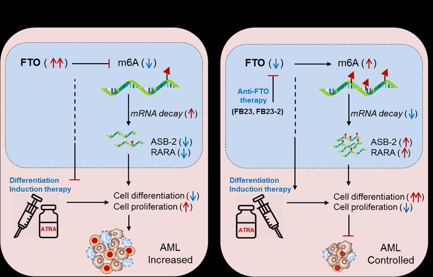
Preprints (www.preprints.org) | NOT PEER-REVIEWED | Posted: 10 May 2021             doi:10.20944/preprints202105.0193.v1

                                 A/C/U) sequences [8-10]. The three major RNA epigenetic modifiers reforming the whole
                                 biological functions are writers, erasers and readers. The detailed mechanism of
                                 m6A-modification and gene regulation is well described in several excellent journals.
                                 However, this review mainly focuses on reforming RNA epigenetic machineries into
                                 druggable form to treat haematological disorders especially AML.

                                 2. AML Therapeutics
                                 AML is a group of neoplastic diseases involving bone marrow with or without the in-
                                 volvement of peripheral blood. It is the most common type of haematological disorder
                                 prevalent in children & adults and globally affecting nearly 3-million people every year
                                 [11]. For some time now, several conventional therapies have been employed to treat
                                 AML including chemotherapy and rarely surgery and radiation [11]. Cytotoxic chemo-
                                 therapy or remission-induction with chemotherapeutic agents (anthracycline and cytar-
                                 abine) followed by consolidation-therapy involving an allogeneic stem cell transfer
                                 (SCT), bone-marrow transplant (BMT) or hematopoietic stem cell transplantation (HSCT)
                                 have been used as a standard therapy regimen for prolonged remission of AML [12].
                                 However, due to the delayed diagnosis of AML and the therapy-related morbidity and
                                 mortality, these conventional means of treating AML have fallen short when it comes to
                                 “do no harm” approach. Recently, with the advancement of NGS and gene-mutational
                                 analysis, several new strategies have been under development to minimize the global
                                 effects of these conventional therapies [13,14]. One of these new strategies includes the
                                 ‘targeted-drug therapy” which is cell, gene or marker specific and allows the option of
                                 treatment in patients where invasive chemotherapy is not feasible. This targeted therapy
                                 includes biological (monoclonal antibodies), epigenetic or combination therapies [15]
                                 (Figure 1, Table 1). Several antigen-specific monoclonal antibodies have now been ap-
                                 proved by the FDA for AML therapy. Among these, CD33-directed Gemtuzumab ozo-
                                 gamicin (GO) mAb was the first to be used for AML therapy[16]. In addition, several
                                 studies have now emerged marking DNA methylation as one of the hallmarks of AML
                                 carcinogenesis     [17]   involving     DNA       methyltransferases    (DNMTs)      and
                                 Ten-eleven-translocation (TET) dioxygenases [18]. Various DNMT inhibitors have been
                                 investigated for AML therapy, however only two of these (azacitidine and decitabine) are
                                 FDA approved [19]. On the other hand, any mutation in the TET family members results
                                 in altered DNA methylation [20]. For example, isocitrate dehydrogenases (IDH1 and
                                 IDH2) can inhibit TET2 and can cause hypermethylation resulting in AML progression
                                 [21]. Therefore, IDH inhibition can resolve this TET2 reduction-induced hypermethyla-
                                 tion [22]. The FDA has approved two oral IDH inhibitors: ivosidenib (AG-120) and
                                 enasidenib (AG-221) for AML treatment [23,24]. In addition to DNA modifiers, histone
                                 modifiers (HATs and HDACs) have also been implicated in AML treatment [25] by reg-
                                 ulating tumor suppressor genes (TSGs) [26,27]. At present, four HDAC inhibitors have
                                 been approved by the US FDA including three as panHDACi: belinostat, vorinostat,
                                 panobinostat, and romidepsin as a selective HDACi [28]. In this review, we have sum-
                                 marized the therapeutic potential of m6A-modifiers in controlling AML. Furthermore,
                                 we have also summarized the current biopharmaceutical companies developing AML
                                 therapeutics as RNA modification drugs (RMDs) [6] Figure 1.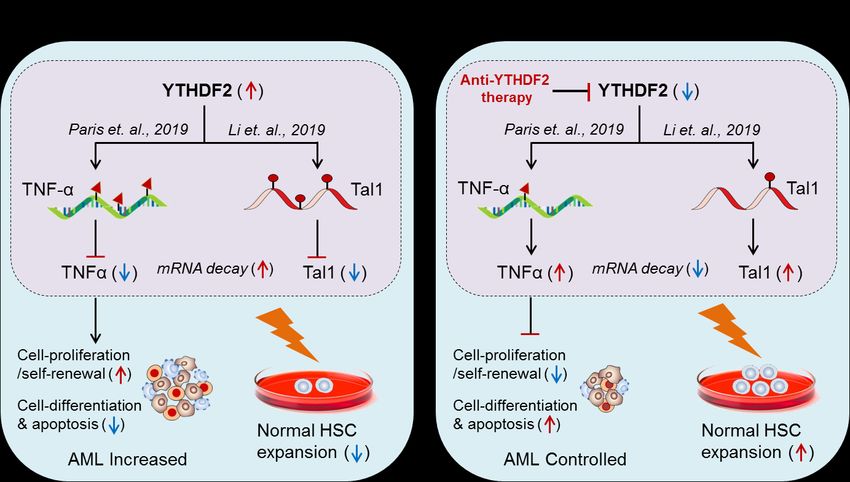
Preprints (www.preprints.org) | NOT PEER-REVIEWED | Posted: 10 May 2021                    doi:10.20944/preprints202105.0193.v1

     Fig 1. Development of AML therapeutics. The major AML therapeutics are depicted in the diagram above which includes; Conven-
     tional therapy (chemotherapy, radiotherapy and surgery), Biological therapy (stem cell therapy or bone marrow transplant), Epi-
     genetic drugs (DNA and histone modifier-based drugs), RNA-modifying drugs (writers/editors, erasers/removers and read-
     ers/effectors), MicroRNAs (miR-29b, miR-34a and miR-150) and Suppressor of cytokine signalling (SOCS1/CISH). Additionally, we
     have mentioned some biopharmaceutical companies developing RMDs and estimated phase-I clinical trials.

                                    3. Epitranscriptomics in AML therapy
                                          The development of advanced molecular techniques potentiates the opportunities to
                                    discover immune cell-based therapeutics over conventional pharmacological-based
                                    treatments. This section describes the strategies to treat haematological disorders, espe-
                                    cially AML by targeting intracellular RNA-modification machineries, like EEE (Edi-
                                    tor/writer: Mettl3, Mettl14; Eraser/remover: FTO, ALKBH5 and Effector/reader:
                                    YTHDF-1, YTHDF-2), microRNAs and Suppressor of Cytokine Signalling family of pro-
                                    teins (SOCS/CISH) and as a personalized medicines (Figure 1).

                                    3.1. Editors (Writers):
                                          3.1.1. Mettl3 in AML: The author has demonstrated the role of m6A writer enzyme
                                    ‘Mettl3’ in AML progression [29] supported by Barbieri et. al.,[30] and enlightened the
                                    molecular mechanism to control leukaemia. He found that ‘Mettl3’ is normally expressed
                                    in CD34+ hematopoietic stem/progenitor cells (HSPCs) but aberrantly expressed in leu-
                                    kemia cells as compared to the other tumor-types. However, Mettl3-silencing (shRNA) in
                                    HSPCs promoted HSPCs differentiation and apoptosis but inhibited self-renewal capac-
                                    ity & proliferation. Conversely, Mettl3 overexpression inhibited cell-differentiation and
                                    apoptosis but allowed cell-proliferation and self-renewal capacities. These results clearly
                                    suggest that Mettl3 facilitates AML cell growth but resist cell-differentiation. Further-
                                    more, it was also reported that the adoptive transfer of Mettl3-deficient (shMettl3) human
                                    AML cell line (MOLM-13) into immunodeficient (recipient) mice induces
                                    cell-differentiation and apoptosis but remarkably delayed leukemia progression. This
                                    result further supports the key role of Mettl3 in AML progression. Mechanistically, he
                                    justified (via m6A-mapping and ribosome profiling) that Mettl3 increases the expression
                                    of c-Myc, Bcl2 (apoptosis regulator) and pTEN (a proto-oncogene or AKT regulator)
                                    genes by m6A-modification in human leukemic MOLM-1 cell-line. Moreover, Mettl3
                                    regulates AKT-RICTOR signalling (PI3K-AKT) pathway and contributes to AML pro-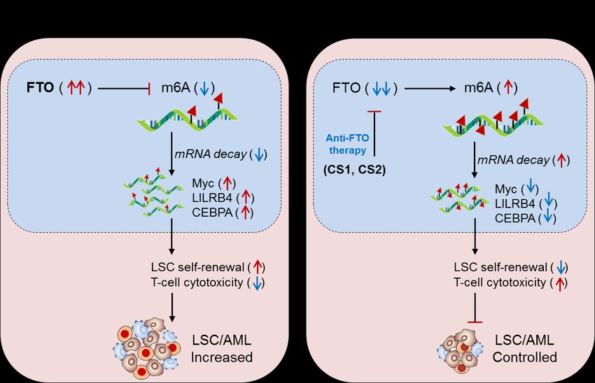
Preprints (www.preprints.org) | NOT PEER-REVIEWED | Posted: 10 May 2021               doi:10.20944/preprints202105.0193.v1

                                 gression by increasing cell-proliferation and decreasing cell-differentiation capacities.
                                 However, the loss of Mettl3 activates pTEN-mediated AKT phosphorylation and controls
                                 AML by reducing cell-proliferation and promoting cell-differentiation, confirmed after
                                 treating AML cells with PI3K-inhibitor in blocking cell-differentiation under
                                 Mettl3-deficient state [29]. Taken together, these investigations confirm the key role of
                                 Mettl3 in facilitating AML growth and survival and potentiate the therapeutic value of
                                 Mettl3 in controlling AML by means of either specific inhibition or selective intracellular
                                 silencing in AML. These findings were further supported by Konstantinos et. al., 2019[31];
                                 by synthesizing two small molecule compounds of Mettl3-inhibitor in inhibiting AML
                                 cell expansion (at 30mg/kg oral/intraperitoneal dose in mouse model with no clinical
                                 toxicity and weight loss) Table 2. Moreover, targeting AKT-RICTOR signalling pathways
                                 by PI3K-inhibitors in combination with other forms of conventional (radio/chemo/stem
                                 cell transplant) treatment procedures could overcome AML progression. These hypoth-
                                 eses were well supported by Batista et. al., 2017 [32] and Omar et. al., 2018 [33].

                                       3.1.2. METTL14 in AML: Weng et. al., 2018 [34]; demonstrated the role of
                                 m6A-writer enzyme ‘Mettl14’ in AML progression, and enlightened the intrinsic mecha-
                                 nism to control AML by targeting Mettl14. The authors have found that ‘Mettl14’ is ab-
                                 errantly expressed in AML patients confirmed by Cancer Genome Atlas
                                 (TCGA)-datasheet, human leukemia cell lines (MM6 and NB4), immortalized
                                 BM-progenitor cells (MA9, AE9a, PML-RARa) and AML cells carrying t(11q23), t(15;17),
                                 or t(8;21) translocation)-mutations, and gradually started decreasing during myeloid
                                 differentiation, suggesting the prominent role of Mettl14-mediated m6A-modification in
                                 leukemia progression. Experimentally he showed that, the in-vivo depletion of Mettl14 in
                                 recipient conditional knockout mice (tamoxifen-induced silencing) Mettl14cKO or
                                 Mettl14fl/fl-CREERT mice decreases cell proliferation and self-renewal capacity of CD45.2+
                                 (of Mettl14cKO) in peripheral blood as compared to the CD45.1 +CD45.2+ (competitor) cells.
                                 However, in-vitro depletion of Mettl14 (‘shM14’ pLKO.1 lentiviral-mediated silencing) in
                                 human HSPCs/-CD34+ promoted cell-differentiation, which was confirmed by increase of
                                 differentiation marker (CD11b-monocyte and CSF1R-macrophage). This suggests that
                                 Mettl14 induces leukemia growth by increasing cell-proliferation and decreasing termi-
                                 nal cell-differentiation. Conversely, the addition of differentiation inducing agent; OP9
                                 culture medium [35], PMA (0.5ng/ml/48h) [36] and all-trans retinoic acid (ATRA
                                 500nM/72h) [37] significantly decreased the expression of Mettl14 and overall
                                 m6A-abundance in AML cells. Reciprocally, Mettl14 overexpression (Mettl14-R298P
                                 vector) inhibits myeloid differentiation as compared to the empty vector. These results
                                 clearly suggest that Mettl14 is playing a significant role in AML progression by decreas-
                                 ing cell-differentiation and increasing cell-proliferation, and thus designating ‘Mettl14’ as
                                 a potential therapeutic target for AML treatment. Mechanistically, they have justified that
                                 (via RNA-sequencing) Mettl14-methylated methylation of Myb and Myc increase mRNA
                                 stability [38] leading to highly upregulated level of these transcription factors (TcFs) in
                                 leukaemia’s and lymphomas [39-41], confirmed by reduced half-life due to decreased
                                 polyribosomes 40S, 60S, 80S (ribosome profiling) and eukaryotic initiation factor-3A
                                 (EIF3A)-binding to the Myb and Myc transcript in Mettl14-silenced (shMettl14) MM6 and
                                 NB4 (human leukemic) cell lines as compared to the control (shCON) transfections.
                                 Furthermore, m6A mapping and immunoprecipitation (miCLIP) revealed increased
                                 m6A-binding to the Myb and Myc transcripts relative to the IgG iso-control, suggesting
                                 the significant role of Mettl14-mediated m6A-modification in increasing Myb and Myc
                                 level and further downstream signalling cascades rather than decaying respective
                                 mRNAs. However, they did not find significant involvement of YTHDF2 reader protein
                                 in regulating TcFs by mRNA-decay mechanism [38] in Mettl14-deficient AML cells. Col-
                                 lectively, these investigations suggests that Mettl14 promotes AML progression by in-
                                 creasing the activity of Myb (in increasing cell proliferation [42]) and Myc (in inhibiting
                                 cell differentiation [43,44]) gene functions. Moreover, they found that PU.1 (Spl1 encoded
                                 transcription factor, also known as master regulator of myeloid cells, or an oncogene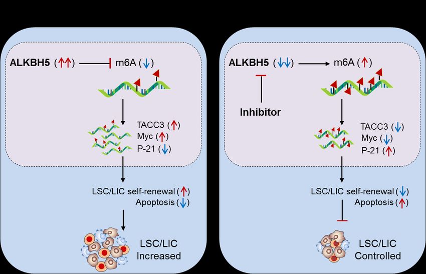
Preprints (www.preprints.org) | NOT PEER-REVIEWED | Posted: 10 May 2021 doi:10.20944/preprints202105.0193.v1
product) binds to Mettl14 at 5kb upstream of TSS and negative regulates its expression,
confirmed by chromatin immunoprecipitation (ChIP-seq) followed by luciferase reporter
assays. Furthermore, Spl1-knockdown increases both mRNA and protein level of
Mettl14. Conversely, Spl1 overexpression decreases Mettl14 expression. These results
further suggests that Mettl14 itself is targeted by ‘Spl1’ and an additional therapeutic
target to control Mettl14 expression. In support of this Konstantinos et. al., 2019; recently
developed a small molecule ‘inhibitor of Mettl3’ by structure-guided medicinal chemis-
try tool and confirmed m6A-inhibition and respective cellular effects comparable to the
Spl1-inhibitor, This study further support the therapeutic potential of Mettl3-inhibitor in
controlling AML progression [31]. Taken together, Mettl14 showed its immunothera-
peutic potential by regulating Spl1-Mettl14-Myb/Myc signalling axis, and therefore in-
tracellular silencing of Mettl14 in combination with standard ATRA (differentiation in-
ducing agent) therapy would be a promising approach to control AML with the other
forms of therapeutic approaches (radio/chemo/BM-transplantation) [34], (Figure 2, Table
2).
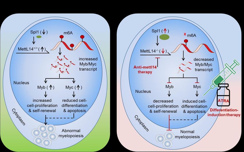
Fig 2. Therapeutic model targeting intracellular m6A-writer enzyme “Mettl14” in controlling AML. (A) Biological mechanism:
Mettl14 methylates and enhances the expression of Myb and Myc transcription factor. The elevated Myb/Myc increases
cell-proliferation and self-renewal capacity and reduces cell-differentiation and apoptotic processes, resulting in abnormal myelo-
poiesis. (B) Therapeutic model: Anti-Mettl14 therapy: intracellular silencing of Mettl14 decreases the expression of Myb/Myc TcFs
resulting in decreased cell-proliferation and improved cell-differentiation and apoptotic processes. Differentiation-induction ther-
apy: all-trans retinoic acid (ATRA) further enhances cell-differentiation and apoptotic processes and thus allows normal myelopoi-
esis. However, targeting other pathways like; Spl1, Myb/Myc, AKT pathways etc. has been also proposed previously [45,46] in
controlling AML.
3.2. Erasers (Removers):
3.2.1. FTO in AML: Li, et. al., 2017 [47]; have demonstrated the oncogenic role of
FTO, an m6A-demethylase, in facilitating leukemogenesis by promoting
cell-proliferation (self-renewal) and reducing cell-differentiation and apoptotic processes,
and enlightened the molecular mechanism to control APL by targeting FTO. They foundPreprints (www.preprints.org) | NOT PEER-REVIEWED | Posted: 10 May 2021 doi:10.20944/preprints202105.0193.v1
that, FTO is aberrantly expressed in a specific subset of hematopoietic stem/progenitor
cells (HSPCs/CD34+CD38-) carrying t(11q23)/MLL-rearrangement and
t(15;17)/PML-RARA mutations, called acute promyelocytic leukemia (APL), confirmed in
one hundred ‘affected’ and nine ‘control’ human samples of (CD34+CD33+ and MNC)
cells with Affymetrix GeneChip and qRT-PCR assays. In order to investigate the causa-
tive mechanism and the potential target of FTO in facilitating leukemogenesis, they
overexpressed FTO in human leukemic ‘MONOMAC-6’ cell lines via retrovirus expres-
sion system (MSCV-PIG-FTO and MSCV-PIG-CON) and voted two stable clones under
0.5µg/ml puromycin selection marker. Before sequencing, they confirmed increase and
decrease of FTO and m6A-abundancy by western blot and dot-blot/methylene blue as-
says. Interestingly, the sequencing result revealed two potential target of FTO, namely
ankyrin repeat and SOCS box protein 2 (ASB2), involved in controlling leukemia [48] by
suppressor of cytokine signalling (SOCS/CISH)-mediated mechanism, and retinoic acid
receptor alfa (RARA), a hallmark feature of APL [49], selected on the basis of MACS and
exomePeak methods [50,51]. Moreover, conserved m6A-methylation motif ‘GGAC’ was
also confirmed enriching >87% CDS as well as near stop codon vicinity. The authenticity
of this potential target gene was validated by dual-luciferase reporter coupled with mu-
tagenesis assays, where, [FTO cloning: pMIRNA1-FTO (wild-type), pMIRNA1-FTO-Mut
(mutant) and pMIRNA1-Ctrl (empty vector); and target gene cloning:
pMIR-REPORT-ASB2-3`UTR containing wild-type ‘GGAC’ and mutant ‘GGTC’ with
four m6A-conserved motifs 464bp; RARA-3`UTR with five conserved motif 474bp and
RARA-5`UTR with six conserved motif 395bp and transfected into HEK-293T cells/24h]
the wild-type FTO significantly decreased the luciferase activity of ASB2 and RARA
fragments containing wild-type m6A-methylated ‘GGAC’ motifs as compared to the
other combinations of mutant and control transfections, indicating the requirement of
m6A-methylation site for the regulation of FTO-mediated demethylase activity [52,53].
Furthermore, the FTO target was again validated by silencing FTO (shFTO) in
MA9/FLT3-ITD leukemic cell lines in hyper-methylating (increased m6A-modification)
ASB2 and RARA genes via m6A-seq and RNA-seq respectively. These results clearly
suggest that (i) FTO regulate its target gene expression by its m6A-demethylase activity
and (ii) the target gene must be m6A-modified. However, under hypo-methylated con-
dition the exact mechanism of FTO-mediated negative regulation of its target gene ex-
pression was further justified by mRNA stability testing, where FTO overexpressing cell
line (5µg/mL actinomycin D for 0, 3 and 6h) showed increased mRNA-decay (i.e. de-
creased half-life t1/2= In2/Kdecay) compared to the FTO silencing, suggesting the mechanism
of target gene control by destabilizing the mRNA stabilities. These investigations clearly
suggests that, under diseased state, the aberrantly expressed FTO negatively regulate its
target gene expression; neither by directly decreasing m6A-level (hypo-methylation) of
the target mRNA nor by m6A-effector protein YTHDF2-mRNA decay-dependent
mechanisms, but by directly affecting the mRNA stability via its demethylase activity [38].
However, the possibility of m6A-reader protein (YTHDF1 and YTHDF2)-mediated reg-
ulation of ASB2 and RARA also cannot be denied completely due to the finding of in-
creased target gene translation after YTHDF1 knockdown. Nevertheless, the
FTO-mediated APL progression is not only by targeting mRNA destabilization, but also
by affecting the efficacy of ‘ATRA-mediated therapy’ [54,55] justified by analysing NB4
cell (human acute promyelocytic leukemia cell line) differentiation marker. Where, the
overexpression of FTO significantly inhibits ATRA-induced cell-differentiation of NB4
cells [56] characterized by decreased (differentiated: CD11b+ CD14+) and increased
(un-differentiated: CD11b- CD14-) markers compared to the FTO-silenced (shFTO) cells in
reversing the both phenotypes. Conversely, the induction of 500mM ATRA significantly
decreased FTO and thereby rescued/increased the expression of its target ASB2 and
RARA genes [57,58]. Moreover, forced expression of ASB2 and RARA also increases
cell-differentiation. Collectively, these results authenticate that FTO is playing a crucial
role in promoting acute promyelocytic leukemia (i) by inhibiting the efficacy of AT-
RA-mediated induction of cell-differentiation and apoptosis, and (ii) by suppressing itsPreprints (www.preprints.org) | NOT PEER-REVIEWED | Posted: 10 May 2021 doi:10.20944/preprints202105.0193.v1
target gene expression. Taken together, these studies revealed the oncogenic property of
FTO in promoting APL. Therefore, targeting FTO would be a recommendable approach
to control APL with other forms of chemotherapeutic drug combinations [47,59] (Figure
3, Table 2, 3).
Fig 3. Therapeutic model targeting intracellular m6A-eraser protein FTO in controlling AML. (A) Biological mechanism: The
m6A-demethylase FTO is abnormally expressed in APL and negatively regulates the expression of its target genes (ASB2 and
RARA) causing decreased ATRA-induced cell differentiation and increased self-renewal capacity of APL cells, resulting in rapid
APL progression. (B) Therapeutic model: Anti-FTO therapy: targeting FTO by means of either selective inhibitor (FB23 and FB23-2)
[60] or intracellular silencing (shFTO) efficiently control APL by inducing m6A-mediated increase of ASB2 and RARA genes, as well
as by enhancing the efficacy of ATRA-mediated differentiation and apoptosis [54].
3.2.2. ALKBH5 in AML: Shen et. al., 2020 [61]; have demonstrated the role of other
m6A-eraser protein ‘α-ketoglutarate-dependent dioxygenase AlkB homolog 5’
(ALKBH5) in facilitating leukemia stem/initiating cells (LSCs/LICs) progression, a sub-
sets of AML, characterized by high self-renewal capacity, and enlightened intrinsic
mechanism to control LSCs by targeting ALKBH5. They found that, likewise the other
m6A-eraser protein FTO [47], ALKBH5 also facilitates leukemogenesis, evidenced by sig-
nificantly higher expression of ALKBH5 in various subtypes of AML cells carrying
t(15;17), Inv(16), t(8;21) and t(11q23) mutations, independent of specific TP53-mutation
(MONOMAC-6/MMC6, NOMO1 and NB4) and TP53-wild type (MA9.3-ITD and
MOLM13) human cell lines, confirmed via GSE42519, GSE13159 Affymetrix,
TCGA-sequencing and western blot analysis, as compared to the normal HSCs. Howev-
er, in vitro silencing (lentiviral shRNA) of ALKBH5 in NOMO1 and MMC6 human cell
lines significantly promotes apoptosis (AnnexinVhiPIhi) and restrict cell
growth/proliferation with overall decreased % survival of TCGA-AML datasheet [62].
Furthermore, ex-vivo conditional knockout of ALKBH5 in MOLM13 (MOLM13-iKD)
cells (doxycycline-inducible conditional knockdown model: where addition of doxycy-
cline depletes ALKBH5, as similar to stable knockout) also promoted apoptosis and re-
duced cell growth/proliferation as compared to the KO (MOLM13-iCas9) cells. Con-
versely, overexpression of wild-type ALKBH5 (A5-WT) reversed this effect and pro-
moted AML-cell progression as compared to the mutant (H204A) ALKBH5, suggesting
the crucial role of ALKBH5 in facilitating leukemogenesis. In addition to this, signifi-
cantly increased abundance of ‘global m6A-level’ was observed in the bone marrow ofPreprints (www.preprints.org) | NOT PEER-REVIEWED | Posted: 10 May 2021 doi:10.20944/preprints202105.0193.v1
ALKBH5-depleted (Exon-1 depletion by CRISPR-Cas9) mice compared to the wild-type
mice. Moreover, marked increase in global m6A-level was also noticed in both in vitro/ex
vivo ALKBH5-depleted cell lines confirmed by LC-MS/MS and dot-blot analysis as well.
These results clearly suggest that (i) the m6A-demethylase ALKBH5 facilitate AML pro-
gression and required for its growth and survival. (ii) ALKBH5-depletion enhances
global m6A-level. Next, to further validate the requirement of ALKBH5 in the promo-
tion/transformation of AML cells, they used MLL-AF9 (MA9)-induced leukemogenesis
model (where, MLL-rearranged fusion protein alone is sufficient to transform normal
HSPCs into leukemic cells [63]) coupled with ALKBH5 knockout (ALKBH5 KO) model.
Interestingly, ALKBH5-depletion significantly inhibited MA9-mediated cell immortali-
zation. On the contrary, forced expression of ALKBH5 (A5-WT) significantly promoted
MA9-mediated cell immortalization compared to the mutant ALKBH5 (A5-Mut), con-
firmed by in vitro colony formation/immortalization assays. Furthermore, adoptive
transfer (or bone marrow transplantation-BMT) of ALHBH5-deficient immune popula-
tion (donor: CD45.2+Lin-) co-cultured with MLL-AF9 cells into recipient (CD45.1) mice
significantly delayed leukemia progression and prolonged survival with decreased
splenomegaly, white blood cells count and immature blast cell (CD11b (Mac-1)+c-Kit+)
populations. Moreover, ALKBH5-deletion inhibited the engraftment of
MA9-transformed donor cells in the peripheral blood as compared to the ALHBH5-wild
type donor under lethal irradiation conditions. These investigations clearly suggest that
‘ALKBH5’ plays a key role in promoting leukemogenesis of a specific subset of AML
cells. The relevant experiments on leukemia xenograft mouse model further potentiate
the involvement of ALKBH5 in self-renewal and maintenance of AML-cells. Next, In
order to understand the molecular mechanism, ALKBH5-depleted MOLM13 and NO-
MO1 cells were sent for RNA sequencing (RNA-seq), which revealed TACC3 (trans-
forming acidic coiled-coil-containing protein 3) as a positive target of ALKBH5. [No-
ticeably, RNA-seq of ALKBH5 [61] revealed: 623 up-regulated and 1237 down-regulated
genes; whereas, RNA-seq of another eraser protein FTO by Li, et. al., 2017 [47] revealed:
2279 up and 888 down-regulated genes. Interestingly, only 251 up-regulated and 119
down-regulated targets were common in both]. Surprisingly, from a total of 18 (12 posi-
tive and 6 negative targets of ALKBH5) only ‘TACC3’ was significant, which was not
in-case of FTO, suggesting different target pattern of these two m6A-demethylaes/eraser
proteins in AML. Furthermore, m6A-sequencing (m6A-seq) revealed enrichment of
m6A-abundance at TACC3 mRNA (from a total of 510 transcripts with maximum en-
richment around protein coding region and 3`-UTR in ALKBH5-depleted AML cells)
confirmed by RNA immunoprecipitation & sequencing (RIP-seq) followed by
gene-specific m6A-qRT-PCR. Mechanistically they proved that, the ALKBH5 silencing
(shALKBH5) decreases the expression of TACC gene, due to hyper-methylation of m6A,
confirmed by decreased half-life of TACC3 (in MOLM13: 2.35h - 1.56h and NOMO1: 3.4h
- 1.55h) compared to control (shNS) transfections. Conversely, forced expression of
ALKBH5 (A5-WT) increases the half-life (3.63h - 7.47h) of TACC3 in NOMO1 cells com-
pared to mutant (A5-Mut) transfections. Moreover, TACC3 has been seen targeting ‘Myc’
and ‘P-21’ genes as well [64,65], confirmed by decreased Myc and increased P-21 protein
levels in ALKBH5-silenced NOMO1 and MMC6 cells (Shen et. al., 2020 figure-6).These
result clearly suggests that (i) TACC3 is the bona fide target of ALKBH5. (ii)
ALKBH5/m6A/TACC3 axis regulates p53 apoptotic pathways in AML. Next, the func-
tional analysis of TACC3 showed similar phenotypic effect like ALKBH5 justified by in-
creased leukemia cell apoptosis (AnnexinVhi PIhi) and decreased cell-proliferation after
silencing TACC3 (shTACC3) in NOMO1 and MMC6 cells confirmed by FACS and colo-
ny-formation and replanting capabilities. Collectively, these studies clearly suggests that
the m6A-eraser protein ALKBH5 as well as also FTO promotes leukemogenesis, and
therefore targeting ALKBH5 by means of either selective inhibitor of specific intracellular
silencing could be a potential approach to control AML in humans (Figure 4, Table 2, 3)
[61].Preprints (www.preprints.org) | NOT PEER-REVIEWED | Posted: 10 May 2021 doi:10.20944/preprints202105.0193.v1
Fig. 4. Therapeutic model targeting intracellular m6A-eraser protein ‘ALKBH5’ in controlling AML. (A) Biological mechanism: The
m6A-demethylase ‘ALKBH5’ is aberrantly expressed in AML, especially in leukemia stem/initiating cells (LSC/LIC) and facilitates
its progression, indicating its requirement for development, self-renewal, maintenance and propagation. Biologically, the enhanced
ALKBH5 in disease-state increases the expression of transforming acidic coiled-coil-containing protein 3 (TACC3) gene expression
by m6A-mediated mechanism, resulting in increased self-renewal capacity and thereby increased progression of LSC/LICs. (B)
Therapeutic model: Anti-ALKBH5 therapy: The selective inhibition of ALKBH5 decreases m6A methylation-mediated expression of
TACC3 gene resulting in decreased self-renewal and increased apoptosis is well-efficient to control AML subsets.
3.3. Effectors (Readers):
3.3.1. YTHDF2 in AML: Paris et. al., 2019 [66]; demonstrated the role of m6A-reader
protein ‘YTHDF2’ in promoting AML, and enlightened the molecular mechanism to
control AML by targeting YTHDF2 in HSPCs. They found that, although, YTHDF2 is
required for the development of leukemia stem cells (LSC) but also initiates AML de-
velopment. However, selective depletion of YTHDF2 inhibits the self-renewal capacity of
leukemic cells and promotes cell-differentiation and apoptosis, suggesting the therapeu-
tic value of YTHDF2 in controlling AML progression. Experimentally, they found that,
YTHDF2 targets tumor necrosis factor-α (TNF-α), required for cell-necrosis and apopto-
sis [67], and inhibits its expression by m6A-mediated mRNA decay mechanism [38].
However, selective removal of YTHDF2 increased the expression of TNF-α by lowering
mRNA decay, confirmed by increased half-life of the target mRNA, and thereby in-
creased terminal cell differentiation and apoptosis. This result clearly suggests that
YHTDF2 plays a crucial role in promoting leukemogenesis [66] was also witnessed by Li
et. al., 2019 [68]. Therefore targeting YTHDF2 would be a potential approach to control
AML by means of either intracellular silencing or via specific inhibitor (Figure 5).
3.3.2. YTHDF2 in stem cell expansion: Li et. al., 2019 [68]; have demonstrated the
therapeutic value of m6A-reader protein ‘YTHDF2’ in treating haematological disorders,
especially AML including other types of cancer. The hematopoietic stem progenitor cell
(HSPC/CD34+CD38-) population is believed to be major limiting issue during stem cell
transplantation therapy, due to a lesser number of HSPCs populations from a single
human umbilical cord blood (hUCB) donor. Therefore, ex-vivo expansion of HSPC pop-
ulation is a major and encouraging challenge for its widespread use. They found that
under steady state, the YTHDF2 sequesters its target gene T-cell acute lymphocytic leu-
kemia 1 (Tal1), which is required for the normal proliferation and self-renewal of HSPCsPreprints (www.preprints.org) | NOT PEER-REVIEWED | Posted: 10 May 2021 doi:10.20944/preprints202105.0193.v1
[69,70] and inhibits its function by m6A-marked mRNA-decay mechanism [38]. Inter-
estingly, the selective depletion of YTHDF2 (YTHDF2 KO) in HSPCs or hUCB-HSCs sig-
nificantly boosted (10-fold increase) the number of HSPCs by rescuing the expression of
Tal1 gene confirmed by increased mRNA stability. This result clearly suggests that
YTHDF2 has therapeutic potential and can be clinically used to expand ex-vivo popula-
tion of normal HSPCs by selective silencing and specific inhibitors of YTHDF2 [68].
However, the requirement of YTHDF2 in the maintenance of HSCs, by targeting
pro-inflammatory cytokines, was also demonstrated by Mapperley et. al., 2020 [71] (Figure
5).
Fig. 5. Therapeutic model targeting intracellular m6A-reader protein ‘YTHDF2’ in controlling AML, and in expanding normal HSCs
for BM-transplant. (A) Biological mechanism: YTHDF2 facilitates leukemia progression by suppressing TNF-α Paris et. al., 2019 [66].
Whereas, hyper-methylation of Tal1 causes HSC-limitations during BM/stem cell transplant Li et. al., 2019 [68]. (B) Therapeutic
model: Anti-YTHDF2 therapy controls AML progression as well as allows normal HSCs expansion in vitro by targeting respective
genes via mRNA-decay mechanism.
4. Therapeutic Strategies
4.1.1 Mettl3 Inhibitor: Recently, Eliza et. al., 2021[72] have discovered the selective
inhibitor of Mettl3 and Mettl14 (STM2457, IC50 = 16.9 nM) in controlling AML via
high-throughput screening of 250,000 drug-like compounds. The binding affinity and
specificity of STM2457 was confirmed by surface plasmon resonance (SPR) and X-ray
crystallography as well as by intraperitoneal injection of STM2457 (50mg/kg) in selec-
tively reducing m6A on poly-A+-enriched mRNA, with no effect on other mRNA modi-
fications. Functionally, they showed significant effect of STM2457 in reducing clonogenic
potential and inducing apoptosis in human and mouse AML model without affecting
normal human cord blood (CD34+/HSPCs) and non-leukemia (HPC7) hematopoietic
cells. Moreover, in vivo studies on 3-human AML patient derived xenograft (PDX) model
and primary murine MLL-AF9/Flt3ltd/+ model showed convincing anti-leukemia effect
compared to control STM2120 inhibitors [72]. Furthermore, the other researchers also
suggested the significant effect of Mettl3/14-inhibitors as anti-leukemia effect (Table 3)
[6,31,33].
4.1.2. FTO Inhibitor: Huang et. al., 2019 [60]; have examined the therapeutic efficacy
of two synthetic small molecule inhibitor of FTO (FB23 and FB23-2) in controlling AML.Preprints (www.preprints.org) | NOT PEER-REVIEWED | Posted: 10 May 2021 doi:10.20944/preprints202105.0193.v1
They found that the m6A-demethylase ‘FTO’ is highly upregulated in certain
AML-subtypes and facilitates leukemogenesis by promoting cell-proliferation and in-
hibiting terminal cell differentiation. The FTO inhibitor especially ‘FB23-2’ dramatically
suppresses cell-proliferation and promotes differentiation and apoptosis of both the cell
lines as well as the primary cells in xeno-transplanted mice. Mechanistically, the synthe-
sized inhibitors directly bind to the FTO and selectively inhibit its m6A-demethylase ac-
tivity, mimicking FTO depletion similar to in vivo knockout. This result suggests that
these FTO inhibitors (FB23 and FB23-2) could be a potential druggable candidate to treat
leukemia [60], supported by Yang et. al., 2019 [7] (Table 3).
4.1.3. FTO in anti-tumor immunity (AML): Su et. al., 2020 [73] have demonstrated
the clinical value of two synthetic small-molecule inhibitor of FTO, designed on the basis
structural guided tool, (CS1 and CS2) in controlling AML, especially leukemia stem cell
(LSC) progression in the aspect of anti-tumor immunity. They found that, the abundantly
expressed FTO facilitates LSCs progression by impairing T-cell activity via increasing the
expression of an immune inhibitory checkpoint molecule/gene ‘LILRB4’. The application
CS1 and CS2 significantly controls LSC progression in mouse model with low drug tox-
icity. Mechanistically, CS1 and CS2 were found to be selectively binds to the FTO and
inhibit its demethylase activity, resulting in decreased expression of ‘LILRB4’ via inhib-
iting YTHDF2-mediated mRNA-decay mechanism. Moreover, increased T-cell cytotoxi-
city and reduced self-renewal abilities of AML cells were noted in the treated mice, sug-
gesting that CS1 and CS2 is a potent inhibitors of FTO and can be developed as a poten-
tial therapeutics against LSC by increasing anti-leukemia T-cell activity [73]. This hy-
pothesis was further supported by Olsen et. al., 2020 [74], (Figure 6). Additionally, some
other FTO-inhibitors are well described in these articles [75,76] and [77,78] (Table 2, 3).
Fig. 6. Therapeutic model targeting m6A-eraser ‘FTO’ in controlling LSC progression by enhancing anti-tumor immunity. (A) Bio-
logical mechanism: FTO has been found to be aberrantly expressed in AML, especially LSC populations, and facilitates its progres-
sion. Mechanistically, the FTO enhance the expression of its target LILRB4 (immune checkpoint) and other Myc and CEBPA genes
via its m6A-demethylase activity by inhibiting m6A-reader protein YTHDF2-mediated mRNA-decay mechanism, causing de-
creased T-cell activity and increased self-renewal capacity, resulting in enhanced LSC progression. (B) Therapeutic model: Anti-FTO
therapy: The two synthetic small-molecule inhibitor of FTO (CS1 and CS2) selectively binds to the FTO domain and inhibits its de-
methylase activity, leading to decreased expression of its target (LILRB4, Myc and CEBPA) mRNA via reducing YTHDF2-mediatedPreprints (www.preprints.org) | NOT PEER-REVIEWED | Posted: 10 May 2021 doi:10.20944/preprints202105.0193.v1
mRNA-stability. The decreased expression of the targeted gene ultimately enhances T-cell cytotoxicity and immune evasion and
thereby reduced self-renewal capacities, resulting in better control over LSC propagation.
5. MicroRNA in AML
5.1.1. MiR-150 in AML: In addition to m6A-modifiers, microRNAs also showed
promising outcome in the treatment of acute myeloid leukemia (AML). Fang and colleague,
2016 [79] have demonstrated the importance of miR-150 in controlling leukemia pro-
gression. They showed that miR-150 is down-regulated in AML and CML patients, but
normalized after complete remission/treatment. However, miR-150 restoration therapy
(miR-150 mimic) significantly inhibited the AML by reducing cell-proliferation and
promoting apoptosis of the leukemia stem cells, leading to reduced tumorigenicity in
xenograft leukemia model. The underlying mechanism evidenced that miR-150 targeted
genes are mainly associated with RNA metabolism (synthesis, export, splicing and sta-
bility), transcriptional regulation, wnt-signalling and mTOR-signalling pathways. Inter-
estingly, knockdown of any of these miR-150 downregulated targets (TET3, EIF4B,
FOXO4B and PRKCA) showed anti-leukemia activity similar to miR-150 restoration
therapy [80]. Conclusively, these results authenticate the druggable value of miR-150 in
treating AML and as a novel candidate for therapeutic drug development [79].
5.1.2. MiR-34a in AML: miR-34a was shown to be another microRNA playing a
crucial role in controlling elderly AML, who were ineligible for conventional chemo-
therapy. The author [81] have found that, the combination of decitabine (dacogen/DAC;
known as 5-aza-2'-deoxycytidine) and all-trans retinoic acid (ATRA; a well-known dif-
ferentiation inducing agent) effectively control AML and prolong overall survival rate by
49.6% in a clinical trial of 36-elderly leukemia patients. The underlying mechanism
demonstrates that the combination of DAC and ATRA inhibited the expression of DNA
methyltransferase 1 (DNMT1) resulting in activation of miR-34a by hypo methylation.
The activated miR-34a further inhibits the expression of Myc [43,44] resulting in cell cycle
arrest and increased apoptosis in vitro. This result suggests that miR-34a could be a
druggable candidate to control AML progression by modulating miR-34a/Myc axis
[81,82].
5.1.3. MiR-29b in AML: Liu et. al., 2019; demonstrated that miR-29b is also playing an
important role in controlling (LSCs) leukemia stem cell progression, a subtype of acute
myeloid leukemia, characterized by increased self-renewal capacity and decreased
apoptosis, by targeting LSC-fucosylation. The author found that fucosyltransferase 4
(FUT4) is overexpressed in LSC population (CD34 +CD38-) compared to the non-LSC
(CD34+CD38+, CD34-CD38+, CD34-CD38-) populations isolated from MOLM13 and KG-1a
cell lines determined by LTL lectin assays. However, selective depletion of FUT4
(shFUT4) significantly decreased cell-proliferation and induced apoptosis of the LSCs
confirmed by reduced sphere formation ability (colony formation assay), reduced Ki67
cell-proliferation (immunofluorescence) and increased LSC apoptosis (TUNEL assays)
indicated by increased cleavage of apoptotic (PARP and caspase3) markers. Moreover,
in-vitro application of chemotherapeutic drug (ADR, Ara-C and Paclitaxel) in combina-
tion with shFUT4-LSC drastically reduced LSC cell-proliferation as compared to shFUT4
and drug effect alone. Furthermore, mouse xenograft studies showed decreased tumour
growth when given in combination with shFUT4 and ADR relative to the individual
treatments. This result clearly suggests that (i) FUT4-mediated fucosylation facilitate
LSCs progression. (ii) FUT4 silencing overcome chemotherapeutic drug resistance in
AML. Mechanistically, he proved that FUT4 is regulated by specificity protein 1 (Sp1)
transcription factor confirmed by Sp1 binding to FUT4 promoter via chromatin im-
munoprecipitation (ChIP) and duel luciferase assays. Interestingly, miR-29b directly
binds with Sp1 (bioinformatics analysis) and inhibits its expression along with
Wnt/β-catenin activation, confirmed by miR29b-mimic in ‘inhibiting’ and anti-miR29b inPreprints (www.preprints.org) | NOT PEER-REVIEWED | Posted: 10 May 2021 doi:10.20944/preprints202105.0193.v1
‘promoting’ Sp1 expression and thus influences FUT4 expression accordingly, was fur-
ther validated by partial restoration of FUT4 expression by co-transfecting anti-miR29b
with siSP1. Moreover, Wnt/β-catenin pathway inhibition by DKK inhibitor induces
apoptosis and reduces cell-proliferation. Taken together, these investigations clearly
suggest that miR-29b could be a potential candidate to control LSC progression by regu-
lating miR-29b/Sp1/FUT4 axis [83].
6. Suppressor of cytokine signalling (SOCS/CISH) in AML
In addition to epigenetic modifiers (RMDs) and microRNAs; the role of cyto-
kine-inducible SH2-domain containing protein (CISH or CIS) as anti-tumor and an-
ti-leukemia activity, also, cannot be denied because of its versatile role in regulating cy-
tokine signalling via sensitizing immune cells. For example, (i) CISH knockdown has
been shown in increasing anti-tumor immunity by enhancing CD8+T-cell effector func-
tion [84]. (ii) CISH-knockdown also increases NK-cell fitness and cytotoxicity [85] and
thus playing a crucial role in protecting tumor metastasis [86]. (iii) More relevantly, CISH
has been shown in maintaining T-cell homeostasis via Mettl3-mediated methylation
mechanism [87,88] and (iv) The m6A-reader protein ‘YTHDF2’ is involved in controlling
NK cell-mediated anti-tumor immunity [89]. Furthermore, the specific function of CISH
in association with acute myeloid leukemia was demonstrated by Zhu et. al., [90], where
the importance of CISH in NK cell-mediated anti-leukemia activity has been described.
They have showed that selective depletion of CISH in NK cells (CISH -/-) derived from
human iPSC (induced pluripotent stem cells) significantly enhance the expansion as well
as survival in the tumor microenvironment (TME), confirmed by improved metabolic
fitness of NK-cells, characterized by increased glycolytic capacity and mitochondrial
respiration (OxPhos activity) via mammalian target of rapamycin (mTOR) signalling
response. Moreover, increased NK-cell cytotoxicity was reported against multiple tumor
cell lines as well as in in-vivo leukemia xenograft model [90]. This result additionally
supports the role of CISH as an alternative approach to control AML by encompassing
immune cell-based therapeutics. Nevertheless, the role of professional antigen presenting
cells like, dendritic cells (DCs) also cannot be circumvent for its proven role in improving
anti-tumor immunity by targeting CISH [91,92]. One more relevant research outcome
showed the involvement of suppressor of cytokine signalling 1 (SOCS1), one of the
members of CISH/SOCS family, in mimicking AML like phenotype. Where, the overex-
pression of SOCS1 in zebrafish model showed increased myelopoiesis with distorted
kidney and splenic morphology, suggesting the therapeutic inference of AML by target-
ing SOCS1 (Figure 1) [93].
7. Conclusions
In this review, we have described the potential of m6A-modifiers in developing
cancer precision medicines and explained how ‘epitranscriptomics’ plays a central role in
regulating the crucial genes associated with AML. Among these, the m6A-writer protein
Mettl3 and Mettl14 has been found to be aberrantly expressed in specific sub-types of
AML and promotes leukemogenesis by regulating Myb, Myc, Bcl2, pTEN and PI3K-AKT
pathways [29,30,34]. Likewise, m6A-methylases (writer); m6A-demethylases FTO and
ALKBH5 (eraser) have also been found to be aberrantly expressed in APL & LSCs/LICs
and promote leukemogenesis by targeting ASB2, RARA, LILRB4, CEBPA, TACC3, Myc
and P-21 gene [47]. Nevertheless, FTO single nucleotide polymorphisms (SNPs) has been
also found to be associated with other cancers [94]. Likewise, writer and eraser; the
reader protein ‘YTHDF2’ has been also found in promoting leukemogenesis by inhibiting
its target Tla1 [68] and TNF-α [66] through universal YTHDF2-mediated mRNA decay
mechanism [38]. These inventions suggest the different mechanism of action ‘of these ep-
igenetics modifiers’ in controlling AML by regulating respective gene targets. However,
still some questions remained unanswered, how all these epigenetic modifiers are
up-regulated simultaneously and facilitates AML development. For example, considering
the balance mechanism of ‘writer’ and ‘eraser’ enzymes in regulating ‘Myc’ in the same cells byPreprints (www.preprints.org) | NOT PEER-REVIEWED | Posted: 10 May 2021 doi:10.20944/preprints202105.0193.v1
Mettl3-methylation as well as by FTO-mediated mechanism remains to be further justified. A
justifiable answer seems to be possible by recruiting different gene set by inviting dif-
ferent RNA-machineries. Nevertheless, The microRNA; miR-150, miR-34a and miR-29b
has also shown significant anti-leukemia effect by targeting wnt-signalling, mammalian
target of rapamycin (mTOR)-signalling pathways [79] Myc [81] and
Sp1/FUT4-fucosylations [83]. In addition to this, the immune-based therapeutics should
also consider in AML treatment by utilizing dendritic cells, T-cells and NK cell-targeting
CISH-mediated signalling [87,89,95]. Therefore, it is very critical to understand the stages
of AML progression and so in implementing these exciting RMDs in eradicating AML
with other forms of standard chemotherapeutic drugs.
8. Future Prospective
Targeting m6A-modification machineries has revolutionized the class of epigenetic
research in discovering novel therapeutic drug targets. Co-targeting of intracellular genes
along with m6A-modification machineries will improve the level of immunotherapy
against those cancers which are resistant to the immune checkpoint-based therapeutics in
patients with blood cancer. The future is not so far where targeted therapy in combina-
tion with other drugs which would become a universal panacea to control many diseases
and other forms of cancer. Moreover, targeting suppressor of cytokine signalling family
of proteins (SOCS/CISH) would further potentiate the immunotherapeutic potential of
RNA modification inhibitors by targeting epigenetic regulator of dendritic cells. Phar-
maceutical approaches pertaining to RNA epigenetic modification machinery are ex-
pected to shed light on the field of cancer immunotherapy for AML and other forms of
blood cancer. Nevertheless, a human clinical trial is required under a medical setup to
access the therapeutic value of these inhibitors by optimizing the initial doses or/if in-
tracellular silencing (gene therapy). However, a proposed clinical trial is underway to
conduct till 2021-2022 by several biopharmaceutical companies mentioned above.
Author Contributions: Conceptualization, S.K.; formal analysis, S.K. and M.U.A.; inves-
tigation, S.K., M.U.A. and A.K.; data curation, S.K.; writing-original draft preparation,
S.K.; writing-review and editing, S.K., Y.-S.B., M.U.A., R.N., and A.K.; visualization,
Y.-S.B., S.K., M.U.A., R.N., and A.K.; supervision, Y.-S.B.; project administration, Y.-S.B.
and S.K.; funding acquisition, S.K. and Y.-S.B. All authors have read and agreed to the
published version of the manuscript.
Funding: This work was supported by the grant of Individual Basic Science and Engi-
neering Research Program (2018R1D1A1B07048567) funded by the Korean National Re-
search Foundation. This work was also supported by a National Research Foundation
(NRF) grant funded by the Korea Ministry of Science and ICT (SRC-2017R1A5A1014560)
and in part by a grant of the Korea Health Technology R&D Project through the Korea
Health Industry Development Institute (KHIDI) funded by the Ministry of Health &
Welfare, Republic of Korea (HI16C1074).
Institutional Review Board Statement: Not applicable.
Informed Consent Statement: Not applicable.
Data Availability Statement: Not applicable.
Acknowledgements: I wish to thanks all the lab members for their support.
Conflicts of Interest: The authors declare no conflict of interest.
Table 1. Therapeutic drug for Acute Myeloid Leukemia (AML).
Conventional drugsPreprints (www.preprints.org) | NOT PEER-REVIEWED | Posted: 10 May 2021 doi:10.20944/preprints202105.0193.v1
Brand/ Clinical
Drug Drug type Ref.
other name trial
Rydapt FDA
Midostaurin Multikinase FLT3 inhibitor [96]
(Novartis) approved
Vyxeos cytarabine and daunorubicin FDA
CPX-351 [97,98]
(Jazz Pharma) combination (5:1 molar ratio) approved
Topoisomerase II inhibitor:
Formerly SNS-595 FDA
Vosaroxin anticancer quinolone [99]
(Sunesis Pharma) approved
derivative (AQD)
ASP2215, Xospata FDA
Gilteritinib Dual inhibitor of FLT3/AXL [100,101]
(Astellas Pharma) approved
Venclexta,
NCT02993523
Venclyxto
Venetoclax Bcl2-inhibitor Phase III NCT03069352
(AbbVie,
[102]
Genentech)
Epigenetic drugs
DNMT inhibitor FDA
Azacitidine Vidaza [19]
(Hypomethylating agent) approved
DNMT inhibitor FDA
Decitabine Dacogen [19]
(Hypomethylating agent) approved
Tibsovo FDA
Ivosidenib IDH1 inhibitor
(AG-120) approved [23,24]
Idhifa FDA
Enasidenib IDH2 inhibitor
(AG-221) approved
Beleodaq
Belinostat Pan-HDAC inhibitor Phase II [103]
(PXD101)
Vorinostat Zolin Pan-HDAC inhibitor Phase I/II [104]
Panobinostat - Pan-HDAC inhibitor Phase I/II [105-107]
Romidepsin Istodax Selective HDAC inhibitor Preclinical [28]
Dinucleotide of decitabine and
Guadecitabine SGI-110 Phase III NCT02348489
deoxyguanosine
Monoclonal Antibodies (mAbs)
GO
Gemtuzumab NCT03374332
(Mylotarg, CD33-targeted Phase 2
Ozogamicin NCT00372593
Wyeth Pharma)
Vadastuximab SGN-CD33A
CD33-targeted Phase I [108]
talirine (Seattle Genetics)
Darzalex Faspro
Daratumumab CD38-targeted Phase II NCT03067571
(Janssen Biotech)
RG7356 CD44-targeted Phase I [109]
Phase III,
Apamistamab Iomab-B CD45-targeted NCT02665065
SIERRAPreprints (www.preprints.org) | NOT PEER-REVIEWED | Posted: 10 May 2021 doi:10.20944/preprints202105.0193.v1
Table 2. RNA epigenetic machineries as a therapeutic target in AML.
RNA Disease Therapeutic
Target Mechanism of action Ref.
modifiers condition strategies
Writers Selective
Myb/Myc, Bcl2, Inhibiting cell differentiation &
Mettl3 Mettl-3/14
up-regulated pTEN , Spl1 apoptosis and promoting cell [29,32]
inhibitor
in AML (PU.1), PI3K-AKT proliferation (self-renewal) [34,72]
Mettl-14 or Targeted
pathway capacity.
therapy
Regulated by FTO related with
Myc, CEBPA [48]
Leukemia
Erasers By inhibiting ASB2 and RARA Targeted
up-regulated ASB2, RARA,
FTO gene targets as well as silencing or
in APL and Myc, LILRB4, [47]
ATRA-induced cell specific
LSC/LICs CEBPA
differentiation and apoptosis FTO/ALKBH5
TACC3, By impairing self-renewal inhibitor
ALKBH5 [61]
Myc, P21 capacities
YTHDF2 inhibit the expression
T-cell acute
Readers up-regulated of essential T-cell acute
lymphocytic [68]
YTHDF2 in AML lymphocytic leukemia 1 (Tal1) Targeted
leukemia (Tal1)
gene. silencing/
YTHDF2 inhibit the expression therapy
Tumor Necrosis
YTHDF2 up-regulated of TNFα required for cell [66]
Factor-alfa (TNFα)
necrosis and apoptosis.
Table 3. Selective inhibitors of m6A-modifiers in AML treatment.
Name Therapeutic application Ref.
Disease Epigenetic Inhibitor Function
By selective inhibition of Mettl-3/14
Writers
AML STM2457 (STC-15) (proposed human clinical trial in 2022 [31,33,72]
by Storm Therapeutics)
Mettl3,
Metabolic SAH Selective inhibition of Mettl-3/14
Mettl14 [110]
disease (S-Adenosyl-homo cysteine) by targeting endogenous metabolite
Inhibitors
Promotes apoptosis
Cancer UZH1/ UZH1a and enhance [31,111]
T-cell anti-tumor activity
FB23 Selective FTO inhibitor works by [60,112]
Erasers
retaining m6A-demethylase activity
FB23-2 [7,60]
in LSCs
FTO Up-regulated
Selective FTO inhibitor
Inhibitors in AML
R-2-hydroxyglutarate targeting m6 A/MYC/ CEBPA
[75]
(R-2HG) signalling and increase anti-tumor
immunityPreprints (www.preprints.org) | NOT PEER-REVIEWED | Posted: 10 May 2021 doi:10.20944/preprints202105.0193.v1
Selective FTO inhibitor targeting
CS1 and CS2 [73,74]
LILRB4 immune checkpoint gene
Non-steroidal
Selective FTO inhibitor
Glioblastoma anti-inflammatory drug,
targeting ADAM19 gene by m6A [113,114]
Stem Cells (MA2) an ethyl ester form of
hyper-methylation
meclofenamic acid
Triple-negative
Selective inhibitor of FTO targeting
inflammatory MO-I-500 [115]
IRX3 gene in SUM149-MA cells
breast cancer
First FTO inhibitor with
BBB-penetrating small
CNS (epilepsy) anticonvulsant activity by targeting [116]
molecule inhibitor of FTO
various microRNAs
Readers BET inhibitor (OTX015) Bromodomain inhibition [117-119]
MB-3
AML
YTHDF2 MB-3 inhibitor of KAT2A Inhibit the expression of KAT2A [120,121]
Inhibitors gene
References
1. Desrosiers R, Friderici K, Rottman F: Identification of methylated nucleosides in messenger rna from
novikoff hepatoma cells. Proc Natl Acad Sci U S A (1974) 71(10):3971-3975.
2. Perry RP, and Kelley, D.E.: Existence of methylated messenger rna in mouse l cells. ( January 1974) Volume
1, Issue 1(Pages 37-42.
3. Jia G, Fu Y, Zhao X, Dai Q, Zheng G, Yang Y, Yi C, Lindahl T, Pan T, Yang YG, He C: N6-methyladenosine in
nuclear rna is a major substrate of the obesity-associated fto. Nat Chem Biol (2011) 7(12):885-887.
4. Jia G, Yang CG, Yang S, Jian X, Yi C, Zhou Z, He C: Oxidative demethylation of 3-methylthymine and
3-methyluracil in single-stranded DNA and rna by mouse and human fto. FEBS Lett (2008)
582(23-24):3313-3319.
5. Shi H, Wei J, He C: Where, when, and how: Context-dependent functions of rna methylation writers,
readers, and erasers. Mol Cell (2019) 74(4):640-650.
6. Cully M: Chemical inhibitors make their rna epigenetic mark. Nat Rev Drug Discov (2019) 18(12):892-894.
7. Van Der Werf I, Jamieson C: The yin and yang of rna methylation: An imbalance of erasers enhances
sensitivity to fto demethylase small-molecule targeting in leukemia stem cells. Cancer Cell (2019)
35(4):540-541.
8. Kane SE, Beemon K: Precise localization of m6a in rous sarcoma virus rna reveals clustering of methylation
sites: Implications for rna processing. Mol Cell Biol (1985) 5(9):2298-2306.You can also read