Roles of Non-Structural Protein 4A in Flavivirus Infection - MDPI
←
→
Page content transcription
If your browser does not render page correctly, please read the page content below
viruses
Review
Roles of Non-Structural Protein 4A in Flavivirus Infection
Paeka Klaitong and Duncan R. Smith *

                                          Institute of Molecular Biosciences, Mahidol University, Nakhon Pathom 73170, Thailand; paeka_p@hotmail.com
                                          * Correspondence: duncan_r_smith@hotmail.com; Tel.: +66-2800-3624-8

                                          Abstract: Infections with viruses in the genus Flavivirus are a worldwide public health problem.
                                          These enveloped, positive sense single stranded RNA viruses use a small complement of only
                                          10 encoded proteins and the RNA genome itself to remodel host cells to achieve conditions favoring
                                          viral replication. A consequence of the limited viral armamentarium is that each protein exerts
                                          multiple cellular effects, in addition to any direct role in viral replication. The viruses encode four
                                          non-structural (NS) small transmembrane proteins (NS2A, NS2B, NS4A and NS4B) which collectively
                                          remain rather poorly characterized. NS4A is a 16kDa membrane associated protein and recent studies
                                          have shown that this protein plays multiple roles, including in membrane remodeling, antagonism
                                          of the host cell interferon response, and in the induction of autophagy, in addition to playing a role in
                                          viral replication. Perhaps most importantly, NS4A has been implicated as playing a critical role in
                                          fetal developmental defects seen as a consequence of Zika virus infection during pregnancy. This
                                          review provides a comprehensive overview of the multiple roles of this small but pivotal protein in
                                          mediating the pathobiology of flaviviral infections.

                                          Keywords: flavivirus; transmembrane protein; autophagy; congenital Zika syndrome; interferon
                                          response; unfolded protein response


Citation: Klaitong, P.; Smith, D.R.
Roles of Non-Structural Protein 4A in     1. Introduction
Flavivirus Infection. Viruses 2021, 13,         The genus Flavivirus of the family Flaviviridae comprises over 50 species of arthropod-
2077. https://doi.org/10.3390/            borne enveloped viruses [1]. Most viruses in this genus are transmitted to vertebrate hosts
v13102077
                                          through the bite of infected hematophagous arthropods, although some have arthropod-
                                          or vertebrate-restricted transmission cycles [2]. Around half of the viruses assigned to a
Academic Editor: Karla Helbig
                                          species in the genus Flavivirus are known human pathogens [3], of which the medically
                                          important viruses causing public health problems worldwide are dengue virus (DENV:
Received: 19 September 2021
                                          DENV 1 to DENV 4), Zika virus (ZIKV), Japanese encephalitis virus (JEV), West Nile
Accepted: 12 October 2021
                                          virus (WNV) and yellow fever virus (YFV), which are transmitted by mosquitoes, and
Published: 15 October 2021
                                          tick-borne encephalitis virus (TBEV) which is transmitted by ticks. Flavivirus infection
                                          of humans causes a variety of manifestations ranging from no symptoms or non-severe
Publisher’s Note: MDPI stays neutral
with regard to jurisdictional claims in
                                          flu-like symptoms to severe or even lethal symptoms such as hemorrhagic fever and shock
published maps and institutional affil-
                                          syndrome for DENV infection, Guillain–Barré syndrome and fetal microcephaly for ZIKV
iations.
                                          infection, meningitis and encephalitis for JEV, WNV, TBEV infections and liver failure
                                          and jaundice for YFV infection. Vaccination is considered a reasonable method to prevent
                                          flavivirus infections. Vaccines for JEV, YFV and TBEV are currently licensed for use in
                                          humans [4], and while a vaccine for DENV is licensed in some countries, the occurrence of
                                          more severe disease in some vaccinated individuals [5] has served to limit its application.
Copyright: © 2021 by the authors.
                                          However, vaccines for the other human pathogenic flaviviruses are either unable to elicit
Licensee MDPI, Basel, Switzerland.
This article is an open access article
                                          broadly protective immune responses or are in varying stages of development. In addition,
distributed under the terms and
                                          to date, there is no specific antiviral drug available to treat any flaviviral infection. Viral
conditions of the Creative Commons
                                          components offer potential drug targets, but as flaviviruses mutate rapidly due to their
Attribution (CC BY) license (https://     low-fidelity replication process, drug-resistant strains can rapidly emerge. Host-oriented
creativecommons.org/licenses/by/          therapeutics have been therefore considered as a potential alternative.
4.0/).

Viruses 2021, 13, 2077. https://doi.org/10.3390/v13102077                                                  https://www.mdpi.com/journal/viruses
Viruses 2021, 13, 2077                                                                                           2 of 19

                               For a successful infection, viruses must manipulate the host cellular environment
                         to establish an optimal platform for their genome replication, protein production, and
                         virion assembly and egress. To this end, viruses utilize both viral and host factors to aid in
                         reorganization of intracellular membranes, manipulation of host signaling and metabolic
                         pathways, and evasion of host immune responses.
                               Flaviviruses possess a non-segmented positive-sense single-stranded RNA genome
                         of 10–11 kb. The genome is modified at the 50 end with a m7GpppAm cap structure and
                         lacks a poly-A tail. The genome contains a long open reading frame (ORF) flanked by a 50
                         untranslated region (UTR) and a 30 UTR of approximately 100 and 400–700 nucleotides,
                         respectively [6]. The 50 and 30 UTR usually form highly conserved secondary and tertiary
                         structures essential for RNA replication and protein translation [7]. The genome is trans-
                         lated in close association with intracellular membranes, giving rise to a single polyprotein
                         precursor. The structural proteins (capsid, precursor membrane (prM) and envelope (E))
                         encompass the 50 one-fourth of the polyprotein, while the non-structural (NS) proteins
                         (NS1, NS2A, NS2B, NS3, NS4A, NS4B and NS5) occupy the remainder of the polyprotein.
                         The polyprotein is co- and post-translationally processed into individual components by
                         cellular proteases and the viral NS2B–NS3 protease complex. While the structural proteins
                         comprise the virion, the NS proteins are primarily responsible for viral RNA replication,
                         virion assembly and modulation of host immune responses [8–14].
                               The role of flavivirus NS1 in viral replication is not fully understood, but it has been
                         shown to function at a very early stage in viral RNA replication [15–18]. NS1 has also
                         been shown to have a role in the modulation of host innate immune response [19–21],
                         and in viral neuroinvasiveness [22]. NS2A and NS4B have been suggested to be involved
                         in anchoring the viral replication complexes to cellular membranes [23] and to act as
                         interferon (IFN) antagonists by blocking IFN-α/β signaling [12]. Furthermore, NS2A is
                         likely to play an important role in coordinating the shift between RNA replication and RNA
                         packaging processes [6,24] and to be involved in virion assembly [25,26]. NS2B forms a
                         stable complex with NS3 and acts as a cofactor for the NS2B-NS3 serine protease [27], which
                         is responsible for viral polyprotein processing. In addition to the protease activity, NS3
                         also possesses RNA-stimulated nucleoside triphosphatase (NTPase), RNA triphosphatase
                         and RNA helicase activity essential for viral RNA replication and capping [28,29]. NS5
                         contains an RNA-dependent RNA polymerase (RdRp) activity responsible for viral RNA
                         replication, and a methyltransferase activity involved in capping of the newly synthesized
                         RNA genome [30,31]. Thus far, however, the functions of NS4A in flavivirus infection
                         remains poorly characterized. The aim of this review is to draw together the knowledge
                         regarding the roles of NS4A in flavivirus infection, and to shed light on the design and
                         development of antiviral therapeutics.

                         2. Roles of NS4A in Flavivirus Replication
                         2.1. NS4A Mediates Flavivirus-Induced Membrane Remodeling
                               Several studies have described ultrastructural changes in cellular membranes, espe-
                         cially in the perinuclear region of cells infected with flaviviruses. In general, the earliest
                         event is the extensive proliferation of endoplasmic reticulum (ER) membranes, followed by
                         the formation of double-membrane vesicles called vesicular packets (VPs) inside the ER
                         lumen [32–35]. The appearance of paracrystalline arrays (PCs) or convoluted membranes
                         (CMs) contiguous with the ER has been described for WNV infection [36,37]. VPs are the
                         sites of viral RNA replication as they contain double-stranded RNA (dsRNA) and the viral
                         NS5 RdRp [17,35,37,38]. CMs have been suggested to be the sites of proteolytic cleavage
                         of the viral polyprotein as evidenced by the fact that these structures are colocalized with
                         the viral NS2B and NS3 (comprising the viral protease) [39]. The flavivirus-induced mem-
                         brane reorganization is therefore thought to give rise to proximal, yet distinct, specialized
                         scaffolds for viral RNA replication versus viral protein translation and processing [40].
                         However, the exact mechanism underlying these virus-induced membrane reorganizations
                         remains unclear.
Viruses 2021, 13, x FOR PEER REVIEW 3 of 19
Viruses 2021, 13, 2077 3 of 19
specialized scaffolds for viral RNA replication versus viral protein translation and pro-
cessing [40]. However, the exact mechanism underlying these virus-induced membrane
reorganizations remains unclear.
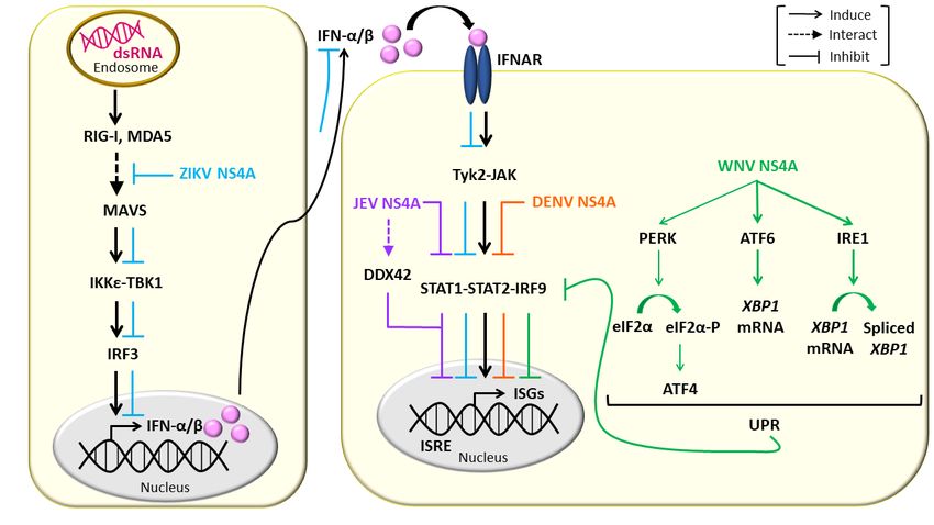
NS4Aisisaa16-kDa
NS4A 16-kDatransmembrane
transmembraneER ERresident
residentprotein
protein consisting
consisting of of an
an N-terminal
N-terminal cy-
cytosolic
tosolic region
region andandfourfour predicted
predicted transmembrane
transmembrane segments
segments (pTMSs)
(pTMSs) [41,42].
[41,42]. The The do-
domains
mains pTMS1
pTMS1 and pTMS3 and pTMS3
span thespanERthe ER membrane,
membrane, whilewhile
pTMS2pTMS2 is embedded
is embedded in the
in the lu-
luminal
minalof
leaflet leaflet
the ER of the ER membrane.
membrane. The C-terminal
The C-terminal pTMS4, pTMS4, referred
referred to astothe
as 2k
thefragment,
2k fragment,spans
spans
the the ER membrane
ER membrane (Figure(Figure
1) and1)acts
andasacts as a signal
a signal peptide peptide forER
for the thelocalization
ER localization of
of NS4B.
NS4B. The 2k fragment is removed from mature NS4A by the NS2B-NS3
The 2k fragment is removed from mature NS4A by the NS2B-NS3 protease [41,43]. It has protease [41,43].
It has
been been shown
shown that allthat
fourallpTMSs
four pTMSs
of DENV of DENV
NS4ANS4A possess
possess membrane
membrane targeting
targeting capa-
capabilities
and are able to mediate membrane association when expressed independently [41].[41].
bilities and are able to mediate membrane association when expressed independently NS4A
NS4A
has been hasshown
been shown
to playto aplay a major
major rolerole in the
in the flavivirus-inducedmembrane
flavivirus-induced membrane remodeling
remodel-
ing (Figure
(Figure 2). Heterologous
2). Heterologous expressionofofWNV
expression WNVNS4ANS4A retaining
retainingthethe2k2kfragment
fragment induced
induced
cytoplasmic membrane remodeling, resembling those events
cytoplasmic membrane remodeling, resembling those events observed upon WNV observed upon WNV infec-
infec-
tion. Removal of the 2k fragment on the other hand, impaired the
tion. Removal of the 2k fragment on the other hand, impaired the ability of WNV NS4A ability of WNV NS4A
to induce membrane remodeling, and resulted in the redistribution of this protein to the
to induce membrane remodeling, and resulted in the redistribution of this protein to the
Golgi apparatus [44]. In contrast to the WNV NS4A, proteolytic removal of the 2k frag-
Golgi apparatus [44]. In contrast to the WNV NS4A, proteolytic removal of the 2k fragment
ment appears to be necessary for heterologously expressed DENV NS4A to induce ER
appears to be necessary for heterologously expressed DENV NS4A to induce ER membrane
membrane remodeling which is similar to that induced by DENV infection [41]. These
remodeling which is similar to that induced by DENV infection [41]. These findings suggest
findings suggest that 2k regulates the function of NS4A in modulating cellular membranes
that 2k regulates the function of NS4A in modulating cellular membranes through distinct
through distinct mechanisms in different flaviviruses.
mechanisms in different flaviviruses.
Figure 1. A
Figure 1. membrane
A membrane topology
topologymodel
modelofofflavivirus
flavivirus NS4As. NS4Aconsists
NS4As. NS4A consistsofofananN-terminal
N-terminal cytosolic
cytosolic region
region andand four
four
predicted
predicted transmembrane
transmembrane segments:pTMS1,
segments: pTMS1,2, 2, 33 and
and 4 with the
the latter
latterreferred
referredtotoasasthe
the2k2kfragment.
fragment.The redred
The andand
yellow
yellow
triangles
triangles mark
mark thethe site
site ofofviral
viralNS2B-NS3
NS2B-NS3protease
proteaseand and host
host signalase
signalasecleavage
cleavagesites,
sites,respectively.
respectively.Thick purple
Thick and
purple dashed
and dashed
green arrows indicate specific amino acid (aa) residues in DENV and WNV NS4A, respectively, contributing to viral rep-
green arrows indicate specific amino acid (aa) residues in DENV and WNV NS4A, respectively, contributing to viral
lication. AH1 is the experimentally determined amphipathic helix 1 of DENV NS4A. The position of critical amino acids
replication. AH1 is the experimentally determined amphipathic helix 1 of DENV NS4A. The position of critical amino acids
is indicated.
is indicated.
TheN-terminus
The N-terminus of WNVWNV NS4ANS4Ahas hasbeen
beenshown
showntotocontribute
contributeto to
thethe
stability of the
stability of the
protein, which is essential for facilitating efficient WNV replication. Mutations P48,
protein, which is essential for facilitating efficient WNV replication. Mutations at P13, at P13,
D49D49
P48, and and
G66 G66
showed variable
showed defects
variable in viral
defects inreplication and membrane
viral replication remodeling
and membrane (Fig-
remodeling
ure 1, Table 1), with the mutations P13A and D49A causing lethal and mild
(Figure 1, Table 1), with the mutations P13A and D49A causing lethal and mild defects, defects, re-
spectively. The highly attenuated mutations P48A and G66A coincidingly
respectively. The highly attenuated mutations P48A and G66A coincidingly showed showed an in-
crease
an in a in
increase specific proteasome-mediated
a specific proteasome-mediated degradation of WNV
degradation of NS4A,
WNV leading to a sub-to a
NS4A, leading
stantial reduction in membrane proliferation, in particular the proliferation
substantial reduction in membrane proliferation, in particular the proliferation of of CM and
CM PCand
structures, and eventually resulting in inefficient viral replication [45]. The residues
PC structures, and eventually resulting in inefficient viral replication [45]. The residues P13,
P48P48
P13, andandG66 G66
of NS4As havehave
of NS4As been been
shown to be highly
shown conserved
to be highly within within
conserved WNV species, and
WNV species,
and between members in the genus Flavivirus [46,47]; thus, these residues are highly likely
to contribute to the protein stability.Viruses 2021, 13,
Viruses 2021, 13, 2077
x FOR PEER REVIEW 64 of
of 19
19
Rolesof
Figure 2. Roles ofNS4As
NS4Asininflavivirus
flavivirusreplication.
replication.NS4As
NS4As induce
induce membrane
membrane remodeling
remodeling similar
similar to that
to that induced
induced by
by fla-
vivirus infection.
flavivirus NS4A
infection. NS4A and
andthe unprocessed
the unprocessedintermediates
intermediatescontaining
containingNS4A
NS4Aare
areessential
essentialcomponents
components of of viral
viral replication
complexes (VRCs), thatthat interacts
interacts with
with host
host factors
factors or
or other
other flavivirus
flavivirus NS
NS proteins
proteins to
to promote
promoteefficient
efficientviral
viralreplication.
replication.
It is still
2.2. NS4A Is anunknown how NS4Aofcontributes
Essential Component to the substantial
the Viral Replication Complex alterations of cellular
membranes, nevertheless, several mechanisms have
Viral RNA replication takes place in viral replication complexesbeen proposed [48,49]. Insertion
(VRCs) of am-
situated in
phipathic α-helix (AH) into one leaflet of membrane bilayers, as
VPs. VRCs are composed of viral dsRNA, viral proteins and essential host factors [52]. well as oligomerization of
membrane the
Although, proteins
exactin or above theofpolar
composition lipid–water
the VRCs is still interface
unknown, areallamong the mechanisms
flaviviral NS proteins
suggested NS4A
including to promotehave thebeeninduction
suggestedof membrane curvature
to be components of [48]. The closely
the VRCs, related hepati-
as co-localization of
tis C virus (HCV; genus Hepacivirus, family Flaviviridae) NS4B contains an N-terminal AH
NS proteins with viral dsRNA in VPs, and interactions among the NS proteins have been
that is able to induce membrane alterations when expressed independently [50]. Homo-
identified [37,39,55,68–72].
oligomerization of HCV NS4B has been reported, and is likely to be required for the
WNV replication has been found to take place in close association with cholesterol-
induction of membrane alterations [51]. A speculative mechanism to account for the cellu-
rich microdomains within the ER membrane [47]. WNV NS4A has been found to contain
lar membrane alterations induced by HCV NS4B is that NS4B induces membrane curvature
a potential cholesterol recognition/interaction amino acid consensus (CRAC) motif (L/V24-
by inserting its AHs into the membranes, and then homo-oligomerization makes large
X(1–5)-Y28-X(1–5)-R/K35) which is a 25L-(X)-29Y-(X)-36K motif, near its N-terminus [47]. This mo-
NS4B complexes that force membrane curvature [52].
tif is highly conserved among members of the Japanese encephalitis subgroup but shows
The cytosolic N-terminal region (amino acids 1 to 48) of DENV NS4A has been an-
low degrees of sequence similarities with the other members in the genus Flavivirus. Mu-
alyzed and found to contain two experimentally determined AHs (AH1: amino acids
tant viruses harboring either mutation of Y/S at position 28 or K/L at position 35 or double
4 to 10 (Figure 1, Table 1); AH2: amino acids 15 to 31) that are separated by an un-
mutation Y/S + K/L showed varying degrees of attenuated phenotypes, with the virus
structured linker [53]. The DENV NS4A (1 to 48) has been shown to bind tightly to
harboring
membranethe doubleparticularly
bilayers, mutation Y/S to +theK/L being extremely
negatively chargedattenuated followedDisruption
bilayer [42,53,54]. by the vi-
ruses harboring single mutation Y/S and K/L, respectively.
of the amphipathic character of AH1 by L6E; M10E mutations reduced the membrane These results suggest that the
CRAC motif within the WNV NS4A plays a significant role
binding of the DENV NS4A (1 to 48) [42,53], indicating that AH1 has a high affinity in facilitating efficient viral
replication.
for membranes. Importantly,
DENV NS4A the Y/S mutation
has also been was showntotoform
shown significantly impair the
homo-oligomers in ability
infected of
the mutant virus to recruit viral components and cellular factors
cells or when expressed independently [42,55]. The AH1 was found to have a significant known to localize to the
VRCs [73] to to
contribution effectively form VRCs at the of
the homo-oligomerization cholesterol-rich
DENV NS4A,microdomains
as the L6E; M10E within the ER
mutations
membrane
in the AH1 reduced homo-oligomerization (Figure 1, Table 1), but did not affect defec-
[47]. Moreover, virus harboring the Y/S mutation was also found to be its lo-
tive in induction
calization [42]. The of the CM/PC
pTMS1 structure
(amino acids[47].
50 Collectively,
to 76) has also these findings
been shownsuggest
to be athat the
major
CRAC motif within
determinant the N-terminus of the
for homo-oligomerization ofWNV
DENVNS4A playsSpecifically,
2 NS4A. a significant role inalone
pTMS1 promot-ex-
ing cytoplasmic membrane remodeling and VRC assembly to specific
hibited homo-oligomerization activity comparable to that of full-length NS4A, while the cholesterol-rich mi-
crodomains within the
cytosolic N-terminal ER (amino
region membrane,acidsthus facilitating
1 to 50) retained efficient
only 20%virus of thereplication. How-
full length NS4A
ever, whether the CRAC motif within WNV NS4A confers
homo-oligomerization activity. Single point mutations E50A and G67A in the pTMS1 direct binding with cellular
chloresterol remains inconclusiveand
decreased homo-oligomerization [47].stability of DENV 2 NS4A [55] (Figure 1, Table 1).
A conserved
While a peptide Pro–Glu–Pro–Glu
(amino acids 1 to (PEPE)
48) of motif
ZIKV in the hydrophobic
NS4A encompassing C-terminus of WNV
the entire cytosolic
NS4A has been
N-terminal regionshown has to be essential
been found tofor VRCa formation
form random coil (Figure 1, Table[56],
in solution 1). Mutations
the peptide in
the PEPE
(amino motif
acids 4 toimpaired VRC most
58) spanning formationof thewhich in turn
cytosolic abolished
N-terminal viraland
region RNA replication
a third of the
pTMS1 has been found to be partially folded in solution [57]. The peptide (amino acidsViruses 2021, 13, 2077 5 of 19
4 to 58) was found to contain two predicted AHs (AH1: amino acids 15 to 33; AH2: amino
acids 38 to 55) and shown to bind membranes, as its helical contents were increased in the
presence of liposomes [57], consistent with previous reports for DENV NS4A [42,53,54].
Moreover, the peptide (amino acids 4 to 58) was also found to form homotrimers even in
the absence of detergents or lipid membranes, suggesting that this part of the protein is
essential for homo-oligomerization of ZIKV NS4A [57] (Table 1).
All of these findings suggest that induction of cellular membrane remodeling by
flavivirus NS4As might be mediated by a mechanism similar to that used by HCV NS4B.
Nevertheless, the predicted membrane topology of DENV NS4A has suggested that the
N-terminal 49 residues (containing AHs [53]) are exposed to the cytosol [41]. In addition,
for a number of viral membrane-bound proteins involved in viral replication such as the
NS4A and NS5A proteins of HCV, GB virus and bovine viral diarrhea virus which contain
AHs [58–60], the AHs have been shown to play a role in mediating membrane association
of these viral proteins [59,61]. Therefore, flavivirus NS4As are more likely to induce
membrane curvature by associating their cytosolic N-terminal AHs to the membranes as
opposed to inserting them directly into the membranes.
Table 1. Contributions of specific amino acid residues within flavivirus NS4As to viral replication.
NS4A Amino Acid Residue Contribution Reference
Membrane binding, homo-oligomerization
L6 and M10 within AH1: aa 4 to 10
and viral replication
[55]
Homo-oligomerization, protein stability and
DENV NS4A E50 and G67
viral replication
Vimentin interaction to mediate the
aa 1 to 50 [62]
anchoring of VRCs to ER membrane
L48, T54 and L60 NS4A-NS4B interaction and viral replication [63]
P13 Viral replication
[45]
P48 and G66 Protein stability
Membrane remodeling, promoting VRC
Potential CRAC motif:
WNV NS4A 25 L-(X)-29 Y-(X)-36 K assembly at cholesterol-rich microdomains [47]
within the ER membrane
VRC formation and promoting the cleavage
P120-E121-P122-E123 (PEPE motif) [46]
of 2k from NS4A
aa 1 to 50 Regulating ATPase activity of NS3 helicase [64]
aa 4 to 58 (containing AH1: aa 15 to Membrane binding and
ZIKV NS4A [57]
33; AH2: aa 38 to 55) homo-oligomerization
Homo-oligomerization of DENV NS4A has been demonstrated to have a biological
importance in viral replication. The reductions in homo-oligomerization of DENV 2 NS4A
caused by either the L6E, M10E mutations in AH1 or the E50A and G67A mutations in
pTMS1, lead to attenuated viral replication [42,55] (Figure 1, Table 1), which was thought
to result from the decreased NS4A protein stability as a consequence of weakened NS4A
homo-oligomerization [55].
The reticulon (RTN) protein family is a group of membrane-bound proteins that are
primarily involved in promoting membrane curvature and vesicle formation [65,66]. Upon
WNV, DENV and ZIKV infection, RTN3.1A has been found to be recruited to the virus-
induced modified ER membranes comprising viral replication complexes. RTN3.1A was
shown to interact with WNV NS4A potentially, through the pTMSs at the N-terminus of
NS4A. Knockdown of RTN3.1A not only reduced WNV, DENV, and ZIKV replication, but
also promoted degradation of viral proteins particularly, NS4A, with the proteasome in part
contributing to the viral protein degradation. In addition, silencing of RTN3.1A affected
virus-induced membrane remodeling; specifically, the numbers and/or sizes of CMs, PCsViruses 2021, 13, 2077 6 of 19
and VPs were reduced, or VPs were aberrantly elongated, coinciding with an increase in
the number of immature virus particles. These findings suggests that RTN3.1A stabilizes
NS4A and functions cooperatively with the membrane-remodeling capability of NS4A
to facilitate virus-induced membrane remodeling for efficient flavivirus replication [67]
(Figure 2).
2.2. NS4A Is an Essential Component of the Viral Replication Complex
Viral RNA replication takes place in viral replication complexes (VRCs) situated in
VPs. VRCs are composed of viral dsRNA, viral proteins and essential host factors [52].
Although, the exact composition of the VRCs is still unknown, all flaviviral NS proteins
including NS4A have been suggested to be components of the VRCs, as co-localization of
NS proteins with viral dsRNA in VPs, and interactions among the NS proteins have been
identified [37,39,55,68–72].
WNV replication has been found to take place in close association with cholesterol-rich
microdomains within the ER membrane [47]. WNV NS4A has been found to contain a
potential cholesterol recognition/interaction amino acid consensus (CRAC) motif (L/V24 -
X(1–5) -Y28 -X(1–5) -R/K35 ) which is a 25 L-(X)-29 Y-(X)-36 K motif, near its N-terminus [47]. This
motif is highly conserved among members of the Japanese encephalitis subgroup but
shows low degrees of sequence similarities with the other members in the genus Flavivirus.
Mutant viruses harboring either mutation of Y/S at position 28 or K/L at position 35 or
double mutation Y/S + K/L showed varying degrees of attenuated phenotypes, with the
virus harboring the double mutation Y/S + K/L being extremely attenuated followed by
the viruses harboring single mutation Y/S and K/L, respectively. These results suggest that
the CRAC motif within the WNV NS4A plays a significant role in facilitating efficient viral
replication. Importantly, the Y/S mutation was shown to significantly impair the ability
of the mutant virus to recruit viral components and cellular factors known to localize to
the VRCs [73] to effectively form VRCs at the cholesterol-rich microdomains within the
ER membrane [47]. Moreover, virus harboring the Y/S mutation was also found to be
defective in induction of the CM/PC structure [47]. Collectively, these findings suggest
that the CRAC motif within the N-terminus of the WNV NS4A plays a significant role in
promoting cytoplasmic membrane remodeling and VRC assembly to specific cholesterol-
rich microdomains within the ER membrane, thus facilitating efficient virus replication.
However, whether the CRAC motif within WNV NS4A confers direct binding with cellular
chloresterol remains inconclusive [47].
A conserved Pro–Glu–Pro–Glu (PEPE) motif in the hydrophobic C-terminus of WNV
NS4A has been shown to be essential for VRC formation (Figure 1, Table 1). Mutations in
the PEPE motif impaired VRC formation which in turn abolished viral RNA replication
and virion production. The PEPE motif was also found to contribute to proteolytic cleavage
to remove the 2k fragment from WNV NS4A, as shown by the fact that the mutations in the
PEPE motif perturbed proteolytic processing at the NS2B-NS3 cleavage site upstream of
the 2k region, specifically at the first proline and downstream glutamic acid residues [46].
The authors of that study were inclined to believe that as the PEPE motif was in close
proximity to NS2B-NS3 cleavage site, the mutations were preventing NS2B-NS3 protease
accessibility and thus activity, resulting in incorrect processing of NS4A which impeded
VRC formation [46].
Upon DENV infection, vimentin, a component of intermediate filaments, is redis-
tributed to the perinuclear site, where it shows co-localization with DENV-induced ER-
derived membranous compartments, and with NS4A representing VRCs. Gene silencing
of vimentin substantially altered the distribution of VRCs in DENV-infected cells, and
the VRCs were diffused and spread throughout the cytoplasm, signifying a structural
contribution of vimentin in anchoring the VRCs to the perinuclear membrane. DENV
NS4A was shown to directly interact with vimentin via a specific region that lies within the
first 50 amino acid residues at the cytosolic N-terminal region of NS4A [62]. Collectively,
these findings suggest that DENV NS4A has a functional role in mediating the anchoring ofViruses 2021, 13, 2077 7 of 19
VRCs to the perinuclear membrane, thus facilitating efficient viral RNA replication (Table 1,
Figure 2).
A nuclear ribonucleoprotein polypyrimidine tract-binding protein (PTB) has been
shown to be involved in pre-mRNA processing [74], polyadenylation regulation [75] and
50 cap-independent translation of viral/cellular RNA mediated by an internal ribosome
entry site [76–79]. PTB has been found to regulate viral RNA transcription, viral protein
translation and viral production of a number of viruses such as picornavirus, coronavirus,
herpes virus [80–82] and hepatitis C virus [83–86]. PTB has been reported to bind to
untranslated regions of flavivirus genomes [87,88]. Interactions of PTB with the DENV
RNA genome and with DENV NS4A have been identified [89], suggesting that DENV NS4A
indirectly binds to DENV RNA genome by associating with PTB. Reducing PTB-DENV
RNA genome binding via knockdown of PTB reduced synthesis of the minus-strand RNA
intermediate which reduced DENV replication, demonstrating the biological significance
of these interactions in the DENV replication cycle [89] (Figure 2).
An unprocessed NS3–NS4A has been detected as a transient intermediate during
flavivirus polyprotein processing [90–92], and is thought to have a possible role as a
protease that is responsible for trans cleavage at the NS4B-NS5 junction [91]. The presence
of an NS3-NS4A intermediate has also led to a hypothesis that NS4A is an essential cofactor
of the NS3 helicase required for unwinding of viral RNA during replication. Based on
the in vitro enzymatic assay of the individual WNV NS3helicase (NS3hel), and a NS3hel
fused with the cytosolic N-terminal residues 1 to 50 of NS4A (NS3hel–NS4A), the NS3hel–
NS4A showed a dramatic decrease in ATPase activity, but a comparable oligonucleotide
duplex unwinding activity as compared to the individual NS3hel. The results showed that
while NS4A had no significant effect on the oligonucleotide duplex unwinding rate of the
NS3hel, the presence of NS4A allowed the NS3hel–NS4A to conserve energy in the course
of oligonucleotide duplex unwinding and enabling the NS3hel to sustain the unwinding
rate of the viral RNA under ATP-deficient conditions. NS4A is therefore suggested to
function as a cofactor that regulates the ATPase activity of NS3hel [64]. This findings
directly complements a study showing that HCV NS4A enhanced the ability of NS3hel to
bind RNA in the presence of ATP, thus acting as a cofactor for HCV NS3hel [93] (Figure 2).
NS1, another component of VRCs [17,37], has an essential but as yet unclear role in
viral RNA replication, as evidenced by the findings that mutations in YFV NS1 profoundly
inhibited RNA replication [18,94]. A YFV genome containing a large in-frame deletion
in the NS1 gene, YF∆SK, has been found to be severely defective in accumulation of
the minus-strand RNA intermediate [16], and was not complemented in trans by DENV
NS1 [95]. However, the RNA replication defect of YF∆SK could be restored by an adaptive
mutation in NS4A [95], indicating that the interaction between NS1 and NS4A is required
for viral RNA replication (Figure 2). DENV NS1 has been shown to physically interact with
the NS4A-2k-NS4B cleavage intermediate, but not with fully processed NS4A or NS4B,
and the interaction was found to play a critical role in viral RNA replication (Figure 2).
However, this interaction is not required for the role of NS1 in VP formation [96].
NS4B is a part of VRCs, as it has been found to colocalize with viral dsRNA and
NS3 in the perinuclear region [70] and NS4A and NS4B have been suggested to function
cooperatively in viral RNA replication based on their functional similarities. In addition to
NS4A, NS4B has been shown to play an important role in cellular membrane reorganization,
thus facilitating efficient viral RNA replication [44]. While WNV NS4A regulates the
ATPase activity of NS3hel [64], DENV NS4B directly interacts with NS3 and enhances the
overall helicase activity of NS3 by dissociating it from ssRNA and thereby enabling it to
bind to a new nucleotide duplex [71]. Similar to NS4A, NS4B has been found to have
a genetic interaction with NS1 to modulate viral RNA replication. The RNA replication
defect of WNV containing NS1 mutations (RQ10NK) could be rescued by a F86E mutation
in NS4B. A novel physical interaction between NS1 and NS4B has been demonstrated and
suggested to be a mechanism by which luminal NS1 conveys signals to the cytoplasm to
regulate RNA replication [97]. An interaction between NS4A and NS4B has been identifiedViruses 2021, 13, 2077 8 of 19
and has been demonstrated to be required for viral RNA replication. A recombinant DENV
1 bearing mutations in the N-terminal cytoplasmic portion of NS4A (in which residues 27 to
34 were replaced by the corresponding region from JEV) is defective in viral replication.
The replication defect can be restored by a non-synonymous mutation in the pTMS3 of
NS4B [98]. NS4A has been shown to directly interact with NS4B in DENV 2 infected cells
and when co-expressed independently. The determinants for the NS4A-NS4B interaction
are amino acids 40 to 76 spanning the pTMS1 (amino acids 50 to 73) of NS4A, and amino
acids 84 to 146 spanning the pTMS1 (amino acids 101 to 129) of NS4B [63]. As pTMS1 of
DENV 2 NS4A is required for both NS4A homo-oligomerization essential for induction
of membrane curvature [55], and the NS4A–NS4B interaction [63], this may suggest that
NS4A regulates the transition from VP formation to VRC formation through the switch
of pTMS1 binding from NS4A to NS4B. Mutations L48A, T54A and L60A in DENV NS4A
that affected the NS4A–NS4B interaction drastically reduced or abolished viral replication
(Figure 1, Table 1). On the other hand, mutations F71A and G75A in NS4A that had no effect
on the NS4A–NS4B interaction only slightly reduced viral replication [63]. These results
suggest a biological significance of the NS4A–NS4B interaction in DENV 2 replication.
3. NS4A Mediates Flavivirus Pathogenesis
3.1. NS4A Antagonizes the Interferon Response and Manipulates the Unfolded Protein Response
Interferon (IFN) response is a crucial innate antiviral mechanism of the host cells [99,100].
It is primarily initiated by the recognition of viral dsRNA intermediates by retinoic
acid-inducible gene I (RIG-I)-like receptors (RLRs), RIG-I and Melanoma differentiation-
associated gene 5 (MDA5), which are members of the DExD/H-box family of RNA helicases.
The recognition leads to the conformational change of RIG-I/MDA5 in such a way that
exposes its N-terminal caspase-recruitment domains (CARD), which are subsequently
bound by the CARD of mitochondrial antiviral adaptor protein (MAVS). MAVS then re-
cruits tumor necrosis factor (TNF) receptor-associated factor (TRAF) 3 and TRAF6 to its
C-terminus and activates downstream signaling molecules of the RIG-I/MDA5 pathway
including inhibitor of kappa-B kinase epsilon (IKKε), TANK-binding kinase 1 (TBK1) and
subsequently, interferon regulatory factor 3 (IRF3), which plays an important role in stimu-
lating the expression of type I IFN (IFN-I) [100–103]. IFN-α/β response occurs upon the
binding of IFN-I to IFN-α/β receptor (IFNAR) and subsequently, through the activation of
the Janus kinase/signal transducer and activator of transcription (JAK/STAT) pathway,
and the transcriptional induction of a number of IFN-α/β-stimulated genes (ISGs) medi-
ated through the IFN-α/β-stimulated response element (ISRE) [104], inducing an antiviral
state. However, flaviviruses have been shown to circumvent IFN antiviral activities, and
establish a successful infection in human [105], with flavivirus NS4As playing a crucial
role in counteracting IFN-I production [11,12,106–109].
ZIKV NS4A has been demonstrated to repress RLR signaling (Figure 3), as evidenced
by the finding that, when expressed independently in the presence of polyI:C (which
stimulates cellular RLR signaling, thus inducing IFN-I expression), ZIKV NS4A reduced
ISRE promoter activity (activated by IRF3/7 or STAT1/2) [108], and mRNA levels of IFN-
stimulated genes, ISG15 [108], interferon induced protein with tetratricopeptide repeats 1 and
20 -50 -oligoadenylate synthetase 1 [106,108]. However, ZIKV NS4A does not interfere with
Toll-like receptor (TLR) signaling as shown by that, when co-expressed with a modified
TLR3 that localizes to the plasma membrane (without stimulation of RLRs by polyI:C),
ZIKV NS4A affected neither the ISRE promoter activity nor the IFN beta 1 and TNF-α
mRNA level [108]. Furthermore, ZIKV NS4A has been demonstrated to suppress IFN-I
induction mediated by ectopic expression of ∆RIG-I (containing only the two CARDs
domain) [108], constitutively active RIG-I-1-228 [109] and MDA5 [108,109], as well as
downstream signaling molecules of the RIG-I/MDA5 pathway, MAVS, IKKε, TBK1, full-
length IRF3 (IRF3-FL) and regulatory domain-deleted IRF3-1-390 [109]. In addition, ZIKV
NS4A has been shown to interact with the CARD domain of MAVS but not RIG-I or
MDA5 [106,108]. ZIKV NS4A competed with RIG-I/MDA5 for the binding to MAVS [108],level [108]. Furthermore, ZIKV NS4A has been demonstrated to suppress IFN-I induction
mediated by ectopic expression of ΔRIG-I (containing only the two CARDs domain) [108],
constitutively active RIG-I-1-228 [109] and MDA5 [108,109], as well as downstream sig-
naling molecules of the RIG-I/MDA5 pathway, MAVS, IKKε, TBK1, full-length IRF3
Viruses 2021, 13, 2077 (IRF3-FL) and regulatory domain-deleted IRF3-1-390 [109]. In addition, ZIKV NS4A 9has of 19
been shown to interact with the CARD domain of MAVS but not RIG-I or MDA5 [106,108].
ZIKV NS4A competed with RIG-I/MDA5 for the binding to MAVS [108], and significantly
decreased the interaction between MAVS and its downstream effectors TRAF6 or TBK1
and significantly decreased the interaction between MAVS and its downstream effectors
[106]. ZIKV NS4A was therefore, suggested to be a dominant negative interactor of RLR
TRAF6 or TBK1 [106]. ZIKV NS4A was therefore, suggested to be a dominant negative
signaling, which competes with RIG-I/MDA5 for binding to the CARD of MAVS, and sub-
interactor of RLR signaling, which competes with RIG-I/MDA5 for binding to the CARD of
sequently modulates the downstream signaling, resulting in the suppression of IFN-I pro-
MAVS, and subsequently modulates the downstream signaling, resulting in the suppression
duction [106]. Interestingly, ZIKV NS4A has also been shown to suppress IFN-I induction
of IFN-I production [106]. Interestingly, ZIKV NS4A has also been shown to suppress IFN-I
upon vesicular
induction uponstomatitis
vesicularvirus (VSV) virus
stomatitis infection
(VSV)and promoteand
infection VSV replication
promote VSVin 293T cells,in
replication
suggesting
293T cells,that the antagonistic
suggesting effect of ZIKV
that the antagonistic NS4A
effect on IFN-I
of ZIKV NS4Aproduction could occur
on IFN-I production in
could
the context of actual viral infection [106].
occur in the context of actual viral infection [106].
NS4Asmodulate
Figure3.3.NS4As
Figure modulatethe theinterferon
interferonresponse.
response.NS4As
NS4Asmanipulate
manipulateRIG-I-like
RIG-I-likereceptor
receptorsignaling,
signaling,DDX42
DDX42helicase,
helicase,
JAK/STAT
JAK/STAT signalingand
signaling andthe
theunfolded
unfoldedprotein
proteinresponse
response(UPR)
(UPR)totocounteract
counteractthe
theinterferon-α/β
interferon-α/β (IFN-α/β)
(IFN-α/β) response.
response.
DENV has been reported to antagonize the IFN response in humans [110], and DENV
DENV has been reported to antagonize the IFN response in humans [110], and DENV
infection has also been shown to counteract the action of IFN in vitro [111]. Potential
infection has also been shown to counteract the action of IFN in vitro [111]. Potential
DENV-derived IFN antagonists have been identified based on the ability of each individual
DENV protein to facilitate the replication of IFN-sensitive Newcastle disease virus (NDV)
in human A549 cells transfected with the plasmids expressing the corresponding DENV
proteins, producing IFN. NDV replication was found to be enhanced in A549 expressing
DENV NS4A, NS4B and NS2A, as compared to the cell transfected with empty plasmid.
DENV NS4A, NS4B and NS2A were shown to reduce the activation of ISRE-54 promoter
(stimulated by IFN-α/β through the activation of the STAT1/STAT2/ISG factor 3 tran-
scription factor) to different extents in Vero cells after stimulation with exogenously added
IFN-α/β, suggesting that these DENV proteins interfere IFN-mediated signaling pathway
(Figure 3). Interestingly, co-expression of these DENV proteins in Vero cells was found to
further enhance their antagonistic effects in the IFN signaling [12]. In contrast, it has been
reported that heterologous expression of DENV NS4A-NS4B fusion protein in Vero cells
did not block IFN signaling unless the fusion protein was processed by the co-expressed
viral peptidase NS2B-NS3, indicating that the IFN-antagonist functions of DENV NS4A
and NS4B required proper viral polyprotein processing [11].
JEV NS4A has been demonstrated to have an antagonistic effect on the IFN-I signaling
by reducing the phosphorylation levels of STAT1 and STAT2, thus blocking the downstream
activation of the JAK-STAT signaling pathway. JEV NS4A was shown to specifically interact
with ATP-dependent RNA helicase DDX42. The DDX42 helicase is a member of the
DExD/H-box family of RNA helicases, like RIG-I and MDA5. Overexpression of DDX42
RNA helicase increased the activation of IFN-I signaling induced by exogenously addedViruses 2021, 13, 2077 10 of 19
IFN-β. These data suggest that DDX42 helicase acts as a dsRNA sensor that activates
the IFN-I response upon flavivirus infection, and the binding of JEV NS4A to the DDX42
helicase could block IFN-I response [107] (Figure 3).
The unfolded protein response (UPR) is an intracellular defense mechanism that is
activated in response to accumulation of unfolded or misfolded proteins in the ER occurring
upon exposure to various internal or external stresses. The UPR acts to increase ER volume
and ER components including chaperones required for protein folding, increase protein
degradation, and inhibit protein translation to decrease protein input. There are three main
branches of the UPR: the protein kinase-like ER resident kinase (PERK), the activating
transcription factor 6 (ATF6) and the inositol-requiring enzyme 1 (IRE1) [112]. Flaviviruses
have been found to up-regulate the UPR and manipulate downstream signaling to favor
their replication [113–116]. The strongly induced UPR observed upon WNV infection was
biased towards the ATF6 and IRE1 branches, as demonstrated by the strong up-regulation
of Xbp-1 expression and splicing, with a low level of PERK activation, as demonstrated
by a modest increase in ATF4 expression [113]. When expressed independently, WNV
NS4A (without or with 2k) was shown to strongly induce Xbp-1 expression and splicing,
coinciding with a reduction in STAT1 nuclear trafficking, an indicator of reduced IFN
signaling (Figure 3). A progressive C-terminal deletion of the hydrophobic regions of
WNV NS4A resulted in a stepwise decrease in Xbp-1 expression and restoration of STAT1
nuclear trafficking, demonstrating a correlation between the UPR and inhibition of IFN
signaling [113]. These findings suggest that the hydrophobicity of WNV NS4A is essential
for WNV to manipulate the UPR and to inhibit the IFN response to facilitate its replication.
3.2. NS4A Modulates Autophagy
Autophagy is an essential mechanism for maintaining cellular homeostasis, by which
unnecessary or dysfunctional cellular components are sequestered in double-membrane
vesicles (autophagosomes), which in turn fused with lysosome (autolysosome) and the
contents in the autolysosome are eventually degraded and recycled. It has been com-
monly found that flaviviruses often persist in the liver and kidney following the acute
phase of infection without cells undergoing induced cell death. Induction of autophagy
has been suggested as a mechanism utilized by flaviviruses to evade the host immune
response to establish a persistent infection. DENV and Modoc infection have been shown
to up-regulate autophagy in MDCK renal epithelial cells and fibroblasts, and subsequently
protect them from death. Inhibition of autophagy by inactivation of phosphoinositide
3-kinases (PI3K) using wortmannin or 3MA reduced protection against death conferred
by DENV and Modoc virus, indicating that protection induced by these viruses is medi-
ated by PI3K-dependent autophagy. In addition, in autophagy-deficient fibroblast cell
lines, Beclin+/− and ATG5−/− , protection conferred by these two viruses was also reduced,
emphasizing an important role of autophagy in flavivirus-induced protection against cell
death. Inhibition of autophagy also attenuated DENV and Modoc virus in MDCK cells,
indicating that autophagy enhances replication of these viruses in such cell type. When
expressed independently, DENV NS4A and Modoc virus NS4A were the only viral proteins
that protected MDCK cells against death in a manner similar to that of the live viruses
and were also shown to induce PI3K-dependent autophagy. These findings suggest that
flavivirus NS4A plays a major role in the up-regulation of PI3K-dependent autophagy in-
duced upon flavivirus infection, which confers protection of cells against death, providing a
well-protected host cell for replication of flaviviruses during their persistent infection [117]
(Figure 4).expressed independently, DENV NS4A and Modoc virus NS4A were the only viral pro-
teins that protected MDCK cells against death in a manner similar to that of the live vi-
ruses and were also shown to induce PI3K-dependent autophagy. These findings suggest
that flavivirus NS4A plays a major role in the up-regulation of PI3K-dependent autoph-
Viruses 2021, 13, 2077 agy induced upon flavivirus infection, which confers protection of cells against 11 death,
of 19
providing a well-protected host cell for replication of flaviviruses during their persistent
infection [117] (Figure 4).
4. NS4As modulate
Figure 4. modulate developmental
developmental processes. NS4As modulate autophagy to facilitate persistent infection and
manipulate
manipulate aa number
numberof ofsignaling
signalingpathways,
pathways,i.e.,
i.e.,Akt-mTOR,
Akt-mTOR,ANKLE2/VRK1,
ANKLE2/VRK1,JAK/STAT
JAK/STATand Notch
and Notchsignaling resulting
signaling in
resulting
in developmental
developmental defects.
defects.
3.3. NS4A
3.3. NS4A Causes
Causes Developmental
Developmental Defects
Defects
ZIKV infection
ZIKV infectionis is
known
knownto cause microcephaly
to cause and other
microcephaly anddevelopmental defects [118–120].
other developmental defects
[118–120]. ZIKV infection has been shown to impair growth and proliferationpluripotent
ZIKV infection has been shown to impair growth and proliferation of induced of induced
stem cells (iPSC),
pluripotent iPSC-derived
stem cells neural stem neural
(iPSC), iPSC-derived cells (NSCs) and human
stem cells (NSCs) fetal neural stem
and human cells
fetal neu-
(fNSCs) [121–123]. Ectopic expression of either ZIKV NS4A or NS4B in human
ral stem cells (fNSCs) [121–123]. Ectopic expression of either ZIKV NS4A or NS4B in hu- fNSCs in-
hibited
man neurosphere
fNSCs inhibitedformation
neurosphere andformation
reduced neurosphere
and reducedsize. Interestingly,
neurosphere size. co-expression
Interestingly,
of ZIKV NS4A and NS4B further reduced neurosphere formation
co-expression of ZIKV NS4A and NS4B further reduced neurosphere formation and average neurosphere
and av-
size. However, co-expression of DENV NS4A and NS4B did not show any significant
erage neurosphere size. However, co-expression of DENV NS4A and NS4B did not show
impairment of neurosphere formation. Individual expression or co-expression of ZIKV
any significant impairment of neurosphere formation. Individual expression or co-expres-
NS4A and NS4B also reduced proliferation rates of fNSCs, and differentiation rates of
sion of ZIKV NS4A and NS4B also reduced proliferation rates of fNSCs, and differentia-
fNSCs into neurons or astrocytes. Furthermore, ZIKV infection was shown to induce
tion rates of fNSCs into neurons or astrocytes. Furthermore, ZIKV infection was shown to
autophagy in fNSCs, which in turn promotes ZIKV replication. Individual expression
induce autophagy in fNSCs, which in turn promotes ZIKV replication. Individual expres-
of either ZIKV NS4A or NS4B showed subtle effects on autophagy induction, whereas
sion of either ZIKV NS4A or NS4B showed subtle effects on autophagy induction,
co-expression of these two ZIKV proteins resulted in a significant up-regulation of au-
whereas co-expression of these two ZIKV proteins resulted in a significant up-regulation
tophagy. ZIKV NS4A was further shown to interact with NS4B in cells, suggesting that
ZIKV NS4A and NS4B function cooperatively to induce efficient autophagy upon ZIKV
infection [122]. Akt-mTOR signaling is essential for neurogenesis by fNSCs and for the
induction of autophagy [124]. Akt, a central signaling molecule in the PI3K pathway
upstream of mTOR, plays crucial roles in brain development [125], and non-functional
Akt mutation leads to microcephaly [126]. Inhibition of mTOR in the developing brain
also causes microcephaly, and inactivation of mTOR by AMPK and p53 signaling induces
autophagy [127–129]. Individual expression of either ZIKV NS4A or NS4B in fNSCs sup-
pressed Thr308 and Ser437 phosphorylations of Akt, whereas co-expression of these two
ZIKV proteins intensified the suppressing effects and consequently led to reduced levels
of mTOR phosphorylation at Ser2448. Overexpression of the constitutively active form
of Akt3 (myr-HA-Akt3 E17K) in fNSCs was shown to down-regulate autophagy induced
by ZIKV infection or NS4A-NS4B co-expression. Collective these findings suggest that
ZIKV NS4A and NS4B impair the neurogenesis of fNSCs and increase autophagy through
inhibition of the Akt-mTOR signaling pathway [122] (Figure 4). However, these results
seem to be in contradiction with a study demonstrating that ZIKV infection in neuronal
and glia cells activated the mTOR complex (mTORC) 1 and mTORC2, which subsequentlyViruses 2021, 13, 2077 12 of 19
suppressed autophagy, resulting in viral protein accumulation and progeny virus produc-
tion [130]. The contradictory findings as to the roles of mTOR and autophagy in ZIKV
infection could be a consequence of different cell types, experimental model systems or the
temporality of the events being evaluated. Moreover, the different molecular tools used
to study mTOR signaling whether phosphorylation status of mTOR at S2448 or of mTOR
substrates, p70S6K, ULK1, and Akt might account for the conflicting findings [130].
Ankyrin repeat and LEM domain containing 2 (ANKLE2) has been shown to be asso-
ciated with hereditary microcephaly, as mutations in ANKLE2 causes microcephaly in
humans [131,132] and Drosophila [133]. The functions of ANKLE2 have also been shown to
be evolutionarily conserved from Drosophila to human, as expression of human ANKLE2
in Drosophila Ankle2 heterozygous hypomorphic mutants (Ankle2A ) rescues the pheno-
type [132]. ZIKV NS4A has been found to physically interact with ANKLE2 in human cells,
and ectopic expression of ZIKV NS4A in Drosophila larval brain resulted in microcephaly,
increased apoptosis, and reduced proliferation of neuroblasts. In comparison with ZIKV
NS4A, DENV 2 NS4A was shown to interact with ANKLE2 with a lower affinity, without
significantly inducing microcephaly, consistent with the fact that DENV does not cause
microcephaly in human. Ectopic expression of ZIKV NS4A in Drosophila Ankle2A mutants
led to a more severe microcephaly phenotype. The microcephaly phenotype caused by
ectopic expression of ZIKV NS4A was found to be rescued by ectopic expression of hu-
man ANKLE2. These data suggest that ZIKV NS4A interacts with the ANKLE2 protein
and inhibits ANKLE2 function, thus contributing to ZIKV-induced microcephaly [134]
(Figure 4).
ANKLE2 has been found to be localized to the ER and nuclear envelope, similar
to ZIKV NS4A. Disruption of Ankle2 resulted in an aberrant nuclear envelope and ER
distribution, leading to the release of a protein kinase Ballchen (ball; Drosophila homolog) or
Vaccina-Related Kinase 1 (VRK1; human homolog) into the cytosol of fly neuroblasts and
human primary fibroblasts, respectively. This was found to be associated with abnormal
localization of Par-complex, i.e., atypical protein kinase C (aPKC), Par-6, Bazooka (Baz), and
Miranda (Mira), which are required for establishing polarity during asymmetric division
of neuroblasts in Drosophila, as well as with spindle orientation defects and reduced
aPKC phosphorylation. Removal of one copy of ball or lethal(2) giant larvae (l(2)gl) in
the Ankle2A mutant rescued the microcephaly phenotype, suggesting that function of
ANKLE2 is modulated by aPKC and l(2)gl levels. Similar to the Ankle2A mutant, ectopic
expression of ZIKV NS4A in Drosophila neuroblasts not only caused microcephaly [134]
but also resulted in an aberrant apical aPKC localization, Mira domain expansion and
spindle orientation defects. These phenotypes induced by ectopic expression of ZIKV
NS4A were rescued by removing a single copy of ball or l(2)gl. These findings suggest that
ZIKV-induced microcephaly is mediated by ZIKV NS4A which hijacks the ANKLE2-ball
(VRK1) pathway and affects asymmetric distribution of cell fate determinants, resulting in
neuroblast division and brain development defects [135] (Figure 4).
Apart from acting as a key regulator of IFN signaling, the JAK/STAT pathway has also
been demonstrated to have a pleiotropic function in regulating tissue development [136].
ZIKV infection in Drosophila has been shown to induce up-regulation of negative regulators
of JAK/STAT signaling, E(bx) and suppressor of cytokine signaling 36E (Socs36E). Eye-
specific overexpression of NS4A resulted in a significant reduction in the developing eye
size [137], a phenotype also observed as a consequence of loss of function of the hopscotch
(hop) gene (encoding JAK) [138,139]. The reduced eye size caused by ZIKV NS4A over-
expression correlated with the reduction in expression levels of the targets of JAK/STAT
signaling, chinmo, Mo25 and domeless, and was linked with a reduced rate of cell prolifer-
ation in the eye imaginal epithelia, although the rate of apoptosis remained unaffected.
Overexpression of ZIKV NS4A together with the dominant-negative form of domeless,
or in combination with STAT1 knockdown, resulted in a synergistic reduction in eye size,
while co-expression of ZIKV NS4A with activated Hop kinase partially rescued the eye
overgrowth. These data demonstrate the interaction between ZIKV NS4A and JAK/STATViruses 2021, 13, 2077 13 of 19
signaling components [137]. Apart from regulating eye development, JAK/STAT signaling
has also been shown to regulate wing development [140,141]. Wing-specific overexpres-
sion of ZIKV NS4A resulted in thickening of the wing vein, a phenotype characteristic
also found upon overexpression of Socs36E (a negative regulator of JAK/STAT signal-
ing), and mutation in Notch signaling [137]. ZIKV NS4A overexpression was shown to
reduce expression of Notch protein as well as Wg and Cut [137], which are targets of
Notch signaling [142,143]. Collectively, these findings suggest that ZIKV NS4A mediates
ZIKV-induced restricted eye and wing growth, through downregulation of JAK/STAT and
Notch signaling, respectively [137] (Figure 4).
4. Conclusions
NS4A is one of the flavivirus NS proteins that remains poorly characterized. Apart
from being known for its role in mediating flavivirus-induced cellular membrane remodel-
ing [41,44], NS4A also act as an essential component of VRCs, that physically interacts with
host factors or other flavivirus NS proteins to promote viral replication [62,63,89,95,98]
(Figures 1 and 2, Table 1). Interestingly, unprocessed intermediates containing NS4As also
have critical roles in viral RNA replication [64,96] (Figure 2). Importantly, NS4A contributes
to the pathogenesis of flaviviruses by counteracting the IFN response, modulating the UPR
and autophagy, as well as causing developmental defects, through hijacking of a number
of cellular signaling pathways [11,12,106–109,113,117,122,134,135,137] (Figures 3 and 4).
These highlight NS4A as a highly promising antiviral drug target. However, more studies
are required to gain further insights into the roles of NS4A in flavivirus infection, as its
critical role in a number of processes suggests that this protein and its interaction may be a
good candidate for the development of effective antivirals.
Author Contributions: Conceptualization, D.R.S.; writing—original draft preparation, P.K.; writing—
review and editing, P.K. and D.R.S. All authors have read and agreed to the published version of the
manuscript.
Funding: Paeka Klaitong is supported by a Mahidol University Post-Doctoral Fellowship (MU-
PD_2021_07). Duncan R. Smith is supported by Mahidol University (NDFR 28/2564) and the
National Research Council of Thailand (NRCT) and Mahidol University (NRCT5-RSA63015-03).
Conflicts of Interest: The authors declare no conflict of interest.
References
1. Simmonds, P.; Becher, P.; Bukh, J.; Gould, E.A.; Meyers, G.; Monath, T.; Muerhoff, S.; Pletnev, A.; Rico-Hesse, R.; Smith, D.B.; et al.
ICTV Virus Taxonomy Profile: Flaviviridae. J. Gen. Virol. 2017, 98, 2–3. [CrossRef] [PubMed]
2. Blitvich, B.J.; Firth, A.E. A Review of flaviviruses that have no known arthropod vector. Viruses 2017, 9, 154. [CrossRef] [PubMed]
3. Gould, E.A.; Solomon, T. Pathogenic flaviviruses. Lancet 2008, 371, 500–509. [CrossRef]
4. Ishikawa, T.; Yamanaka, A.; Konishi, E. A review of successful flavivirus vaccines and the problems with those flaviviruses for
which vaccines are not yet available. Vaccine 2014, 32, 1326–1337. [CrossRef]
5. Halstead, S.B. Dengvaxia sensitizes seronegatives to vaccine enhanced disease regardless of age. Vaccine 2017, 35, 6355–6358.
[CrossRef]
6. Lindenbach, B.; Thiel, H.J.; Rice, C.M. Flaviviridae: The viruses and their replication. Fields Virol. 2007, 1, 1101–1151.
7. Brinton, M.A.; Basu, M. Functions of the 30 and 50 genome RNA regions of members of the genus Flavivirus. Virus. Res. 2015, 206,
108–119. [CrossRef]
8. Dalrymple, N.A.; Cimica, V.; Mackow, E.R. Dengue virus NS proteins inhibit RIG-I/MAVS signaling by blocking TBK1/IRF3
phosphorylation: Dengue virus serotype 1 NS4A is a unique interferon-regulating virulence determinant. mBio 2015, 6, e00553-e15.
[CrossRef]
9. Liu, W.J.; Chen, H.B.; Wang, X.J.; Huang, H.; Khromykh, A.A. Analysis of adaptive mutations in Kunjin virus replicon RNA
reveals a novel role for the flavivirus nonstructural protein NS2A in inhibition of beta interferon promoter-driven transcription. J.
Virol. 2004, 78, 12225–12235. [CrossRef]
10. Liu, W.J.; Wang, X.J.; Mokhonov, V.V.; Shi, P.Y.; Randall, R.; Khromykh, A.A. Inhibition of interferon signaling by the New York 99
strain and Kunjin subtype of West Nile virus involves blockage of STAT1 and STAT2 activation by nonstructural proteins. J. Virol.
2005, 79, 1934–1942. [CrossRef]
11. Muñoz-Jordán, J.L.; Laurent-Rolle, M.; Ashour, J.; Martínez-Sobrido, L.; Ashok, M.; Lipkin, W.I.; García-Sastre, A. Inhibition of
alpha/beta interferon signaling by the NS4B protein of flaviviruses. J. Virol. 2005, 79, 8004–8013. [CrossRef] [PubMed]You can also read