Mechanisms for Maintaining Eukaryotic Replisome Progression in the Presence of DNA Damage
←
→
Page content transcription
If your browser does not render page correctly, please read the page content below
REVIEW
                                                                                                                                            published: 06 July 2021
                                                                                                                                   doi: 10.3389/fmolb.2021.712971

                                           Mechanisms for Maintaining
                                           Eukaryotic Replisome Progression in
                                           the Presence of DNA Damage
                                           Thomas A. Guilliam *
                                           Division of Protein and Nucleic Acid Chemistry, Medical Research Council Laboratory of Molecular Biology, Cambridge,
                                           United Kingdom

                                           The eukaryotic replisome coordinates template unwinding and nascent-strand synthesis
                                           to drive DNA replication fork progression and complete efficient genome duplication.
                                           During its advancement along the parental template, each replisome may encounter an
                                           array of obstacles including damaged and structured DNA that impede its progression and
                                           threaten genome stability. A number of mechanisms exist to permit replisomes to
                                           overcome such obstacles, maintain their progression, and prevent fork collapse. A
                                           combination of recent advances in structural, biochemical, and single-molecule
                          Edited by:       approaches have illuminated the architecture of the replisome during unperturbed
                Christopher Cooper,
                                           replication, rationalised the impact of impediments to fork progression, and enhanced
           University of Huddersfield,
                     United Kingdom        our understanding of DNA damage tolerance mechanisms and their regulation. This review
                        Reviewed by:       focusses on these studies to provide an updated overview of the mechanisms that support
              Nicholas Edward Dixon,       replisomes to maintain their progression on an imperfect template.
   University of Wollongong, Australia
                        Rodrigo Reyes,     Keywords: DNA replication, replisome, DNA damage tolerance, translesion synthesis, repriming, recoupling,
             McGill University, Canada     replication fork
                     Michael Trakselis,
      Baylor University, United States
                         Aga Gambus,       INTRODUCTION
             University of Birmingham,
                       United Kingdom      In eukaryotes, DNA replication is initiated from multiple origins across the genome (Robinson and
                  *Correspondence:
                                           Bell, 2005). Here, MCM double hexamers—loaded onto duplex DNA in G1 phase of the cell
                   Thomas A. Guilliam      cycle—are activated upon entry into S phase in a highly regulated manner, facilitated by the
         guilliam@mrc-lmb.cam.ac.uk        recruitment of numerous “firing factors” (Douglas et al., 2018). Many other origins remain dormant
                                           and only fire in response to replication impediments (Courtot et al., 2018). During activation, the
                   Specialty section:      lagging strand is ejected from each MCM ring to initiate unwinding of the parental duplex (Langston
        This article was submitted to      et al., 2017; Douglas et al., 2018), culminating in the formation of two active CMG (Cdc45-MCM-
                    Structural Biology,    GINS) holo-helicases encircling their respective leading-strand templates (Lewis and Costa, 2020).
               a section of the journal    Each CMG helicase translocates 3′–5′ on the leading strand to catalyse bidirectional fork propagation
   Frontiers in Molecular Biosciences
                                           and additionally acts as a hub to coordinate the replicative enzymes and binding partners that
            Received: 21 May 2021          collectively compose the replisome (Gambus et al., 2006; Fu et al., 2011; Pellegrini and Costa, 2016;
           Accepted: 25 June 2021
                                           Georgescu et al., 2017). Unwinding of the parental duplex provides a template for synthesis of
           Published: 06 July 2021
                                           nascent strands by the replicative polymerases (Pols) α, δ, and ε (Guilliam and Yeeles, 2020a). Since
                              Citation:    the DNA double helix is antiparallel and all DNA polymerases catalyse synthesis in the 5′–3′
  Guilliam TA (2021) Mechanisms for
                                           direction, the nascent leading strand is synthesised mostly continuously in the same direction as
   Maintaining Eukaryotic Replisome
       Progression in the Presence of
                                           replisome progression. Conversely, the lagging strand is replicated discontinuously in the opposite
                        DNA Damage.        direction through repeated priming and extension to form Okazaki fragments. These are
         Front. Mol. Biosci. 8:712971.     subsequently ligated together to generate a continuous daughter-strand molecule (Kainuma-
   doi: 10.3389/fmolb.2021.712971          Kuroda and Okazaki, 1975; Burgers and Kunkel, 2017).

Frontiers in Molecular Biosciences | www.frontiersin.org                         1                                             July 2021 | Volume 8 | Article 712971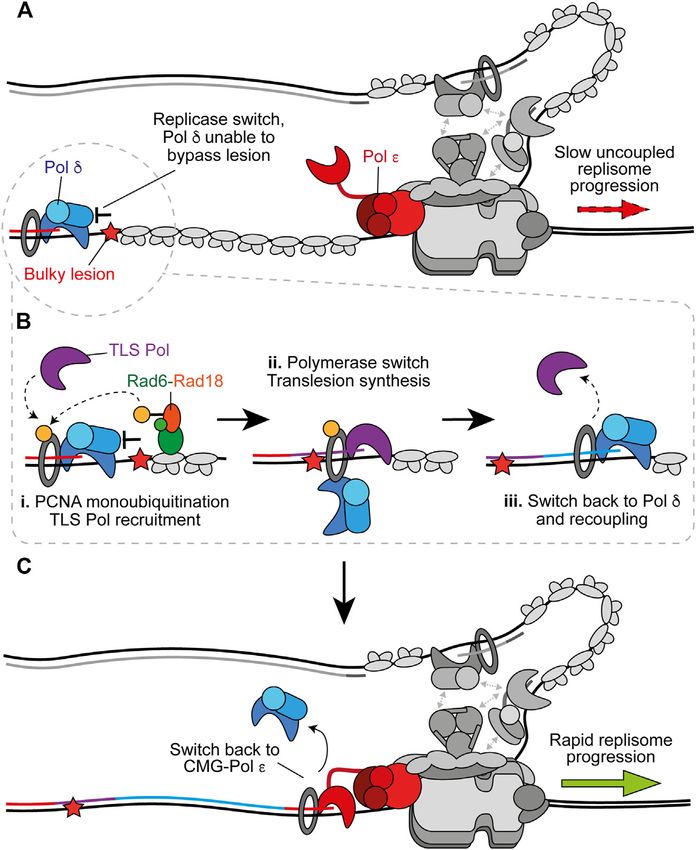
Guilliam                                                                                                       Mechanisms Maintaining Eukaryotic Replisome Progression

  FIGURE 1 | Replisome architecture during unperturbed progression. CMG (Cdc45-GINS-MCM2-7) tracks along the leading strand to unwind the parental duplex.
  Pol ε synthesises the leading strand continuously in complex with CMG. Pol α initiates discontinuous lagging-strand synthesis through regular priming. Primers are
  extended by Pol δ to form Okazaki fragments that are ligated together to produce a continuous daughter strand. Possible physical links between Pol α, Pol δ, Ctf4 (And-
  1), and CMG are indicated by gray arrows. Both Pol ε and Pol δ associate with PCNA during synthesis and RPA binds any exposed ssDNA. Csm3-Tof1(Tipin-
  Timeless) bind ahead of CMG and contact the parental duplex. Mrc1 (Claspin) may bind across one side of CMG spanning the N and C tiers. Synthesis by Pol ε, Pol δ,
  and Pol α is shown in red, blue, and green respectively.

   To efficiently complete DNA replication, yeast replisomes                              restart efficient CMG progression following slowing or stalling of
must traverse inter-origin distances up to 100 kilobases (kb)                            the replisome.
before fork convergence and termination (Sekedat et al., 2010;
Dewar and Walter, 2017). To cover such distances within S
phase, replisomes progress at an average rate of 1.5–2 kb min−1                          REPLISOME ARCHITECTURE AND
(Conti et al., 2007; Sekedat et al., 2010; Yeeles et al., 2017).                         UNPERTURBED PROGRESSION
However, genotoxic stress including DNA damage, DNA
secondary structures and repeat sequences, interstrand and                               The driving force of replisome progression is the ATP-dependent
DNA-protein crosslinks, and limiting nucleotide levels can                               DNA unwinding activity of MCM, a two-tiered hetero-hexameric
slow or stall replisome progression by inhibiting template                               ring of Mcm2–7 subunits. One tier is composed of amino-
unwinding and/or DNA synthesis (Zeman and Cimprich,                                      terminal domains (N tier) and the other of carboxy-terminal
2014). Since CMG encircles and tracks along the leading                                  AAA + ATPase domains (C tier), with ATP-binding sites located
strand, bulky obstacles located only in the lagging-strand                               at the subunit interfaces, which serve as the motor to power DNA
template are more readily bypassed by the replisome (Fu                                  template unwinding (Bell and Botchan, 2013). Loading of Cdc45
et al., 2011). Similarly, during lagging-strand replication,                             and GINS—a hetero-tetramer of Psf1–3 and Sld5—on to MCM
polymerase stalling at one Okazaki fragment does not affect                              during initiation forms the CMG holo-helicase and stabilises the
synthesis of subsequent fragments or replisome progression,                              N tier (Figure 1) (Costa et al., 2011; Yuan et al., 2016). Following
resulting only in the generation of a single-stranded (ss) DNA                           the reconstitution of origin-dependent eukaryotic DNA
gap (Taylor and Yeeles, 2018). Here, DNA damage tolerance                                replication with purified yeast proteins (Yeeles et al., 2015),
(DDT) occurs in a postreplicative gap-filling manner to                                   2D single-particle electron microscopy (EM) studies of
complete synthesis of the daughter strand molecule. In                                   reconstituted CMG revealed that the helicase translocates with
contrast, bulky leading-strand obstacles can block CMG                                   an N tier first orientation (Douglas et al., 2018). This is consistent
translocation while cessation of leading-strand synthesis                                with the orientation suggested by a previous 3D cryo-EM
significantly slows the replisome (Fu et al., 2011; Taylor and                            structure (Georgescu et al., 2017) and that observed in more
Yeeles, 2018, 2019). This necessitates the coordination of DDT                           recent studies (Goswami et al., 2018; Eickhoff et al., 2019; Yuan
mechanisms with the replisome to maintain canonical                                      et al., 2020a; Baretić et al., 2020).
replication fork progression in the presence of leading-strand                               Helicase translocation results from the allosteric coupling of
template aberrations.                                                                    ATP hydrolysis to movement of DNA-binding elements in each
   In recent years, structural, biochemical, and single-molecule                         subunit lining the centre of the ring (Enemark and Joshua-Tor,
approaches have begun to decipher the architecture and                                   2006). Structures of homo-hexameric helicases support a rotary
mechanism of unperturbed replisome progression, the effect of                            translocation mechanism (Enemark and Joshua-Tor, 2006;
impediments on this, and the details of DDT mechanisms used to                           Thomsen and Berger, 2009; Itsathitphaisarn et al., 2012; Gao
maintain progression. This review highlights these recent studies                        et al., 2019; Meagher et al., 2019). Here, five-subunits form a
to provide an overview of DDT mechanisms which maintain or                               right-handed staircase surrounding the DNA with the sixth

Frontiers in Molecular Biosciences | www.frontiersin.org                             2                                                July 2021 | Volume 8 | Article 712971
Guilliam                                                                                      Mechanisms Maintaining Eukaryotic Replisome Progression

disengaged. Sequential ATP binding, hydrolysis, and product                (Figure 1) and grip the parental DNA duplex ahead (Baretić et al.,
release facilitate subunit movement to escort DNA between                  2020). Fork rate enhancement by Csm3-Tof1 is dependent on
each subunit and through the staircase (reviewed in Enemark                Mrc1 (Yeeles et al., 2017) which appears to bind across one side of
and Joshua-Tor, 2008). However, studies of CMG translocation               CMG, spanning the N and C tiers (Baretić et al., 2020). The
are complicated by the observation that not all ATPase domains             mechanism by which Mrc1 stimulates fork rates is not currently
are equally important among the MCM subunits (Ilves et al.,                clear, however it also interacts with the flexible catalytic domain
2010; Eickhoff et al., 2019). Insight into this process has been           of Pol ε (Lou et al., 2008), potentially aiding its correct positioning
offered by high resolution cryo-EM structures of Drosophila                behind CMG (Zhou et al., 2017). Pol ε—comprised of the
CMG in three translocation states during unwinding (Eickhoff               catalytic subunit Pol2 and three non-catalytic subunits Dpb2,
et al., 2019). This study proposed an asymmetric hand-over-hand            Dpb3, and Dpb4—is a core component of the replisome required
rotational mechanism of CMG translocation in which binding of              for origin firing (Sengupta et al., 2013; On et al., 2014). It binds
Mcm5-Mcm3 to the ATPase staircase occurs as a block,                       stably to CMG via interactions between the noncatalytic
promoting a double step. This could explain the functional                 C-terminal domain of Pol2 with Mcm2 and Mcm5 (Goswami
asymmetry of ATPase domains in the MCM ring (Eickhoff                      et al., 2018) and, additionally, between Dpb2—the accessory
et al., 2019). A recent cryo-EM structure of human CMG                     subunit of Pol ε—with Mcm3 and the GINS subunit Psf1
bound to fork DNA is consistent with a sequential rotary                   (Sengupta et al., 2013; Sun et al., 2015; Goswami et al., 2018;
model of translocation, but the ATP-hydrolysis status of                   Yuan et al., 2020b). Dpb2 may also serve to direct the leading
Mcm3 differs from the asymmetric translocation model                       strand from CMG to the Pol ε active site (Yuan et al., 2020b).
proposed by Eickhoff et al. (2019). Further studies are                    These interactions place Pol ε in complex with and behind CMG
necessary to resolve if these differences represent species-               to couple leading-strand synthesis to template unwinding
specific variation in the CMG translocation mechanism.                      (Figure 1) (Guilliam and Yeeles, 2020a), which is essential for
    Regardless of the precise mechanistic details, the C tier motor        maximum fork rates (Yeeles et al., 2017; Taylor and Yeeles, 2018,
ring pulls DNA through the N tier to facilitate strand separation          2019). The homo-trimeric eukaryotic sliding clamp processivity
and replisome translocation. In contrast to classic strand-                factor, PCNA, additionally helps tether the flexible catalytic
exclusion models, where one strand is sterically excluded from             domain of Pol2 to the nascent leading strand, further
the central channel of the helicase while the other is pulled              contributing to maximum replisome progression rates in vitro
through, duplex DNA enters the N tier of yeast CMG a short                 (Yeeles et al., 2017).
distance (Georgescu et al., 2017) and, moreover, the central                  Unlike leading-strand synthesis, which is mostly continuous,
channel is wide enough to completely accommodate duplex                    lagging-strand replication requires repeated priming by Pol α and
DNA (Wasserman et al., 2019). The N tier is subdivided into                extension by Pol δ to generate Okazaki fragments (Figure 1)
a zinc finger (ZF) ring at the front of CMG followed by an                  (Guilliam and Yeeles, 2020a). How Pol α is recruited to CMG to
oligonucleotide-binding (OB) fold ring. A recent structure of              initiate primer synthesis is not currently understood. One
yeast CMG revealed that separation of the parental duplex occurs           potential mechanism is through Ctf4 (And-1), a trimeric hub
at the bottom of the ZF sub-ring (Yuan et al., 2020a). Here, the           that binds to CMG at the Cdc45-GINS interface (Yuan et al.,
lagging strand is diverted by the OB hairpin loops of Mcm3,                2019; Baretić et al., 2020; Rzechorzek et al., 2020) and interacts
Mcm4, Mcm6, and Mcm7, with a possible exit channel between                 with a range of proteins including Pol α (Gambus et al., 2009;
the ZF domains of Mcm3 and Mcm5. This mechanism, referred                  Simon et al., 2014; Villa et al., 2016; Guan et al., 2017; Kilkenny
to as the dam-and-diversion model, differs from the separation             et al., 2017). However, Ctf4 is not required for DNA replication
pin model, whereby a specific structural element separates the              in vitro (Yeeles et al., 2015, 2017; Kurat et al., 2017) and only
two strands. However, a putative strand-separation pin has been            minimally affects retention of Pol α at the replisome in yeast cells
identified in a subsequent structure (Baretić et al., 2020). This is       (Kapadia et al., 2020). Instead, the Ctf4-Pol α interaction may be
provided by a conserved phenylalanine in the N-terminal hairpin            more important for recycling of parental histones on to the
loop of Mcm7 that abuts the final base pair before strand                   lagging strand, than for primer synthesis (Evrin et al., 2018;
separation (Baretić et al., 2020). Whether this conserved                 Gan et al., 2018). Curiously, a recent cryo-EM report
residue is required for efficient unwinding remains to be                   demonstrated that Ctf4 can link two CMGs into a single
determined. In both structures, the path of the DNA template               “replication factory” (Yuan et al., 2019). However, further
is the same and therefore separation may be achieved by a                  studies are required to confirm whether this occurs during
combination of the two mechanisms.                                         active replication.
    Although activated CMG helicases reconstituted from an                    Until recently, Pol δ was assumed to function disconnected
origin in vitro are sufficient for robust DNA synthesis in the              from the replisome, synthesising each Okazaki fragment while
presence of Pol α and Pol ε, replication fork speeds are much              bound to PCNA before dissociating from DNA to permit ligation
slower than those observed in vivo in the absence of critical              (Bell and Labib, 2016; Lancey et al., 2020a). However, two single-
accessory proteins (Yeeles et al., 2015, 2017). These include the          molecule studies, one in vitro (Lewis et al., 2020) and one in vivo
“fork protection complex” (FPC) comprised of Mrc1 (Claspin)                (Kapadia et al., 2020), have challenged this assumption by
and Csm3-Tof1 (Tipin-Timeless). A recent high resolution cryo-             demonstrating that Pol δ is retained at the replisome for
EM structure of the FPC and CMG on forked DNA revealed that                multiple rounds of Okazaki fragment synthesis. It was
Csm3-Tof1 bind to MCM via Tof1 at the front of the replisome               suggested that Pol δ may interact with the replisome via Pol α

Frontiers in Molecular Biosciences | www.frontiersin.org               3                                         July 2021 | Volume 8 | Article 712971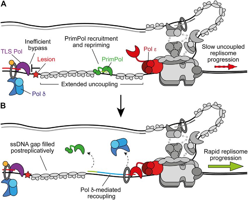
Guilliam                                                                                      Mechanisms Maintaining Eukaryotic Replisome Progression

(Huang et al., 1999; Johansson et al., 2004; Lewis et al., 2020).           CMG (Yang and Gao, 2018). Similarly, limiting nucleotide levels
However, in vivo the residency time of Pol δ at the replisome               and repetitive DNA sequences can stall synthesis without directly
exceeded that of Pol α, indicating that it interacts with CMG               stopping CMG translocation (Zeman and Cimprich, 2014;
independently, potentially through Ctf4 (Figure 1) (Bermudez                Devbhandari and Remus, 2020).
et al., 2010; Kapadia et al., 2020). Since lagging-strand synthesis             Stalling of Pol δ on the lagging strand does not affect ongoing
occurs in the opposite direction to CMG progression, these                  replisome progression or the synthesis of subsequent Okazaki
findings additionally posit the existence of lagging-strand                  fragments because priming occurs continually downstream, with
loops (Figure 1). Unlike Pol ε, Pol δ is not required for                   respect to helicase translocation direction (Figure 2A) (Taylor
maximum rates of replisome progression, but can support                     and Yeeles, 2018). This leaves a short ssDNA gap which can be
leading-strand synthesis, albeit more slowly, in the absence of             filled in behind the fork by classic DDT mechanisms (Leung et al.,
active Pol ε and is essential for complete lagging-strand synthesis         2019). Moreover, Pol δ is not required for maximum fork rates
(Yeeles et al., 2017). Consequently, although emerging evidence             (Yeeles et al., 2017). In contrast, stalling of Pol ε causes
supports Pol δ as a stable component of the replisome, coupling of          uncoupling of leading-strand synthesis from template
Pol δ to CMG does not appear to be important for maximum fork               unwinding, resulting in slow uncoupled fork progression
rates, likely because lagging-strand synthesis occurs in the                (Figure 2B). Here, template unwinding and lagging-strand
opposite direction to replisome translocation. However, recent              synthesis continue in the absence of leading-strand synthesis
biochemical and cellular studies revealed that Pol δ also plays a           but at a much-reduced rate (Taylor and Yeeles, 2018, 2019).
key role in the initiation of leading-strand synthesis. Here, it            Uncoupled forks can progress multiple kilobases (Taylor and
extends the first lagging-strand primer, generated by the                    Yeeles, 2018, 2019), generating long stretches of RPA-coated
oppositely translocating replisome, back across the origin to               ssDNA on the leading strand, as observed in UV-irradiated
couple synthesis to CMG-Pol ε and establish rapid fork                      yeast cells (Lopes et al., 2006). It is likely that uncoupling
progression (Garbacz et al., 2018; Aria and Yeeles, 2019).                  occurs as a result of the dissociation of the flexibly tethered
                                                                            catalytic domain of CMG-bound Pol ε from the 3′ end of the
                                                                            nascent leading strand and PCNA (Figure 2B). This has been
UNCOUPLING OF LEADING-STRAND                                                proposed to also occur spontaneously in the absence of genotoxic
SYNTHESIS AND FORK SLOWING                                                  stress to populate the leading strand with PCNA for nucleosome
                                                                            assembly and mismatch repair (Georgescu et al., 2017; Zhou et al.,
Since the central channel of CMG is wide enough to                          2017).
accommodate duplex DNA, many DNA lesions can readily                            If not rapidly resolved, the RPA-coated ssDNA accumulated
pass through the helicase, stalling DNA synthesis instead of                from uncoupling can activate the DNA replication checkpoint by
CMG translocation (Zeman and Cimprich, 2014). Damaged                       providing a platform for recruitment of Mec1-Ddc2 in yeast, or
nucleobases, abasic sites, and more bulky adducts such as                   ATR-ATRIP in vertebrates (Byun et al., 2005; Pardo et al., 2017).
pyrimidine dimers can be accommodated in the central                        This subsequently activates the checkpoint effector kinase Rad53
channel of CMG and therefore do not pose a steric block to                  in budding yeast, or CHK1 in vertebrates, to elicit the cellular
unwinding even when present in the leading-strand template                  stress response. In reconstituted DNA replication experiments,
(Taylor and Yeeles, 2018, 2019; Guilliam and Yeeles, 2021).                 Rad53 was recently shown to further slow uncoupled CMG
However, the replicative polymerases are typically considered               progression (Devbhandari and Remus, 2020). This may help
to be intolerant to DNA lesions and mismatched 3′ termini (Sale             prevent RPA depletion when uncoupling occurs for an
et al., 2012). The general structure of replicative polymerases is          extended period of time (Toledo et al., 2013). This mechanism
likened to a right hand; the catalytic site sits in the palm domain         was shown to be important in response to hydroxyurea (HU)
while the finger and thumb domains grip the template and primer              which inhibits ribonucleotide reductase (RNR), resulting in
(Sale et al., 2012). The intolerance of replicative polymerases is in       dNTP depletion (Gan et al., 2017). In rad53-1 mutant yeast
part due to “induced fit” conformational changes during catalysis,           cells, HU treatment caused asymmetric DNA synthesis
whereby the finger domain moves to switch the polymerase from                whereby extension proceeded much further along the lagging
an open to a closed conformation only when the incoming dNTP                strand than the leading strand as a result of uncoupling. This may
correctly pairs with the templating base (Johnson, 2008;                    be due to a requirement for higher dNTP levels for leading-strand
Freudenthal et al., 2013; Yang and Gao, 2018). The recent                   synthesis by Pol ε than lagging-strand synthesis by Pol δ.
structure of the Pol ε holoenzyme revealed that the finger                   Uncoupling in rad53-1 cells was suppressed by elevated dNTP
domain tilts 27° upon DNA-dNTP binding, changing the                        levels (Gan et al., 2017). Importantly, checkpoint kinases
enzyme from a gapped circle conformation to one that                        upregulate dNTP levels in response to replication stress
completely encircles DNA ready for catalysis (Yuan et al.,                  (Yeeles et al., 2013). Rad53 may therefore be important for
2020b). The compact catalytic site prevents the                             limiting uncoupling by both directly preventing excessive
accommodation of bulky DNA adducts, while 3′ mismatches                     template unwinding by CMG and promoting recoupling by
weaken DNA binding to the active site triggering relocation to the          elevating dNTP levels. Recent biochemical data demonstrate
3′–5′ proofreading exonuclease domain (Reha-Krantz, 2010).                  that direct slowing of replication forks by Rad53 is at least in
These features greatly improve fidelity and processivity but                 part mediated by Mrc1 (McClure and Diffley, 2021, Preprint).
also make the enzyme intolerant to lesions which pass through               Although Mrc1 functions upstream of Rad53 following

Frontiers in Molecular Biosciences | www.frontiersin.org                4                                        July 2021 | Volume 8 | Article 712971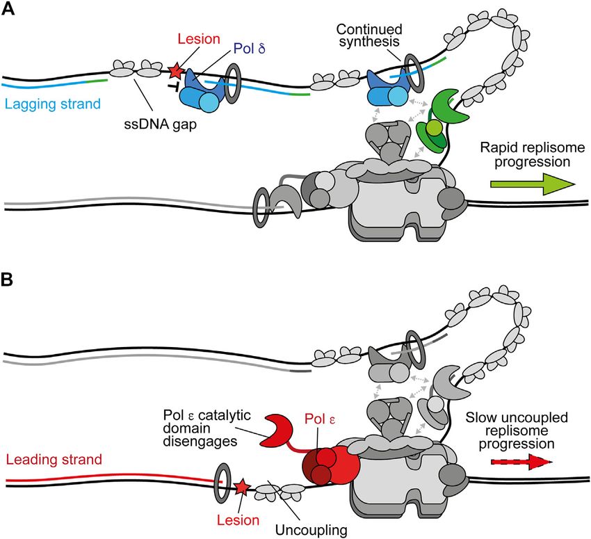
Guilliam                                                                                                        Mechanisms Maintaining Eukaryotic Replisome Progression

  FIGURE 2 | Polymerase stalling on the leading and lagging strands. (A) Stalling of Pol δ at a DNA lesion during synthesis of one Okazaki fragment does not affect
  synthesis of subsequent fragments or ongoing replication fork speeds. Instead, a persistent ssDNA gap is formed. Synthesis by Pol δ and Pol α are shown in blue and
  green respectively. Other replisome components are shown in gray for clarity (B) Stalling of Pol ε by a DNA lesion during leading-strand synthesis causes uncoupling due
  to disengagement of the flexible catalytic domain from the nascent strand and PCNA. Here, template unwinding and lagging-strand synthesis continue but at a
  much-reduced rate, resulting in the generation of RPA-coated ssDNA on the leading strand. Synthesis by Pol ε is shown in red.

replication stress, it is also a target for phosphorylation by Rad53                      (Figure 3A) (Guilliam and Yeeles, 2020b). Once recruited, Pol δ
and Mec1 (Alcasabas et al., 2001). Phosphorylation of Mrc1                                can readily recouple the leading strand to CMG-Pol ε whenever
prevents it from stimulating CMG, thereby slowing template                                uncoupling occurs spontaneously or due to factors which are more
unwinding (McClure and Diffley, 2021, Preprint).                                           readily tolerated by Pol δ than Pol ε (Figure 3B) (Guilliam and
   Interestingly, inhibition of RNR by HU also elevates reactive                          Yeeles, 2020b). Indeed, we recently showed that Pol δ can efficiently
oxygen species (ROS) in human cells (Somyajit et al., 2017). This                         bypass the oxidative single base lesions, thymine glycol and 8-
can cause replication fork slowing via a checkpoint-independent                           oxoguanine, to recouple uncoupled forks in vitro (Guilliam and
mechanism. Here, peroxiredoxin 2 (PRDX2) acts as a ROS                                    Yeeles, 2021). Similarly, Pol δ promotes recoupling following
sensor. At low ROS levels it binds to Timeless in an                                      stalling of leading-strand synthesis at hairpin-forming sequences
oligomeric state. However, elevated ROS levels disrupt                                    (Casas-Delucchi et al., 2021, Preprint). In conjunction, studies of
oligomerised PRDX2, causing its dissociation and the                                      vertebrate cells support a role for Pol δ in bypassing some lesions at
displacement of Timeless from the replisome, consequently                                 the replication fork (Hirota et al., 2015, 2016). This replicase switch
slowing fork progression (Somyajit et al., 2017).                                         mechanism allows the replisome to quickly resume rapid progression
                                                                                          following uncoupling, limiting ssDNA exposure on the leading strand
                                                                                          while maintaining synthesis by the high-fidelity replicative
REPLICASE SWITCHING AND                                                                   polymerases; in turn avoiding potentially mutagenic DDT
RECOUPLING                                                                                mechanisms or checkpoint activation. Similarly, Pol δ can excise
                                                                                          and correct errors made by Pol ε, whereas Pol ε can only correct its
Accumulating evidence suggests that, in addition to its role as the                       own misincorporations (Flood et al., 2015; Bulock et al., 2020). This
lagging-strand replicase, Pol δ replicates the leading strand whenever                    suggests that misincorporated nucleotides that are not removed by
synthesis is not coupled to CMG (Guilliam and Yeeles, 2020a).                             Pol ε may cause uncoupling, facilitating a switch to Pol δ before
Consistent with this, Pol δ acts as a “first responder” to stalling of                     proofreading of the error and recoupling of leading-strand synthesis.
leading-strand synthesis by outcompeting other polymerases,                               A recent cryo-EM structure of the yeast Pol δ holoenzyme revealed a
including free Pol ε, for the uncoupled nascent strand                                    high degree of flexibility between the Pol3 catalytic and Pol31–Pol32

Frontiers in Molecular Biosciences | www.frontiersin.org                              5                                               July 2021 | Volume 8 | Article 712971
Guilliam Mechanisms Maintaining Eukaryotic Replisome Progression
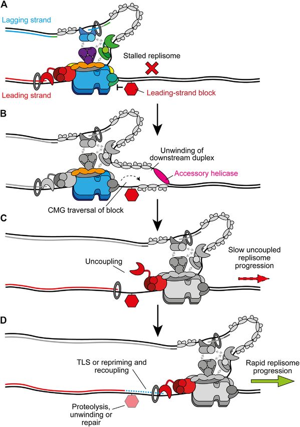
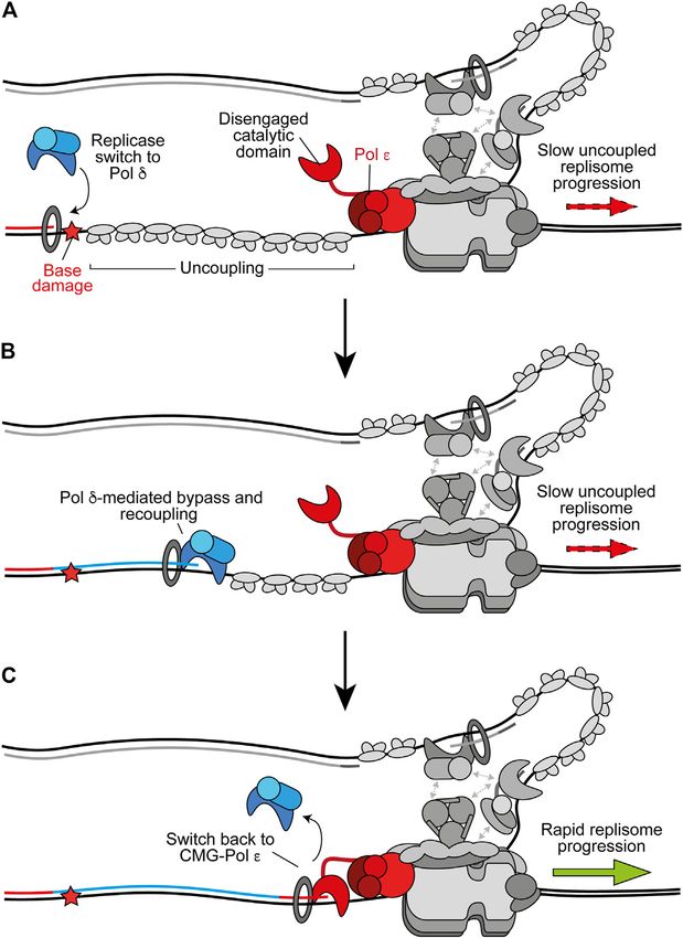
FIGURE 3 | Replicase switching and recoupling of leading-strand synthesis. (A) Upon uncoupling, a replicase switch occurs. Here, Pol δ is recruited to the
uncoupled nascent leading strand. Other replisome components are shown in gray for clarity. (B) When stalling of Pol ε occurs due to any factor that is more readily
tolerated by Pol δ, such as certain oxidative lesions, Pol δ directly extends the nascent leading strand through the impediment to recouple synthesis. (C) Upon recoupling
the nascent leading strand is handed back to CMG-bound Pol ε to restore rapid replisome progression. Synthesis by Pol ε and Pol δ is shown in red and blue
respectively.
regulatory subunits, which may aid proofreading and facilitate instead mediated by water, allowing PCNA to function as a
binding to a wider variety of DNA substrates (Jain et al., 2019). “water skate” to permit rapid and processive DNA synthesis
Pol δ is likely favored for recoupling due to its faster rate of while generating minimal resistance. Likewise, PCNA was
synthesis than the rate of uncoupled CMG progression, higher recently shown to enhance nucleotide incorporation rates by
affinity than free Pol ε for PCNA, and its ability to processively Pol δ from 40 nucleotides per second to more than 350 per
synthesise kilobases of DNA on an RPA-coated template in the second (Mondol et al., 2019). This rate of nucleotide
presence of PCNA (Langston and O’Donnell, 2008; Georgescu incorporation would allow the polymerase to rapidly catch up
et al., 2014; Yeeles et al., 2017; Sparks et al., 2019). Interestingly, a with uncoupled CMG, which has been estimated to unwind DNA
structure of Pol δ-PCNA recently demonstrated that the at ∼55 base pairs per minute (Sparks et al., 2019; Devbhandari
polymerase holds the DNA substrate such that it is positioned and Remus, 2020). It is not currently understood how the nascent
through the centre of the PCNA clamp without direct PCNA- leading strand is transferred from Pol δ back to CMG-Pol ε
DNA contacts (Zheng et al., 2020). DNA-clamp interactions are (Figure 3C). However, unlike free Pol ε, CMG-Pol ε does
Frontiers in Molecular Biosciences | www.frontiersin.org 6 July 2021 | Volume 8 | Article 712971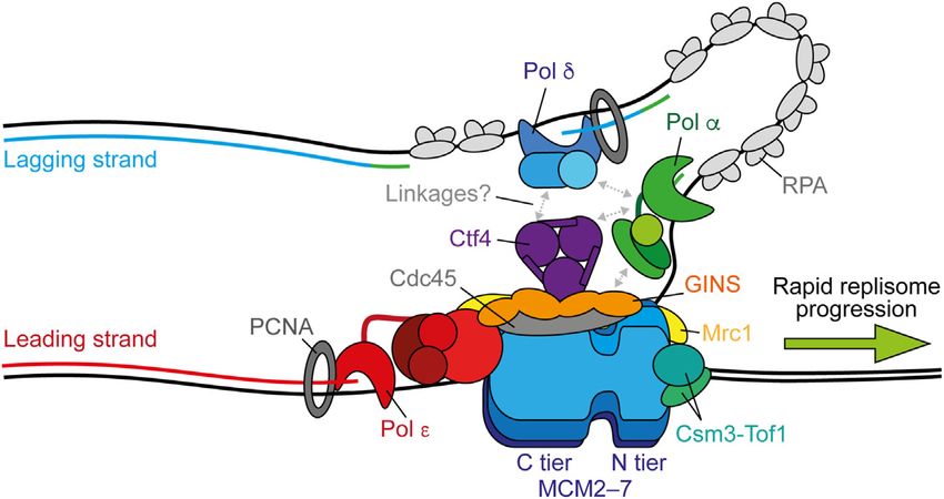
Guilliam                                                                                                    Mechanisms Maintaining Eukaryotic Replisome Progression

  FIGURE 4 | On the fly translesion synthesis can promote recoupling. (A) When Pol δ is recruited to the leading strand following uncoupling, but is unable to bypass
  the impediment, TLS is required prior to recoupling. Other replisome components are shown in gray for clarity. (B) i. RPA-coated ssDNA recruits Rad6-Rad18 which
  catalyse monoubiquitination of PCNA. This stimulates recruitment of a TLS polymerase. ii. The TLS polymerase extends the nascent strand past the impediment. This
  could occur without complete dissociation of Pol δ from PCNA. iii. After bypass a switch back to Pol δ facilitates rapid recoupling. (C) Pol δ hands the nascent
  leading strand back to Pol ε to restore canonical fork rates. Synthesis by Pol ε, Pol δ, and the TLS polymerase is shown in red, blue, and purple respectively.

outcompete Pol δ for the leading strand (Georgescu et al., 2014).                      encountering more bulky or distorting lesions such as a
It is possible handover occurs through collision release in which                      cyclobutane pyrimidine dimer (CPD), a well-characterised
Pol δ is ejected from PCNA upon running into uncoupled CMG,                            UV-induced photoproduct (Taylor and Yeeles, 2018; Guilliam
similar to the mechanism proposed to occur during Okazaki                              and Yeeles, 2020b). In these instances, specialised translesion
fragment synthesis (Langston and O’Donnell, 2008; Schauer and                          synthesis (TLS) polymerases are required for direct synthesis
O’Donnell, 2017). Regardless, rapid fork rates are reinstated                          across damage prior to recoupling (Guilliam and Yeeles, 2020b).
following Pol δ-mediated recoupling, suggesting CMG-Pol ε                              TLS that functions to promote recoupling is termed “on the fly”
efficiently resumes leading-strand synthesis downstream                                 TLS (Figure 4) to distinguish it from gap-filling TLS. The main
(Guilliam and Yeeles, 2020b).                                                          TLS polymerases in eukaryotes are those of the Y-family; Pol η,
                                                                                       Pol ι, Pol κ, and Rev1, in addition to the B-family polymerase Pol
                                                                                       ζ (Sale et al., 2012). Of these, only Pol η, Rev1, and Pol ζ are
TRANSLESION SYNTHESIS                                                                  present in budding yeast. The Y-family polymerases have
POLYMERASES                                                                            spacious solvent accessible active sites formed between the
                                                                                       palm and finger domains that make few, mostly non-specific,
Despite a greater ability to tolerate certain single-base lesions than                 contacts with the DNA template, permitting the accommodation
Pol ε (Guilliam and Yeeles, 2021), Pol δ also stalls upon                              of bulky lesions and template distortions (Fleck and Schär, 2004;

Frontiers in Molecular Biosciences | www.frontiersin.org                           7                                              July 2021 | Volume 8 | Article 712971
Guilliam Mechanisms Maintaining Eukaryotic Replisome Progression
Sale et al., 2012). They also possess an additional “little finger” their ubiquitin-binding ZF (Pol η and Pol κ) or helical
domain, not present in other polymerase families, which together ubiquitin-binding motifs (Pol ι and Rev1), in addition to
with the thumb domain grips duplex DNA and may play a role in canonical PCNA interacting peptide (PIP) box motifs in their
lesion specificity (Boudsocq et al., 2004; Fleck and Schär, 2004; non-catalytic C-terminal extensions (Leung et al., 2019). Rev1
Wilson et al., 2013). Compared to the replicative polymerases, the lacks a PIP box and instead interacts with PCNA via its
finger domain of the Y-family polymerases is stubbier and N-terminal BRCT and little finger domains (Guo et al., 2006;
remains in a closed conformation irrespective of DNA-dNTP Sharma et al., 2011).
binding, thereby removing the “induced fit” mechanism of dNTP The ubiquitin moiety is located on the back side of PCNA
selection (Vaisman and Woodgate, 2017). They also lack (Freudenthal et al., 2010). This may allow TLS polymerases to be
proofreading exonuclease activity and therefore the ability to recruited to the back face of monoubiquitinated PCNA while Pol
excise mispaired nucleotides. Consequently, the Y-family δ remains bound at the front (Freudenthal et al., 2010). This
polymerases gain an increased tolerance of template lesions at toolbelt model, in which multiple binding partners can occupy
the expense of fidelity and processivity. PCNA monomers not bound by Pol δ, is supported by the recent
Pol ζ is thought to function as an extender when two structure of Pol δ-PCNA-FEN1 in complex on DNA (Lancey
polymerases are required for lesion bypass. Here, a Y-family et al., 2020a) which suggests a toolbelt mechanism for flap
polymerase first incorporates nucleotides opposite the damage, cleavage during Okazaki fragment maturation. This study also
before Pol ζ takes over to catalyse extension beyond the lesion in a revealed that PCNA can tilt up to 20°, disrupting interactions
process that is typically error-prone (Johnson et al., 2000). Only between the catalytic subunit of Pol δ and PCNA that are critical
recently was the structure of Pol ζ determined (Malik et al., 2020), for DNA synthesis, while the polymerase remains bound to
revealing that it adopts a pentameric ring conformation PCNA via the PIP box located in its thumb domain (Lancey
composed of the catalytic subunit Rev3, two Rev7 subunits, et al., 2020a). The same group recently solved the structure of Pol
and the non-catalytic Pol31 and Pol32 subunits which are κ-PCNA-DNA and stalled Pol δ-PCNA-DNA to propose a
shared with Pol δ (Johnson et al., 2012; Makarova et al., mechanism of polymerase switching (Lancey et al., 2020b,
2012). Similar to the replicative polymerases, the finger Preprint). Therein, Pol δ releases the primer-template from
domain transitions from an open to a closed conformation the active site upon stalling, Pol κ binds to an exposed PCNA
upon binding the correct dNTP, conferring higher fidelity protomer in a flexible state and either actively displaces Pol δ, or
during nucleotide selection compared to the Y-family Pol δ independently dissociates, from DNA to form the final Pol κ
polymerases (Malik et al., 2020). However, key differences holoenzyme. Importantly, tilting of PCNA provides enough room
between Pol ζ and Pol δ explain the former’s penchant for to accommodate active Pol κ and retain Pol δ on PCNA via its
extending aberrantly paired 3′ ends (Johnson et al., 2000). In thumb domain PIP box. Pol δ might therefore hand over
Pol δ, the linker between the NTD and palm domain contacts the synthesis to the TLS polymerase without dissociating from
terminal base pair to prevent efficient extension of a mismatch. PCNA to allow a rapid switch back after lesion bypass
However, these contacts are not present in Pol ζ and although the (Figure 4B), before subsequent recoupling (Figure 4C).
polymerase contains an exonuclease domain, it is inactive. These Importantly, evidence supports a toolbelt model of polymerase
features make Pol ζ well suited to fulfill an extender role during switching in E. coli (Indiani et al., 2005; Kath et al., 2016) and
TLS and reveal why, despite having higher fidelity than the archaea (Cranford et al., 2017), giving credence to the prospect
Y-family polymerases (McCulloch and Kunkel, 2008), it is that a similar mechanism occurs in eukaryotes. However, it is also
involved in almost all damage-induced mutagenesis (Lawrence, possible that Pol δ fully dissociates from PCNA and DNA to
2002). permit TLS before reassociating to recouple synthesis.
In addition to PCNA, the C-terminal domain of Rev1 interacts
with the other Y-family TLS polymerases through PIP-like motifs
THE POLYMERASE SWITCH DURING TLS (Boehm and Washington, 2016), and binds the Rev7 subunit of
Pol ζ (Murakumo et al., 2001; Zhao and Washington, 2017).
On the fly TLS can rescue uncoupled forks when Pol δ alone is Numerous studies support a non-catalytic role for Rev1 in the
unable to recouple the leading strand (Figure 4A) (Guilliam and recruitment of other TLS polymerases (Nelson et al., 2000;
Yeeles, 2020b). However, Pol δ outcompetes TLS polymerases for Haracska et al., 2001; Ross et al., 2005; Edmunds et al., 2008).
binding to stalled nascent 3′ ends and in doing so limits One proposed mechanism is through the formation of “Rev1
mutagenesis by negatively regulating TLS when it is not bridges” in which a TLS polymerase is linked to PCNA via Rev1
required for recoupling (Guilliam and Yeeles, 2020b). RPA- without interacting directly with the clamp. Both PCNA toolbelts
coated ssDNA, generated by persistent stalling, recruits the and Rev1 bridges have been observed in single-molecule studies
E2–E3 ubiquitin ligase complex, Rad6–Rad18 (Watanabe et al., and they can interchange dynamically without dissociation
2004; Davies et al., 2008), which facilitates monoubiquitination of (Boehm et al., 2016). The relative contribution of PCNA
PCNA to promote a switch from Pol δ to TLS polymerase to monoubiquitination and Rev1 in recruiting and coordinating
facilitate lesion bypass (Figure 4B) (Hoege et al., 2002; Stelter and TLS is likely to be lesion specific (Wang and Xiao, 2020).
Ulrich, 2003; Bienko et al., 2005; Garg and Burgers, 2005; Plosky Indeed, accurate CPD bypass by Pol η does not require Rev1
et al., 2006; Guilliam and Yeeles, 2020b). Y-family TLS or Pol ζ but is dependent on PCNA monoubiquitination
polymerases are recruited to monoubiquitinated PCNA via (Kannouche et al., 2004; Bienko et al., 2005; Andersen et al.,
Frontiers in Molecular Biosciences | www.frontiersin.org 8 July 2021 | Volume 8 | Article 712971Guilliam Mechanisms Maintaining Eukaryotic Replisome Progression
2011; Guilliam and Yeeles, 2020b), whereas BaP-dG (N2-benzo[a] et al., 2013; Wan et al., 2013; Guilliam et al., 2015b; Guilliam
pyrene-dG) bypass by Pol κ is dependent on its interaction with and Doherty, 2017). PrimPol interacts directly with RPA and this
Rev1 (Ohashi et al., 2009). The requirement for Rev1 is more interaction is required for recruitment of the primase to the
important for two-polymerase TLS that also requires Pol ζ for ssDNA generated by uncoupling (Figure 5A) (Guilliam et al.,
extension and is typically error-prone (Livneh et al., 2010). 2015a, 2017; Šviković et al., 2019). The naming of PrimPol reflects
Moreover, the affinity of different TLS polymerases for Rev1 its ability to perform both DNA primase and polymerase
varies, in some cases being relatively weak and allowing activities, and similar to the TLS polymerases it can directly
competition (Pustovalova et al., 2016). Consequently, Rev1 bypass a number of lesions in vitro (Bianchi et al., 2013; Keen
may help select a different polymerase if the first cannot et al., 2014b). However, the catalytic domain of PrimPol does not
bypass the lesion following recruitment via monoubiquitinated resemble a canonical polymerase fold, completely lacks a thumb
PCNA, switching TLS from a one to a two-polymerase mode subdomain, makes almost no contacts with the primer strand, has
where necessary. In addition to Rev7, Rev1 was more recently very low inherent processivity, and does not noticeably interact
found to also interact with the PolD3 (Pol32 in yeast) subunit of with PCNA (Keen et al., 2014b; Guilliam et al., 2015a; Rechkoblit
human Pol ζ/Pol δ with higher affinity than it does with other et al., 2016). Likewise, although PrimPol can bypass a (6–4) T–T
Y-family polymerases (Pustovalova et al., 2016). This interaction photoproduct, its active site is not able to accommodate this
may help displace the “inserter” polymerase from Rev1 and lesion and it is likely bypassed by a looping out mechanism that
monoubiquitinated PCNA to facilitate extension by Pol ζ generates deletions (Bianchi et al., 2013; Rechkoblit et al., 2016;
beyond the lesion (Pustovalova et al., 2016). The sharing of Guilliam and Doherty, 2017). Moreover, the primase but not
Pol31-Pol32 between Pol δ and Pol ζ led to the proposal that polymerase activity of PrimPol is dependent on its noncatalytic
removal of Pol3 and recruitment of Rev3-Rev7 at the site of C-terminal ZF domain, which binds and selects the first 5′-
damage coordinates a switch between the two (Baranovskiy et al., nucleotide of the nascent primer strand (Keen et al., 2014b;
2012). In support, Pol3 is degraded in response to damage Martínez-Jiménez et al., 2018). An intact ZF is essential for
(Daraba et al., 2014). However, Pol ζ exists as a stable four- PrimPol’s function in vivo (Mourón et al., 2013; Keen et al.,
subunit complex (Rev3–Rev7–Pol31–Pol32) in all stages of the 2014a, 2014b; Kobayashi et al., 2016; Schiavone et al., 2016;
cell cycle irrespective of damage, potentially arguing against Šviković et al., 2019). Consequently, the available evidence
subunit exchange with Pol δ (Makarova et al., 2012). strongly suggests the primary function of PrimPol in vivo is as
a primase not a TLS polymerase (Guilliam and Doherty, 2017).
In avian cells, loss of PrimPol causes damage sensitivity,
REPRIMING replication fork slowing and growth arrest after damage
(Bailey et al., 2016; Kobayashi et al., 2016). In human cells,
Aside from TLS, uncoupled replication forks can be rescued by deletion of PrimPol does not affect survival after damage but
reinitiating leading-strand synthesis through the generation of a does result in delayed recovery, increased mutagenesis, and sister
new primer downstream of the impediment in a process termed chromatid exchanges (Bailey et al., 2019). However, loss of
repriming. Similar to ongoing Okazaki fragment synthesis after PrimPol in human cells lacking Pol η or Pol ζ significantly
Pol δ stalling on the lagging strand, this leaves behind a ssDNA increases sensitivity to damage (Kobayashi et al., 2016; Bailey
gap which can be filled by TLS or template switching. In et al., 2019). This suggests that PrimPol may function to rescue
reconstitution experiments with yeast proteins, Pol α could uncoupled forks when Pol δ alone or TLS cannot readily bypass a
readily synthesise primers on the lagging strand but was leading-strand obstacle (Figure 5A). In support of this, PrimPol
inefficient at repriming the leading strand due to inhibition by is required for the tolerance of impediments not efficiently
RPA (Taylor and Yeeles, 2018). This disparity between leading bypassed by TLS, including chain-terminating nucleosides
and lagging-strand priming efficiency suggests these two (Kobayashi et al., 2016), DNA secondary structures (Schiavone
processes are mechanistically distinct. It is likely that lagging- et al., 2016), R-loops (Šviković et al., 2019), cisplatin-induced
strand priming is facilitated by the position of Pol α in the adducts and hydroxyurea (Quinet et al., 2020), bulky DNA
replisome, while leading-strand repriming uses free Pol α and adducts that induce recombination (Piberger et al., 2020), and
is therefore sensitive to RPA concentrations. Coupling of leading- interstrand crosslinks (ICLs) (González-Acosta et al., 2020,
strand synthesis during initiation occurs via extension of a Preprint). Indeed, although Rev1 is involved in bypass of G4-
lagging-strand primer back across the origin by Pol δ quadruplexes (G4s)—DNA secondary structures formed by
(Garbacz et al., 2018; Aria and Yeeles, 2019), further arguing Hoogsteen base-pairing between guanines—containing long
against an efficient Pol α-mediated leading-strand priming loops (Schiavone et al., 2014), PrimPol is required for
mechanism in unperturbed conditions. However, it is possible tolerance of G4s that are more thermodynamically stable
that leading-strand repriming by Pol α occurs under conditions of (Schiavone et al., 2016). Moreover, on the fly TLS by Pol η
RPA exhaustion, or requires factors or modifications not present was recently found to be the primary mechanism to recouple
in the reconstituted system (Lopes et al., 2006; Fumasoni et al., replisomes in response to UV damage in human fibroblasts, while
2015; Toledo et al., 2017; Taylor and Yeeles, 2018). PrimPol compensated for loss of Pol η, with the resulting ssDNA
In higher eukaryotes, leading-strand repriming is catalysed by gaps filled in by template switching (Benureau et al., 2020,
a second archaeo-eukaryotic primase named PrimPol that is Preprint). Therefore, the relative efficiency of TLS bypass of a
absent in budding yeast (Bianchi et al., 2013; García-Gómez given impediment vs. repriming by PrimPol may dictate pathway
Frontiers in Molecular Biosciences | www.frontiersin.org 9 July 2021 | Volume 8 | Article 712971Guilliam Mechanisms Maintaining Eukaryotic Replisome Progression
FIGURE 5 | PrimPol-mediated repriming restarts leading-strand synthesis. (A) When replicase switching and on the fly TLS are inefficient at bypassing the leading-
strand impediment, extended uncoupling occurs. This stimulates the recruitment of PrimPol to RPA-coated ssDNA downstream and subsequent repriming. Other
replisome components are shown in gray for clarity. (B) Following repriming, Pol δ may extend the nascent primer to recouple synthesis to Pol ε and restore rapid
replisome progression rates. This leaves behind a ssDNA gap at the site of the lesion which may be filled in postreplicatively by TLS or template switching. Synthesis
by Pol ε, Pol δ, and PrimPol are shown in red, blue, and green respectively.
choice. Although importantly, TLS can also function to fill in the replication fork through fork reversal (Berti et al., 2020). Here, the
ssDNA gap generated after repriming (Figure 5B) (Daigaku et al., fork regresses by annealing the nascent daughter strands,
2010; Karras and Jentsch, 2010). generating a four-way “chicken foot” structure (Figure 6A).
Following repriming, the nascent primer is likely extended by This may allow extension of the nascent leading strand by
Pol δ to promote recoupling, as observed following TLS using the undamaged nascent lagging strand as a template
(Figure 5B) (Guilliam and Yeeles, 2020b). Interestingly, (Figure 6B), or place the damaged parental strand in a
PolDIP2 (Pol δ-interacting protein 2, PDIP38) interacts with dsDNA context to permit canonical repair before fork restart
Pol δ, PCNA, and PrimPol, enhancing the polymerase activity of (Figure 6C) (Berti and Vindigni, 2016).
the latter (Maga et al., 2013; Guilliam et al., 2016; Kasho et al., Fork reversal requires Rad51 which binds ssDNA to promote
2021). PolDIP2 might therefore promote primer extension by strand invasion during HR (Scully et al., 2019) and partially
PrimPol before also coordinating a switch to Pol δ for recoupling. replaces RPA on the ssDNA exposed by uncoupling, or nascent
A number of TLS polymerases also interact with PolDIP2 and loss strand resection, to promote reversal (Berti et al., 2020). How
of the protein causes a decrease in TLS in vivo (Tissier et al., 2010; these short stretches of Rad51 separated by RPA—termed
Maga et al., 2013; Tsuda et al., 2019). However, whether PolDIP2 metastable Rad51 filaments—are able to promote fork reversal
coordinates a switch back to Pol δ or another aspect of TLS or is not currently clear (Berti et al., 2020). However, intriguingly
repriming in higher eukaryotes remains to be determined. this is not dependent on the enzymatic strand exchange activity of
Rad51 (Mason et al., 2019). Additionally, following
monoubiquitination by Rad6-Rad18, PCNA can be
FORK REVERSAL polyubiquitylated (Ripley et al., 2020). In yeast, Rad5 serves as
the E3 ubiquitin-ligase (Hoege et al., 2002) and also has helicase
Template switching is a homologous recombination (HR)-like activity that can directly reverse forks (Blastyák et al., 2007).
mechanism that promotes damage tolerance by using the Humans have two Rad5-related proteins, HLTF and SHPRH,
undamaged nascent strand of the sister chromatid as a both of which can polyubiquitylate monoubiquitinated PCNA,
template (Branzei and Szakal, 2016). It can function with HLTF shown to catalyse fork reversal in vitro (Unk et al.,
postreplicatively to fill ssDNA gaps on the lagging strand, or 2006; Blastyák et al., 2010; Kile et al., 2015). Two additional
following repriming on the leading strand by PrimPol (Piberger translocases also mediate fork reversal in humans, ZRANB3 and
et al., 2020). However, template switching may also occur at the SMARCAL1, which are recruited by polyubiquitylated PCNA
Frontiers in Molecular Biosciences | www.frontiersin.org 10 July 2021 | Volume 8 | Article 712971Guilliam Mechanisms Maintaining Eukaryotic Replisome Progression FIGURE 6 | Fork reversal may maintain or restart replisome progression past leading-strand impediments. (A) Following uncoupling, fork reversal can be catalysed by Rad51, ZRANB3, SMARCAL1, and HLTF to generate a four-way chicken foot structure in a process which is not currently well understood. The fate of CMG here is not clear, one possibility is that it transitions on to duplex DNA downstream of the lesion. (B) The nascent lagging strand could serve as a template for extension of the nascent leading strand. Subsequent reversed branch migration, catalysed by RECQ1, may support fork restart if the extended 3′ end of the nascent leading strand is relocated downstream of the lesion. CMG may then transition back onto ssDNA to restart canonical replication. (C) Alternatively, fork reversal may place the lesion in a dsDNA context to permit repair, before resetting of the fork by RECQ1 or resection of nascent strands by DNA2-WRN. Again, CMG may transition back onto ssDNA to restart rapid fork progression. (D) Another possibility is that accessory helicases unwind the parental duplex downstream of the impediment following fork reversal. This may allow the establishment of a new replication fork which would require repriming of the leading strand. This would leave the chicken foot structure behind the new fork to be resolved following postreplicative repair of the lesion. and RPA-ssDNA respectively (Ciccia et al., 2012; Bétous et al., Reversed forks have been directly detected using transmission 2013; Kolinjivadi et al., 2017; Taglialatela et al., 2017). These electron microscopy (TEM) in human cells treated with a range of translocases fulfill non-redundant roles and might therefore genotoxic agents, suggesting it is a universal response to contribute to different steps or be required in different replication stress (Zellweger et al., 2015). In support, fork contexts (Berti et al., 2020). reversal has been observed as an ATR-dependent global Although the generation of reversed forks does not require response to ICLs, even at forks not directly challenged by stable Rad51 filaments, BRCA2-mediated stable Rad51 damage, to slow replication and promote repair (Mutreja filaments are required to protect reversed forks from et al., 2018). However, ATR phosphorylation of SMARCAL1 nucleolytic degradation following their formation (Schlacher has also been shown to limit fork remodeling (Couch et al., 2013). et al., 2011; Berti et al., 2020). Forks can subsequently be Moreover, a recent study of human fibroblasts failed to detect any restarted by unwinding and controlled resection by WRN- evidence of fork reversal by TEM in response to UV damage, even DNA2 (Thangavel et al., 2015) or through reversed-branch in the absence of Pol η, with repriming instead compensating for migration by RECQ1 (Figures 6B,C) (Berti et al., 2013). loss of TLS (Benureau et al., 2020, Preprint). In yeast, reversed Alternatively, a protected reversed fork could await forks were observed in repriming (Fumasoni et al., 2015) and resolution by the arrival of the convergent fork. However, checkpoint (Sogo et al., 2002; Lopes et al., 2006) mutants in prolonged fork reversal or de-protection can lead to response to bulky DNA lesions. A recent report showed that processing by structure specific endonucleases, producing a ATR-dependent up-regulation of PrimPol in cancer cells broken fork (Berti et al., 2020). This can be rescued by following multiple doses of cisplatin treatment suppresses fork break-induced replication, whereby strand invasion by the reversal, revealing a competition between the two mechanisms broken parental strand restarts replication but in an error (Quinet et al., 2020). This may be because PrimPol limits prone and conservative manner that does not rely on a uncoupling and RPA-ssDNA is a prerequisite for fork reversal canonical replisome (Kramara et al., 2018). (Zellweger et al., 2015). Therefore, fork reversal and repriming Frontiers in Molecular Biosciences | www.frontiersin.org 11 July 2021 | Volume 8 | Article 712971
Guilliam                                                                                                       Mechanisms Maintaining Eukaryotic Replisome Progression

  FIGURE 7 | Bypass of CMG-blocking impediments on the leading strand. (A) Bulky leading-strand impediments including DPCs, ICLs, and preformed G4s can
  pose a block to CMG translocation, causing stalling of the replisome. (B) CMG traversal of the impediment is aided by transient opening of the MCM2-7 ring and
  accessory helicases which unwind the downstream parental duplex. The specific accessory helicase involved depends on the nature of the lesion, see text for details.
  Note that the accessory helicase may act on either the leading or lagging strand. Other replisome components are greyed out for clarity. (C) Following CMG
  traversal of the impediment, the replisome is uncoupled and fork progression is slow. (D) Recoupling first requires resolution of the impediment and/or extension of the
  leading strand past the damage site. Alternatively, leading-strand synthesis may be reinitiated downstream by repriming before postreplicative repair of the damage. In
  the case of DPCs, proteolysis occurs before TLS and recoupling. G4s are unwound by FANCJ to promote bypass and recoupling. ICL bypass requires PrimPol-
  mediated repriming before recoupling, leaving an X-shaped structure for postreplicative repair. Upon recoupling, rapid fork rates resume. Recoupling is indicated by a
  blue dotted line and Pol ε synthesis is shown in red.

likely compete when on the fly TLS cannot promote efficient                                 dissociate to permit remodeling of the fork (Manosas et al., 2012).
bypass, with cell type, the nature of the replication impediment,                         However, in eukaryotes reloading of MCMs is inhibited in S
and complex ATR-orchestrated responses potentially dictating                              phase to prevent re-replication (Chen and Bell, 2011; Frigola
which pathway is favored.                                                                 et al., 2013). To function as a mechanism to restart or maintain
    The precise details of fork reversal remain to be determined. In                      progression of a replisome, fork reversal must not cause
particular, it is not clear what happens to the replisome during                          dissociation of CMG. One possibility is that uncoupled CMG
this process. In bacteriophage T4, the replicative helicase must                          traverses onto dsDNA downstream of the stalled fork junction,

Frontiers in Molecular Biosciences | www.frontiersin.org                             12                                               July 2021 | Volume 8 | Article 712971
Guilliam Mechanisms Maintaining Eukaryotic Replisome Progression
before moving back onto ssDNA to restart replication when the by Mcm10 (Figure 7B) (Sparks et al., 2019; Wasserman et al.,
canonical fork is restored (Figures 6A–C). Single-molecule 2019). Single-molecule experiments confirmed that CMG bypass
analyses recently demonstrated that MCM can transiently occurs before DPC degradation by the specialised protease SPTRN/
open to transition from ssDNA to dsDNA, before diffusing DVC1 (Wss1 in yeast) (Sparks et al., 2019). Interestingly, upon
back again to reform a functional replisome (Wasserman collision the replisome facilitates ubiquitylation of the DPC by the
et al., 2019). This is not dependent on the Mcm2–5 gate used E3 ubiquitin ligase TRAIP to promote proteolysis after CMG
for loading during origin licensing, but does require Mcm10 bypass. Ubiquitylation of the DPC also aids CMG bypass
which is essential for CMG activation. Here, Mcm10 may prevent (Sparks et al., 2019). TRAIP is proposed to bind the leading
dissociation of CMG upon opening of the ssDNA gate. However, edge of the replisome to fulfill this role which also positions it
Mcm10 also inhibits fork reversal by SMARCAL1 in vitro (Mayle to ubiquitylate an adjacent CMG after fork convergence at an ICL
et al., 2019). Moreover, in Xenopus egg extracts when CMG runs (Wu et al., 2019, 2021). Following bypass, CMG progression is
onto dsDNA at a lagging-strand nick it is ubiquitylated and initially slow due to uncoupling (Figure 7C). TLS after DPC
removed by the same pathway used during termination (Vrtis proteolysis subsequently recouples the fork (Figure 7D) (Duxin
et al., 2021). It is currently unclear how CMG encircling dsDNA et al., 2014; Sparks et al., 2019).
to permit fork reversal would resist unloading by the same ICLs also represent a block to replisome progression. In Xenopus
mechanism. Alternatively, CMG may rapidly reengage ssDNA extracts, ICL repair occurs following fork convergence at the lesion
downstream of the stalled fork junction, aided by accessory (Zhang et al., 2015). Here, TRAIP-dependent ubiquitylation of CMG
helicases (Figure 6D). Such a mechanism would require stimulates recruitment of the NEIL3 glycosylase to resolve psoralen
repriming and leave behind the chicken foot structure for and abasic ICLs (Wu et al., 2021). If NEIL3 fails to unhook the
resolution in a postreplicative manner (Berti et al., 2020). In crosslink, continued ubiquitylation of CMG by TRAIP triggers
either case, it is unclear what would trigger uncoupled CMG to replisome disassembly to permit repair of the ICL by the Fanconi
transition from ssDNA to dsDNA. Indeed, in reconstitution anemia pathway (Wu et al., 2021). Since both pathways depend on
experiments uncoupled CMG continues to unwind the fork convergence, they cannot be considered as mechanisms to
parental template for multiple kb downstream of a CPD maintain progression of individual replisomes. However, studies in
(Taylor and Yeeles, 2018, 2019). avian and mammalian cells suggest CMG can also traverse ICLs and
maintain progression via an alternative mechanism (Huang et al.,
2013, 2019; Ling et al., 2016; Mutreja et al., 2018). Here, FANCM
TRAVERSAL OF IMPEDIMENTS TO CMG associates with CMG in a FANCD2 and ATR-signalling dependent
TRANSLOCATION manner which triggers release of GINS from the replisome (Huang
et al., 2019). FANCM translocase activity and loss of GINS may then
Instead of causing uncoupling, bulkier replication impediments promote opening of the Mcm2-7 ring and allow traversal of the ICL.
can directly block CMG translocation (Figure 7A). One example FANCM also interacts with BTR (BLM/TOP3A/RMI1–2), and BLM
are DNA-protein crosslinks (DPCs), chromatin-bound proteins helicase activity is important for ICL traverse (Ling et al., 2016). BLM
which are covalently crosslinked to DNA by chemotherapeutics might therefore play an analogous role to RTEL1 during DPC bypass
or endogenous aldehydes (Stingele et al., 2017). Since dsDNA by unwinding the parental strands downstream of the ICL (Ling
enters CMG a short distance, with strand separation occurring at et al., 2016). BTR also interacts with both RPA (Wu et al., 2018) and
the bottom of the ZF sub-ring (Yuan et al., 2020a; Baretić et al., PrimPol (González-Acosta et al., 2020, preprint). Recent work
2020), DPCs might be expected to stall replisomes regardless of demonstrated that PrimPol participates in ICL bypass, likely by
which template strand they are located in. However, in Xenopus reinitiating leading-strand synthesis to restore rapid fork rates
extracts replisome progression is stalled much more significantly (González-Acosta et al., 2020, preprint). Traversal would leave
by a streptavidin block in the leading strand compared to the the X-shaped ICL structure behind the re-established replication
lagging strand (Fu et al., 2011). Purified yeast CMG is stalled by a fork to be repaired by the Fanconi anaemia pathway in a
streptavidin block located in either strand (Langston and postreplicative manner (Lopez-Martinez et al., 2016).
O’Donnell, 2017), but can bypass the lagging-strand block in Accessory helicases also assist the replisome in bypassing
the presence of Mcm10 (Langston et al., 2017). Here, Mcm10 is preformed G4 structures that can stall CMG when present in
hypothesised to bind the N tier of CMG to isomerise it to a steric the leading-strand template (Lerner and Sale, 2019). Experiments
exclusion model capable of bypassing impediments on the lagging in Xenopus extracts have recently delineated a mechanism for G4
strand (Langston et al., 2017). It is unknown if Mcm10 remains bypass similar to that employed for DPC traversal (Sato et al.,
stably bound to the replisome following initiation or if it interacts 2020, preprint). Following CMG collision with a G4, the
dynamically when required. accessory helicase DHX36 unwinds the parental duplex
Recent experiments in Xenopus extracts have revealed that the downstream of the structure. This allows CMG to bypass the
accessory helicase RTEL1 also stimulates bypass of a lagging strand G4 without unwinding through opening of the Mcm2-7 ring
DPC, although bypass still occurs in its absence (Sparks et al., (Sparks et al., 2019; Wasserman et al., 2019). A second helicase,
2019). Meanwhile, CMG stalling at a leading strand DPC is more FANCJ, then assists G4 unwinding to permit recoupling of the
extended and more sensitive to loss of RTEL1. Here, RTEL1 leading strand to the replisome. There is partial redundancy
generates ssDNA downstream of stalled CMG to facilitate DPC between these two helicases for CMG bypass and notably the
bypass by transient opening of the Mcm2-7 ring, potentially aided requirement for both is abolished by a convergent fork (Sato et al.,
Frontiers in Molecular Biosciences | www.frontiersin.org 13 July 2021 | Volume 8 | Article 712971You can also read