Interfaces between cellular responses to DNA damage and cancer immunotherapy - Genes Dev
←
→
Page content transcription
If your browser does not render page correctly, please read the page content below
Downloaded from genesdev.cshlp.org on August 1, 2021 - Published by Cold Spring Harbor Laboratory Press

REVIEW

Interfaces between cellular responses to
DNA damage and cancer immunotherapy
Domenic Pilger,1 Leonard W. Seymour,2 and Stephen P. Jackson1
1
 Wellcome Trust/Cancer Research UK Gurdon Institute, Department of Biochemistry, University of Cambridge, Cambridge CB2
1QN, United Kingdom; 2Department of Oncology, University of Oxford, Oxford, Oxford OX3 7DQ, United Kingdom

The DNA damage response (DDR) fulfils essential roles to                    trolled activity of DNA processing and repair factors can
preserve genome integrity. Targeting the DDR in tumors                      themselves generate DNA damage, thereby posing threats
has had remarkable success over the last decade, exempli-                   to genome integrity and cell survival. Consequently, dys-
fied by the licensing of PARP inhibitors for cancer thera-                  regulation and mutations in such DDR factors and their
py. Recent studies suggest that the application of DDR                      regulators have implications for human health and dis-
inhibitors impacts on cellular innate and adaptive im-                      ease (Jackson and Bartek 2009). Moreover, in recent years,
mune responses, wherein key DNA repair factors have                         DDR components have become accepted as attractive
roles in limiting chronic inflammatory signaling. Antitu-                   therapeutic targets for cancer therapy, with numerous
mor immunity plays an emerging part in cancer therapy,                      small molecule inhibitors targeting DNA repair enzymes
and extensive efforts have led to the development of                        now being explored clinically and with inhibitors of the
immune checkpoint inhibitors overcoming immune sup-                         DNA-repair enzyme PARP being approved for treating
pressive signals in tumors. Here, we review the current                     various tumor types (Piliè et al. 2019). Due to the frequent
understanding of the molecular mechanisms underlying                        loss or deregulation of DDR mechanisms and high levels
DNA damage-triggered immune responses, including cy-                        of DNA replication stress caused by oncogene activation,
tosolic DNA sensing via the cGAS/STING pathway. We                          cancer cells often display elevated levels of endogenous
highlight the implications of DDR components for thera-                     DNA lesions while simultaneously relying on certain
peutic outcomes of immune checkpoint inhibitors or                          DNA repair pathways for survival (Hanahan and Wein-
their use as biomarkers. Finally, we discuss the rationale                  berg 2011). This enhanced dependence on certain DDR
for novel combinations of DDR inhibitors with antago-                       components—sometimes via the concept of synthetic le-
nists of immune checkpoints and current hindrances lim-                     thality, where loss of one cellular pathway leads to reli-
iting their broader therapeutic applications.                               ance on an alternative pathway—came into the spotlight
                                                                            with the development of poly-(ADP-ribose) polymerase
                                                                            (PARP) inhibitors as precision medicines for certain can-
                                                                            cers harboring defects in the DDR mechanism of homolo-
                                                                            gous recombination (Lord and Ashworth 2017). PARP
Targeting DNA damage response factors for cancer                            enzymes (mainly PARP1 and PARP2) fulfill various func-
therapy                                                                     tions during DNA repair, but in particular, PARP1 is im-
                                                                            portant for the effective repair of DNA single-strand
The genome of every cell is constantly exposed to endog-
                                                                            breaks (SSBs). Notably, alongside inhibition of SSB repair,
enously-arising and exogenous sources of DNA damage.
                                                                            PARP inhibitors compete with the cofactor NAD+, there-
To ensure genome stability and faithful replication and
                                                                            by preventing poly-ADP-ribosylation (PARylation) of
transmission of the genetic material, various DNA repair
                                                                            PARP1 itself and various other proteins. Because auto-
pathways have evolved to allow cellular and organism sur-
                                                                            PARylation promotes the release of PARP1 from DNA,
vival. This complex network of DNA damage sensor, me-
                                                                            PARP inhibitors result in PARP1 becoming “trapped”
diator, and effector proteins is known as the DNA damage
                                                                            on DNA. Importantly, much of the cytotoxicity of
response (DDR) (Ciccia and Elledge 2010). The DDR ex-
                                                                            PARP inhibitors has been attributed to when DNA repli-
hibits tight spatiotemporal control, ensuring the precise
                                                                            cation forks encounter these trapped PARP–SSB complex-
and proper actions and coordination of repair enzymes
                                                                            es, leading to replication fork collapse and generation of
in a DNA lesion-specific manner. Unscheduled or uncon-
                                                                            DNA double-strand breaks (DSBs), specifically single-end-
                                                                            ed DSBs (a DSB end with no associated DNA end to be li-
[Keywords: DNA damage response; DNA repair; immunotherapy; PARP             gated to). The potential to exploit this scenario became
inhibitors; PD-1; PD-L1; STING; cGAS]
Corresponding author: s.jackson@gurdon.cam.ac.uk, dlp37@cam.ac.uk
Article published online ahead of print. Article and publication date are
online at http://www.genesdev.org/cgi/doi/10.1101/gad.348314.121. Free-     © 2021 Pilger et al. This article, published in Genes & Development, is
ly available online through the Genes & Development Open Access             available under a Creative Commons License (Attribution 4.0 Internation-
option.                                                                     al), as described at http://creativecommons.org/licenses/by/4.0/.

                  GENES & DEVELOPMENT 35:1–17 Published by Cold Spring Harbor Laboratory Press; ISSN 0890-9369/21; www.genesdev.org               1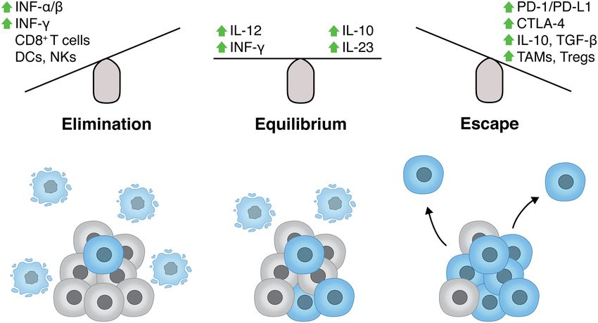
Downloaded from genesdev.cshlp.org on August 1, 2021 - Published by Cold Spring Harbor Laboratory Press

Pilger et al.

apparent when two independent studies reported dra-                   tion is dispensable for cellular survival in HR-proficient
matic cytotoxicity of PARP inhibitors in the context of de-           cells, it becomes extremely toxic in the context of
ficiencies in the tumor suppressor genes BRCA1 and                    BRCA1/2 deficiency, highlighting the key role of HR in re-
BRCA2 (breast cancer susceptibility genes 1 and 2) (Bryant            pairing PARP inhibitor-induced damage (Lord and Ash-
et al. 2005; Farmer et al. 2005). BRCA1 and BRCA2 have                worth 2016). So far, four different PARP inhibitors
crucial functions during homologous recombination                     (olaparib, niraparib, rucaparib, and talazoparib) have
(HR)-mediated repair of DSBs (Fig. 1). While PARP inhibi-             been approved by the FDA for use as single-agent chemo-
                                                                      therapeutics (Table 1).
                                                                         The clinical successes of PARP inhibitors have nur-
                                                                      tured efforts to target other components of the DNA re-
                                                                      pair network for therapeutic applications. Thus, the
                                                                      three apical PI3K-like kinases ATM, ATR, and DNA-PK,
                                                                      in addition to the S-phase checkpoint kinase CHK1 and
                                                                      G2 checkpoint kinase WEE1, have emerged as promising
                                                                      targets for small molecule inhibitors, with several of these
                                                                      compounds currently undergoing clinical development
                                                                      (Blackford and Jackson 2017; Forment and O’Connor
                                                                      2018). The justification for targeting ATR in cancer cells
                                                                      centers around ATR’s functions in protecting stalled
                                                                      DNA replication forks, regulating replication origin firing,
                                                                      and controlling the transition of cells from G2 phase into
                                                                      mitosis by enforcing the G2/M cell cycle checkpoint (Sal-
                                                                      divar et al. 2017). Due to their proliferative nature and be-
                                                                      ing subject to oncogenic forces that either lead to
                                                                      heightened levels of reactive oxygen species or deregulat-
                                                                      ed S-phase entry and/or progression, cancer cells are par-
                                                                      ticularly vulnerable to ATR inhibition, experiencing
                                                                      extensive DNA and chromosomal damage. Furthermore,
                                                                      it has been found that ATR inhibitors are selectively toxic
Figure 1. DDR inhibitors and their impacts on DSB repair and          to cancer cells harboring mutations in the ATM tumor
cell cycle progression. In mammalian cells, four main DSB path-       suppressor, in part because ATM and ATR share some
ways exist, which operate dependent on the stage of the cell cycle:   overlapping targets (Kantidze et al. 2018). Additionally,
nonhomologous end-joining (NHEJ), alternative end-joining (alt-       ATM loss impairs DSB repair by HR and also weakens
EJ), homologous recombination (HR), and single-strand annealing       the G1/S cell cycle checkpoint, thereby generating more
(SSA) (Scully et al. 2019). TOP2i induced DSBs are predominantly      replicative stress (Lecona and Fernandez-Capetillo 2018).
repaired via NHEJ, which while being described as “error-prone,”
                                                                      Several ATR inhibitors are currently undergoing clinical
ensures effective repair of broken DNA ends particularly during
G0 and in the G1 phase of the cell cycle. DNA-PK is crucial for
                                                                      development, with their application mainly aiming to ex-
effective NHEJ, and DNA-PK inhibitors impair DSB repair via           ploit high levels of replication stress in tumors. Since
NHEJ. TOP1 inhibitors generate single-ended DSBs (seDSBs) in          CHK1 is the effector kinase of ATR, several cellular func-
S phase, which require HR for accurate repair. HR is a form of        tions are shared between these kinases, and the rationales
DNA recombination where DNA homology and synthesis can ac-            behind exploring CHK1 inhibitors for cancer therapy are
curately regenerate the sequence surrounding the DSB, facilitated     hence similar to those for ATR inhibitors (Table 1; For-
by a sister chromatid as template and therefore restricted to S or    ment and O’Connor 2018).
G2 (Karanam et al. 2012). DNA damage arising from PARP inhi-             WEE1 is involved in responses to DNA damage, where-
bition also requires HR, since spontaneously occurring SSBs are       by it enforces a cell cycle arrest/checkpoint at the transi-
converted into seDSBs during DNA replication upon PARP inhib-
                                                                      tion from S phase into M phase in response to DNA
itors (PARPis). In addition, PARPi cause mitotic catastrophe and
induce DNA replication stress through altering DNA replication
                                                                      damage or replication stress. WEE1 phosphorylates and in-
fork speed. A fundamental step during HR-mediated repair is           hibits cyclin-dependent kinases (CDKs) CDK1 and CDK2,
DNA end resection, generating ssDNA overhangs that are rapidly        thus counteracting cell cycle progression and unscheduled
covered by RPA and consequently by RAD51. ATR is activated by         replication-origin firing (Elbæk et al. 2020). One of the con-
ssDNA as a result of DNA end resection or DNA polymerase un-          cepts for the clinical application of WEE1 inhibitors is that
coupling from helicase activity during DNA replication. Conse-        WEE1 inhibition should potentiate the effects of certain
quently, ATR inhibitors primarily have impacts during S and           other DNA damage-inducing chemotherapeutics, since
G2 phases of the cell cycle. Additionally, ATRi overrides the         cells undergoing WEE1 inhibition would enter mitosis
G2/M cell cycle checkpoint, therefore causing premature entry         with DNA lesions or underreplicated DNA, thereby caus-
into mitosis. Importantly, ATM inhibition affects efficient HR,
                                                                      ing mitotic catastrophe and ensuing cell death. Indeed, the
alongside its impact on G1/S and G2/M cell cycle checkpoints
in response to DNA damage. Analogous to ATRi and ATMi,
                                                                      efficacy of WEE1 inhibition in combination with other
WEE1 inhibitors affect the G2/M cell cycle checkpoint. More-          DNA-damaging agents such as platinum drugs or irradia-
over, WEE1i cause replication stress through dysregulated origin      tion has been observed in preclinical model systems (Hirai
firing and cleavage of DNA replication forks, resulting in DSBs.      et al. 2009; PosthumaDeBoer et al. 2011).

2        GENES & DEVELOPMENT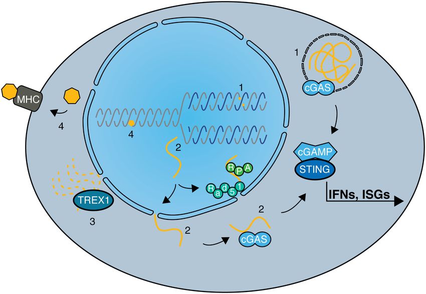
Downloaded from genesdev.cshlp.org on August 1, 2021 - Published by Cold Spring Harbor Laboratory Press

                                                                                     Interfaces between DDR and cancer immunotherapy

Table 1. Inhibitors targeting the DNA damage response and their clinical development

Cellular                           Stage of clinical
target          Compound            development                                        Disease setting

PARP          Niraparib             FDA approved         Ovarian cancer (HRD)
              Olaparib              FDA approved         Breast cancer (gmBRCA), ovarian cancer, pancreatic cancer, and prostate
                                                           cancer (sm/gmBRCA)
              Rucaparib             FDA approved         Ovarian cancer (gmBRCA) and prostate cancer (sm/gmBRCA)
              Talazoparib           FDA approved         Breast cancer (gmBRCA)
ATR           VX970                 Phase II             Solid tumors
              AZD6738               Phase II             Solid tumors
              BAY-1895344           Phase I              Solid tumors, lymphomas
CHK1          LY2606368             Phase II             Solid tumors (HRD or CCNE1 amplification), SCLC
              SRA737                Phase I/II           Solid tumors, non-Hodgkin’s lymphoma
              MK-8776               Phase I              Solid tumors, lymphomas
              GDC-0575              Phase I              Solid tumors, lymphomas
WEE1          AZD1775               Phase II             Uterine carcinoma, acute myeloid leukaemia, solid tumors (SETD2-
                                                           deficient)
ATM           AZD1390               Phase I              Brain cancers in combination with RT
              AZD0156               Phase I              Solid tumors in combination with conventional chemotherapy
DNA-PK        M3814                 Phase I/II           Pancreatic cancer and prostate cancers (in combination with RT)
              AZD7648               Phase I              Solid tumors

   DNA-PK and ATM are key protein kinases that func-                Covey et al. 1989; Pommier 2009). In the context of
tion in the DDR to promote DNA nonhomologous end-                   TOP1, trapped TOP1cc becomes particularly toxic in rep-
joining (NHEJ) and HR, respectively, with ATM also play-            licating cells when DNA replication forks encounter
ing major roles in regulating signaling cascades in re-             these lesions, leading to replication fork arrest, fork col-
sponse to irradiation and other DNA-damaging agents                 lapse, and eventually DNA DSB formation (Hsiang et al.
(Blackford and Jackson 2017). Because their absence or in-          1989; Strumberg et al. 2000; Furuta et al. 2003). Although
hibition sensitizes cells to irradiation, PARP inhibitors,          camptothecin was among the first TOP1 inhibitors to be
and various DNA-damaging chemotherapeutic agents,                   identified, its clinical application was not pursued further,
there is the potential for using ATM or DNA-PK inhibi-              and the camptothecin derivatives topotecan and irinote-
tion to enhance antitumor efficacies of radiotherapy and            can instead gained regulatory approval (Table 1). Clinical-
certain chemotherapies, although their potential to also            ly approved TOP2 inhibitors, such as etoposide and
accentuate toxic side effects in patients will have to be           doxorubicin (Table 1), function as interfacial inhibitors
carefully managed in such contexts (Table 1).                       by trapping the TOP2ccs on DNA and preventing religa-
   In contrast to DDR enzyme inhibitors, other classes of           tion of the DNA backbone, analogous to the actions of
drugs possess the potential to generate DNA damage                  camptothecin derivatives on TOP1 (Wu et al. 2011,
and therefore are classed as genotoxic agents. Among                2013a). However, due to TOP2’s dimeric mode of action,
them, topoisomerase inhibitors are well-established che-            key aspects of TOP2 inhibitors’ mechanism are their abil-
motherapeutics for a variety of cancers. The topoisomer-            ity to directly induce DSBs and consequently yield such
ase enzymes TOP1 and TOP2 generate transient DNA                    toxic lesions in all stages of the cell cycle (Holm et al.
breaks in order to resolve topological stresses during              1989).
DNA replication and transcription, using transesterifica-              Platinum-based chemotherapeutics represent another
tion reactions to break the DNA phosphodiester backbone             class of genotoxic agents, which have found broad applica-
while at the same time forming a transient covalent bond            tion in cancer therapy. Initially, cisplatin was FDA ap-
between the enzyme’s catalytic tyrosine (Tyr) and the               proved for the treatment of testicular and ovarian
DNA, known as the topoisomerase cleavage complex                    cancers in 1978, whereas nowadays cisplatin and its deri-
(TOPcc). Exploiting the potential of topoisomerases to in-          vates carboplatin and oxaliplatin are established chemo-
duce DNA damage, especially in highly replicating and               therapeutic agents for various tumor types (Kelland
transcribing cells, underpinned the development of sever-           2007). Platinum-based agents create DNA monoadducts
al classes of TOP1 and TOP2 inhibitors. Among others,               and DNA cross-links, which impair cellular processes
molecules that reversibly stabilize TOPccs to block the             such as DNA replication and transcription and require
religation reaction have had dramatic impacts as chemo-             specific DNA repair pathways for their resolution. As a
therapeutic agents. Since these molecules act as interfa-           consequence, platinum agents are strong inducers of cell
cial inhibitors (functioning via stabilizing a reaction             cycle arrest and apoptosis (Rottenberg et al. 2021). Mech-
intermediate), DNA damage arises from TOP1 or TOP2                  anistically, cisplatin is activated intracellularly through
that has become trapped in the state where the DNA back-            the aquation of the chloride group(s) and subsequently
bone is already cut and the enzyme cross-linked to DNA,             chemically cross-link DNA molecules, preferably at N7
but the reverse reaction is abrogated (Hsiang et al. 1985;          or O6 in purines. Notably, platinum agents can cross-

                                                                                                    GENES & DEVELOPMENT            3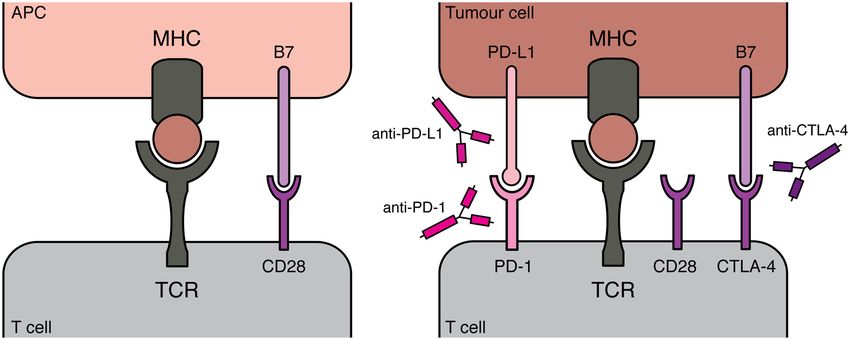
Downloaded from genesdev.cshlp.org on August 1, 2021 - Published by Cold Spring Harbor Laboratory Press

Pilger et al.

link proteins to DNA, although the occurrence of this               ic DNA into IRF3 and NFκB transcriptional programs,
type of lesion is much rarer compared with DNA intra-               which control the balance between cellular survival and
strand and inter-strand cross-links (Siddik 2003). Along-           controlled cell death (Fitzgerald et al. 2003; Ishikawa and
side topoisomerase inhibitors and platinum drugs, radia-            Barber 2008; Ishikawa et al. 2009; Tanaka and Chen 2012).
tion therapy can be classified as a genotoxic treatment                In order to prevent the accumulation of cytosolic DNA
and represents the most frequently applied cancer therapy           and potentially persistent proinflammatory signaling,
aside from surgery. The idea of radiation therapy is based          several enzymes and factors constantly survey and elimi-
on the locally targeted induction of DNA damage in order            nate nucleic acids appearing in the cytosol. One of these,
to deprive cancer cells of their proliferative potential by         TREX1, is a 3′ –5′ exonuclease that acts on either single-
forcing them into senescence, apoptosis, or other forms             stranded DNA (ssDNA) or dsDNA and comprises the pre-
of cell death (Bernier et al. 2004). Since radiation therapy        dominant enzyme that degrades cytosolic DNA in mam-
induces a variety of DNA lesions, including DSBs, SSBs,             malian cells (Yang et al. 2007; Stetson et al. 2008; Lindahl
base damage, and cross-links, the application of various            et al. 2009). Mutations in TREX1 result in dysregulation of
DDR enzyme inhibitors can further potentiate the cyto-              type I IFN production and are associated with the autoim-
toxic effects of radiation therapy (Moding et al. 2013).            mune disorders Aicardi-Goutières syndrome (AGS) and fa-
                                                                    milial chilblain lupus (Crow et al. 2006a; Rice et al. 2007).
                                                                    Type I IFN responses observed in TREX1-deficient cells
Sensing damaged DNA by components of the innate                     are dependent on the cGAS/STING system, which pre-
immune system                                                       sumably recognizes elevated cytosolic DNA arising in
                                                                    the absence of its TREX1-mediated clearance. In line
Alongside the immediate cytotoxic effects and persistent            with these observations, inactivation of cGAS/STING ab-
DNA damage caused by certain DDR inhibitors (DDRis),                rogates type I IFN signaling in TREX1-deficient cells and
induction of innate immune responses has frequently                 rescues the embryonic lethality of Trex1−/− mice (Stetson
been observed in such settings. Unrepaired DNA lesions              et al. 2008; Ablasser et al. 2014; Gray et al. 2015). Notably,
and/or impaired chromosome segregation during mitosis               individuals with mutations in the RNase H2 complex,
contribute to the formation of endogenous cytosolic                 consisting of RNASEH2A, RNASEH2B, and RNASEH2C,
DNA, often in the form of micronuclei. Although their or-           show clinical pathologies that resemble those observed in
igin is distinct, DNA damage-induced cytosolic DNA                  TREX1 mutated patients, including aberrant type I IFN
shares common downstream effects and cellular responses             signaling along with AGS and systemic lupus erythemato-
with those observed upon viral or bacterial infections. In          sus. Although this maladaptive immune response upon
mammalian cells, multiple factors and enzymes have the              impaired RNase H2 function relies on cGAS/STING, as
capability to sense cytosolic DNA to activate appropriate           is the case for TREX1 dysfunction, it is unlikely that RN-
immune responses. The induction of innate immune re-                ase H2 directly eliminates cytosolic DNA (Crow et al.
sponses by the presence of cytosolic DNA not only is cru-           2006b; Mackenzie et al. 2016). Rather, RNase H2 func-
cial for the host organism’s first-lines of defense against         tions inside the nucleus by resolving DNA–RNA hybrids
invading pathogens but also fulfils key oncosuppressive             and removing ribonucleotides during DNA replication,
functions through elimination of damaged cells (Van-                thereby preserving genome integrity by restricting cyto-
pouille-Box et al. 2018). Intensive research in recent years        solic accumulation of broken DNA molecules (Reijns
has led to the discovery and characterization of the cGAS/          et al. 2011, 2012). Notably, DNA–RNA hybrids harbor
STING system as a major component of cells’ intrinsic im-           the potential to directly stimulate cGAS and, consequent-
mune response to the occurrence of cytosolic oligonucleo-           ly, cGAMP production in vitro (Mankan et al. 2014). Al-
tides. cGAS is a cytosolic nucleotidyltransferase that,             though the induction of cGAMP synthesis in the
upon binding DNA, catalyzes the synthesis of cyclic                 presence of DNA–RNA hybrids is orders of magnitude
GMP-AMP (cGAMP) from ATP and GTP, which subse-                      lower compared with cGAMP synthesis upon dsDNA
quently acts as a messenger molecule for the adaptor pro-           stimulation, it may be that DNA–RNA hybrids represent
tein STING (Ablasser et al. 2013; Sun et al. 2013; Wu et al.        direct stimuli for IFN responses upon RNase H2 dysfunc-
2013b). cGAS binds double-stranded DNA (dsDNA) in a                 tion in certain settings. Recently, it became apparent that
sequence-nonspecific manner through interaction with                the simple model of cGAS being exclusively localized in
the sugar-phosphate backbone, although a preference for             the cytoplasm might not represent the full picture. Sever-
specific DNA length (>45 bp) has been observed (Civril              al reports indicated nuclear functions for cGAS, suggest-
et al. 2013; Li et al. 2013; Herzner et al. 2015; Zhou et al.       ing that a subfraction of the cellular cGAS pool resides
2018). Upon cGAMP binding, STING, originally located                within the nucleus (Liu et al. 2018a; Jiang et al. 2019).
in the endoplasmic reticulum (ER) membrane, translo-                The concomitant conflict of autoreactivity against self-
cates to the Golgi apparatus via the ER–Golgi intermediate          DNA is reported to be overcome by the tethering of
compartment, while activating at least three distinct ki-           cGAS to the acid patch of nucleosomes via its DNA bind-
nases—TBK1, IKK, and NIK (TANK-binding kinase 1,                    ing domains, thus blocking DNA binding and activity to-
IκB kinase, and NFκB-inducing kinase, respectively)—dur-            ward chromatinized DNA (Boyer et al. 2020; Kujirai et al.
ing this process. The activation of these kinases and con-          2020; Michalski et al. 2020; Pathare et al. 2020; Zhao et al.
comitant phosphorylation of interferon regulatory factor            2020). This mechanism may also give a plausible explana-
3 (IRF3) and IκBα transduces the initial stimulus of cytosol-       tion to the long-standing question of how cGAS activity is

4        GENES & DEVELOPMENT
Downloaded from genesdev.cshlp.org on August 1, 2021 - Published by Cold Spring Harbor Laboratory Press
Interfaces between DDR and cancer immunotherapy
suppressed in mitosis, when the nuclear envelope breaks
down and chromosomes are exposed to cytosolic cGAS
(Zierhut et al. 2019).
Impacting cellular immune responses through DDR
inhibitors
The importance of cytosolic DNA-stimulated immune re-
sponses in the context of cancer therapy is highlighted by
the contribution of cGAS/STING to antitumor immunity
in response to radiotherapy. Longstanding reports have in-
dicated that, besides generating cytotoxic DNA damage,
radiotherapy also induces a tumor-specific immune re-
sponse that contributes to the efficacy of this therapeutic
modality (McBride et al. 2004). Secretion of inflammatory
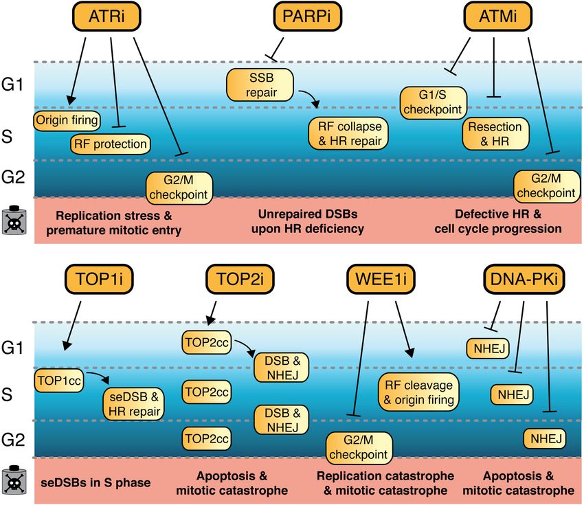
cytokines such as TNF-α, IL-1α, and IL-6 has been ob- Figure 2. DNA repair defects and their impacts on cellular im-
mune responses. Defects in DNA repair pathways or DDR com-
served in response to ionizing radiation, which ultimately
ponents affect innate and adaptive immune responses in
leads to adaptive immune responses via CD8+ T cells (Hal- various ways. (1) The induction of DSBs via chemotherapeutics
lahan et al. 1989; McBride et al. 2004; Lee et al. 2009; Di or irradiation can lead to micronuclei formation and consequent
Maggio et al. 2015). Recent reports have established that recognition of cytosolic DNA by cGAS/STING. Deficiencies in
cGAS/STING plays a key role in linking innate and adap- genes encoding proteins such as BRCA2, RNase H2, or ATM fur-
tive immune responses after irradiation. Specifically, ther augment these effects. (2) Cytosolic DNA can also be a result
cGAS/STING mediates increased IFN-β production in of aberrant processing of DNA replication intermediates, with
the tumor environment to promote antitumor responses several DDR factors limiting either the generation (SAMHD1)
upon irradiation. Impaired type I IFN signaling resulting or the translocation (RAD51 and RPA) of cytosolic DNA from
from cGAS/STING inactivation can be bypassed by exog- the nucleus, or degrading DNA once it is present in the cytosol
(TREX1). (3) Activation of the cGAS-cGAMP-STING cascade
enous supplementation of cGAMP to induce the antitu-
leads to IRF3 and NFκB transcriptional programs, resulting in ex-
mor efficacy of radiotherapy, further supporting the idea pression of IFN and ISGs, therefore inducing strong innate im-
that in response to radiotherapy, cGAS/STING is an ini- mune responses. (4) In contrast, MMR defects can lead to
tial sensor and signal transducer of antitumor immune re- adaptive immune responses through increased somatic muta-
sponses (Burnette et al. 2011; Woo et al. 2014; Wu et al. tions and consequent synthesis of neoantigens. When presented
2014; Deng et al. 2014b). From the above, one can assume by MHC molecules at the cell surface, neoantigens elicit a strong
that the stimulus that activates cGAS/STING after irradi- T-cell response, dependent on the immunogenicity of the
ation must originate from cytosolic DNA. Indeed, micro- neoantigen.
nuclei, resulting from irradiation and ensuing incomplete
DNA repair, are recognized by cGAS/STING to activate
type I IFN signaling (Fig. 2). Importantly, micronuclei for- somes, resulting in dampened cGAS/STING activation
mation requires progression through mitosis following and IFN-β production in DCs (Diamond et al. 2018).
DNA-damage induction, wherein the dissolution and en- Besides radiation-induced antitumor responses, conven-
suing reformation of the nuclear membrane end up en- tional chemotherapy has the potential to stimulate the in-
closing DNA fragments in a micronuclear envelope nate and adaptive immune systems. As described above,
(Harding et al. 2017; Mackenzie et al. 2017). Although HR deficiency (HRD) is a predictor of PARP inhibitor effi-
this envelope shares similarities with the normal nuclear cacy and PARP inhibitors are particularly cytotoxic in tu-
envelope, its assembly remains rudimentary, reflecting its mors displaying mutations in BRCA1/2. In this regard, it is
fragility that frequently results in the breakdown of enve- worthwhile mentioning that beside the generation of
lope integrity and cytosolic exposure of genomic DNA DNA damage, induction of a STING-dependent antitumor
(Liu et al. 2018b). This phenomenon is particularly impor- immunity is a considerable feature of PARP inhibitor can-
tant in explaining cGAS/STING activation in response to cer therapy. Specifically, PARPi-mediated trapping of
micronucleation, since cGAS recognizes cytosolic DNA PARP on DNA lesions appears to be influential for the in-
rather than the outer structure of a micronucleus (Mac- nate immune response, as the extent of PARP trapping cor-
kenzie et al. 2017). Interestingly, TREX1 antagonizes im- relates with the magnitude of innate immune signaling
munogenicity of cancer cells following radiation via its (Kim et al. 2020). PARP inhibitor-mediated immunogenic-
own up-regulation and the consequent enhanced degrada- ity depends on the activation of cGAS/STING to elicit a
tion of cytosolic DNA. However, continuous low-dose ir- cytotoxic T-cell response. Intriguingly, this immune re-
radiation circumvents TREX1 engagement and promotes sponse is augmented upon BRCA deficiency, since
cGAS/STING-dependent IFN-β secretion for antitumor Brca1-proficient mouse tumors show mitigated immuno-
immunity (Vanpouille-Box et al. 2017). In addition, genicity in response to PARP inhibitors as compared with
TREX1 inhibits the transfer of dsDNA from irradiated tu- Brca1 mutated ones (Ding et al. 2018; Pantelidou et al.
mor cells to dendritic cells (DCs) via tumor-derived exo- 2019). In line with these observations, PARP inhibition
GENES & DEVELOPMENT 5Downloaded from genesdev.cshlp.org on August 1, 2021 - Published by Cold Spring Harbor Laboratory Press Pilger et al. led to a strong induction of interferon-stimulated genes Consequences of DDR defects for immune responses (ISGs) in BRCA2-deficient cells compared with their BRCA2-proficient counterpart (Reislander et al. 2019). The involvement of DDR factors and their inhibition in However, it has been reported that PARP inhibitors have the induction of innate immune responses has been high- the potential to initiate cGAS/STING-dependent type I lighted by the consequences of RNase H2 dysfunction in IFN production in human cancer cells independent of their the autoimmune disorder AGS. While certain DDR fac- BRCA gene status (Shen et al. 2019), presumably due to the tors such as TREX1 participate in the sensing of extranu- mitotic defects or increased replication stress caused by clear DNA, others influence cellular immunity via more PARPi affecting replication fork speed (Maya-Mendoza indirect mechanisms. The tumor suppressor BRCA2 has et al. 2018; Slade 2019). Future studies are required to illu- a pivotal role in HR-mediated repair and in the protection minate the underlying determinants for the differential re- of stalled replication forks, while its absence is accompa- sponses in BRCA-proficient and BRCA-deficient contexts. nied by genome instability and chromosome breakage Other chemotherapeutic agents that generate DNA (Prakash et al. 2015). Because BRCA2 loss is highly detri- damage also affect cellular immune responses in various mental to cell viability, BRCA2-deficient cancer cells ways, often depending on the DNA damage-induced re- have invariably undergone adaptation processes in order lease of cytosolic DNA. For example, the topoisomerase to survive, exemplified by the inactivation of p53 func- II inhibitor teniposide induces STING-dependent type I tions. Importantly, alongside rewiring of DNA repair IFN signaling and NF-κB activation in a mouse tumor processes, the absence of BRCA2 causes enhanced phos- model, with the consequent DC and T-cell activation pro- phorylation and therefore activation of STAT1-IRF3, moting antitumor responses, including increased im- followed by concomitant up-regulation of ISGs. Mecha- mune cell infiltration (Wang et al. 2019). Furthermore, nistically, this adaptive immune response is dependent the TOP2 inhibitors doxorubicin and daunorubicin in- on cGAS/STING, with BRCA2-deficient cells exhibiting duce IFN-β production in human cancer cell lines via a an increase in cGAS-positive micronuclei compared mechanism shown to be dependent on STING function with BRCA2-proficient cells, presumably as result of the (Luthra et al. 2017). Notably, doxorubicin treatment en- DNA repair defects and chromosome instability observed hances CD8+ T-cell amplification and infiltration, as in the absence of BRCA2 function (Reislander et al. 2019). well as IFN-γ production, in tumor environments in In line with these observations, elevated secretion of mice (Mattarollo et al. 2011). Similar to TOP2 inhibitors, proinflammatory cytokines, such as TNF-α, arises as a topoisomerase I inhibitors trigger potential immunoge- consequence of BRCA2 loss, resulting from cytosolic nicity, exemplified by DC and CD8+ T-cell activation in DNA sensing by cGAS/STING and ensuing interferon re- mice treated with topotecan (Kitai et al. 2017). sponses (Heijink et al. 2019). Additionally, it has been Despite lacking clear evidence for affecting the cellular shown that in the absence of BRCA2, RNase H2 recruit- immune system, ATR inhibitors have been shown to po- ment to DSBs is impaired, resulting in increased DNA– tentiate immune stimulations in response to radiothera- RNA hybrid formation and thus providing and additional py. Thus, combinatorial treatment of radiotherapy and explanation for cGAS/STING activation in BRCA2-defi- ATR inhibition was found to induce type I/II IFN-based cient cells (D’Alessandro et al. 2018). Importantly, the ab- gene expression changes and CD8+ T-cell infiltration in rogation of TNF-α signaling improves viability of BRCA2- a manner dependent on cGAS/STING (Vendetti et al. deficient cells, indicative of TNF-α signaling promoting 2018; Dillon et al. 2019; Sheng et al. 2020). While ATR in- cell death when BRCA2 function is impaired (Heijink hibitors do not damage DNA directly, it can be assumed et al. 2019). Besides BRCA2 deficiency causing induction that the increased immunogenicity in irradiated tumors of interferon responses and an increase in TNF-α signaling in the context of ATRi results from overriding the G2/M per se, it also further sensitizes cells to autocrine TNF-α cell cycle checkpoint, with an increased proportion of signaling. cells with unrepaired DNA lesions entering mitosis, lead- Notably, depletion of BRCA1 or the inter-strand DNA ing to DNA fragmentation and micronuclei formation ca- cross-link (ICL) repair factor FANCD2 sensitizes cells to pable of triggering innate immune responses (Ruiz et al. recombinant TNF-α, suggesting that the general impair- 2016; Harding et al. 2017). In line with this model, it has ment of HR repair or replication stress arising from com- been observed that inhibition of the ATR effector kinase promised ICL repair harbors the potential to make cells CHK1 abrogates the G2/M checkpoint post irradiation, more susceptible to interferon responses (Heijink et al. leading to micronuclei formation and type I IFN signaling 2019). The idea of unprotected replication forks compris- in cancer cells (Chao et al. 2020). Moreover, increased ing an entry point for aberrant DNA processing and conse- CD8+ T-cell infiltration and tumor volume reduction quent leakage into the cytosol is further supported by the was observed in mice treated with a combination of radio- report that mutations in SAMHD1 are causative for the au- therapy and the CHK1 inhibitor AZD7762 compared with toimmune disorder AGS (Crow and Manel 2015). Besides treatments with these agents individually (Chao et al. its role as a dNTPase (deoxynucleotide triphosphohydro- 2020). Similar to ATR inhibitors, pharmacologic inhibi- lase), SAMHD1 promotes the controlled degradation of tion of ATM in combination with radiotherapy was found newly synthesized DNA at stalled replication forks via to induce type I IFN signaling, which notably occurred in- the exonuclease MRE11. In the absence of SAMHD1, na- dependent of cGAS/STING but was reliant on TBK1 scent DNA at stalled replication forks is displaced by the (Zhang et al. 2019). RECQ1 helicase and translocates to the cytosol, where it 6 GENES & DEVELOPMENT
Downloaded from genesdev.cshlp.org on August 1, 2021 - Published by Cold Spring Harbor Laboratory Press

                                                                                     Interfaces between DDR and cancer immunotherapy

activates the cGAS/STING pathway and ensuing type I                 Mismatch repair deficiency and adaptive immunity
IFN responses (Coquel et al. 2018).
   In order to avoid chronic proinflammatory signaling as           Deficiencies in DNA repair pathways can also affect adap-
occurs in AGS patients, several cellular mechanisms                 tive immune responses, which are key for immunogenic-
have evolved to counteract or minimize leakage of DNA               ity of tumors and have clinical implications in the context
fragments into the cytosol. The DNA repair proteins                 of cancer immunotherapy. Additionally, DNA damage re-
RPA and RAD51 both have the capability to directly                  sponses are crucial during the development of the im-
bind ssDNA, which is crucial for their functions during             mune system and maturation of immune cells, as
DNA replication and DNA repair (Bhat and Cortez 2018).              exemplified by the controlled induction and concomitant
In addition, they prevent the accumulation of cytosolic             repair of DSBs during V(D)J recombination and class
DNA by binding and therefore retaining ssDNA within                 switch recombination in lymphocytes (for an extensive
the nucleus, thereby working in cooperation with nuclear            review, see Bednarski and Sleckman 2019). Furthermore,
membrane-bound TREX1, which normally swiftly de-                    over recent years, a connection between defects in the
grades any DNA leaking into the cytosol (Fig. 2). Exhaus-           DNA mismatch repair (MMR) pathway and tumor immu-
tion of the available RPA/RAD51 pool in the absence of              nogenicity has been observed both in preclinical model
TREX1, or upon depletion of RPA or RAD51, results in                systems and in cancer patients. The MMR machinery de-
cGAS/STING-dependent type I IFN signaling (Wolf et al.              tects and replaces base mismatches resulting from errone-
2016). Additionally, RAD51 protects newly synthesized               ous DNA replication or repair and, in particular, plays key
DNA from aberrant processing by MRE11 and consequent                roles in correcting small insertion or deletions (indels)
cytosolic DNA translocation and ensuing cGAS/STING                  arising at repetitive sequences in the genome, so-called
activation (Bhattacharya et al. 2017). Notably, the func-           microsatellite instability (Kunkel and Erie 2015). Tumors
tions of MRE11 as a nuclease that processes DNA replica-            harboring mutations in genes encoding the core MMR fac-
tion fork intermediates can be seen as a double-edged               tors MLH1, MSH2, MSH6, or PMS2 are characterized by
sword. On one hand, MRE11 prevents cGAS/STING acti-                 microsatellite instability (MSI), a hypermutator pheno-
vation through trimming of replication intermediates,               type specified by large numbers of single-nucleotide vari-
stimulated by SAMHD1. On the other hand, excessive pro-             ants (SNVs) and indels, thus leading to a high mutational
cessing of unprotected, stalled replication forks in the ab-        burden. Additionally, specific DNA proofreading muta-
sence of RAD51 by MRE11 generates cytosolic DNA and                 tions in the replicative DNA polymerases POLE and
consequently stimulates cGAS/STING. Whether or in                   POLD1 drive hypermutation phenotypes in some cancers
which form RAD51 cooperates with SAMDH1 for fine-                   without causing MSI (Campbell et al. 2017). MSI predom-
tuning of this process is an interesting area for future re-        inantly occurs in endometrial, gastric, and colorectal can-
search. Besides MRE11-dependent degradation of nascent              cers, suggesting that such tissue types or the tumor
DNA at stalled or collapsed replication forks, other DNA            environment and extracellular influences affect MSI de-
nucleases can process DNA structures in such contexts,              velopment (Hause et al. 2016; Cortes-Ciriano et al.
subsequently leading to activation of innate immune re-             2017). The correlation between MSI colorectal tumors
sponses via cytosolic DNA sensing. In particular, the ac-           and high numbers of tumor-infiltrating lymphocytes has
tions of the structure-specific endonuclease MUS81 can              long been acknowledged. Colorectal MSI tumors fre-
bring about the accumulation of cytosolic DNA via                   quently display high infiltration of CD8+ cytotoxic T
MUS81-dependent cleavage of stalled replication forks,              cells, type 1 helper (Th1) cells, and memory T cells, along-
leading to cGAS/STING activation and induction of type              side the up-regulated expression of interferon γ (IFN-γ) and
I IFNs, as observed in various prostate cancer cell lines           immune checkpoint molecules PD-1, PD-L1, and CTLA4
(Ho et al. 2016). Moreover, the MUS81–STING axis is re-             (Fig. 3; Llosa et al. 2015; Mlecnik et al. 2016).
sponsible for a prostate cancer cell-specific T-cell response          MSI endometrial tumors also exhibit increased numbers
in mice, thus highlighting a likely role for MUS81 in pro-          of tumor-infiltrating lymphocytes and elevated expression
moting antitumor immunity. In addition, endogenously                of PD-1/PD-L1 compared with microsatellite stable (MSS)
arising DSBs are capable of stimulating innate immune re-           tumors (Howitt et al. 2015). Recently, this connection has
sponses in situations where accurate DNA repair is com-             been attributed to the increase in neoantigens due to the
promised. Accordingly, deficiency in ATM induces type I             high mutational burden in these settings (Tougeron et al.
IFNs in unchallenged conditions, indicative of spontane-            2009; Maby et al. 2015). To a certain degree, it seems rea-
ously arising DNA DSBs being the stimulus of this im-               sonable to assume that the more mutations in the genome,
mune response. Furthermore, in the absence of ATM,                  the more likely it is that neoantigens are formed and in due
ssDNA resulting from unrepaired DNA lesions accumu-                 course recognized by the immune system (Fig. 2; Turajlic
lates in the cytoplasm and activates cGAS/STING-depen-              et al. 2017). In line with this idea, increased mutational
dent type I IFN signaling (Härtlova et al. 2015). Notably,          burden correlates with improved survival in colorectal
this DDR defect primes type I IFNs to enhance the innate            cancer patients (Giannakis et al. 2016). Interestingly, addi-
immune response toward invading pathogens, highlight-               tional tumor types such as melanomas and lung cancers,
ing the fact that damaged DNA serves as a danger signal             despite being microsatellite stable (MSS), exhibit an in-
for cellular homeostasis and prepares the innate immune             creased number of overall mutations compared with oth-
system for a rapid response in the face of bacterial challeng-      ers, most likely due to exposure to exogenous mutagens,
es (Härtlova et al. 2015).                                          including ultraviolet light and tobacco smoke

                                                                                                    GENES & DEVELOPMENT           7
Downloaded from genesdev.cshlp.org on August 1, 2021 - Published by Cold Spring Harbor Laboratory Press
Pilger et al.
Figure 3. Innate and adaptive immune responses in
cancer. In recent years, the concept of cancer immu-
noediting evolved from traditional views of the im-
mune system constantly surveying and eliminating
transformed cells in order to counteract cancer devel-
opment (immunosurveillance) (Keast 1970). Cancer
immunoediting unifies observations of the immune
system promoting tumor outgrowth with reports of
immunosurveillance, highlighting the dual functions
of the immune system during tumor development
(Schreiber et al. 2011). The cancer immunoediting
concept consists of three phases: elimination, equi-
librium, and escape. In the elimination phase, com-
ponents of both the innate and adaptive immune
response recognize and destroy cells undergoing on-
cogenesis. Elimination is promoted by a number of
signaling molecules such as type I and type II IFNs
and is executed via the interplay of a subset of immune cells such as CD8+ T cells, dendritic cells (DCs), natural killer cells (NKs), natural
killer T cells (NKTs), proinflammatory (“M1”) macrophages, and others (Mittal et al. 2014). Notably, the DDR participates in this process,
since DNA damage induction in tumors cells results in up-regulation of ligands for the receptors NKG2D and DNAM-1, therefore stim-
ulating cytotoxicity of NK and CD8+ T cells in addition to IFN-γ secretion (Gasser et al. 2005; Croxford et al. 2013). Moreover, radiother-
apy-induced DNA damage, and consequent cell death due to uncomplete DNA repair, stimulates cross-presentation by dendritic cells and
increased lymphocyte influx, thus further contributing to cancer cell elimination (Deng et al. 2014b; Samstein and Riaz 2018; Cornel et al.
2020; Cheng et al. 2021). Paradoxically, TNF-α has both antitumor and tumor-promoting activity. When secreted by macrophages and
innate immune cells, TNF-α induces cancer cell elimination, whereas chronic inflammation promoted by TNF-α signaling can drive car-
cinogenesis (Balkwill 2009; Charles et al. 2009). In the equilibrium phase, the adaptive immune system holds the tumor in a dormant state
with cancer cells resisting constant immune recognition through genetic and epigenetic changes in antigen presentation and immuno-
suppressive pathways. Cancer cells achieve immune evasion by various mechanisms, including loss of tumor antigens or factors involved
in antigen presentation, such as type I HLA (MHC) function, expression of inhibitory ligands (e.g., PD-L1 and CTLA-4), secretion of im-
munosuppressive cytokines (IL-10, TGF-β), and recruitment of tumor-associated macrophages (TAMs) and regulatory T cells (Tregs).
These scenarios result in the inability of innate and adaptive immune cells to recognize and appropriately respond to oncogenic cells,
therefore facilitating tumor progression (escape phase) (Vinay et al. 2015).
components, respectively, suggesting that these may also therapy or classical chemotherapies (Fig. 4; Ribas and
promote antitumor immune responses (Alexandrov et al. Wolchok 2018). In particular, their potential to counteract
2013). In accord with this idea, a recent study by Bardelli immune suppressive signals in the tumor microenviron-
and colleagues (Germano et al. 2017) demonstrated that ment to overcome T-cell exhaustion has been proven to
colorectal, breast, and pancreatic mouse cancer cell lines, be beneficial in clinical settings (Fig. 3). ICBs target im-
where MMR was genetically inactivated, grew signifi- mune checkpoints, which in normal settings are impor-
cantly slower when transplanted into immunocompetent tant to accurately regulate T-cell activation and T-cell
mice compared with the isogenic MMR-proficient cancer receptor signaling, thus preventing chronic (or inappropri-
cell lines, indicating rejection by the host immune system. ate) immune responses. In cancers, these checkpoints, of-
Enhanced immunosurveillance was accompanied by accu- ten engaged via cell surface ligands or receptors, are
mulation of neoantigens over time in MMR-deficient repurposed to dampen antitumor immune responses and
cells, wherein the amount of neoantigens remained stable create an immune-suppressive tumor microenvironment.
in MMR-proficient cells, implicating neoantigens generat- ICB agents, in the form of antibodies, binding to these li-
ed by MMR deficiency as a direct cause for immune sys- gands/receptors (anti-CTLA-4 and anti-PD-1/anti-PD-L1)
tem-mediated elimination of cancer cells (Germano et al. can overcome inhibitory signaling and reactivate T-cell
2017). Importantly, fully established MMR-deficient tu- engagement toward the tumor (Fig. 4). In order to broaden
mors are often sensitive to immune checkpoint inhibitors the spectrum of applications for ICB, extensive efforts
in people, which points toward MMR deficiency as an im- have centered around the identification of suitable bio-
portant determinant of immune checkpoint blockade (Fig. markers for predicting ICB efficacy, as well as exploring
4) efficacy, at least in certain cancer types (Le et al. 2015). the potential of combination therapies with conventional
irradiation or chemotherapy. Increased mutational load is
a promising indicator of ICB responsiveness, and indeed,
Combined targeting of DDR and immune checkpoints patients harboring mutations in MMR genes and display-
in cancer therapy ing MSI showed strong responsiveness to PD-1 antago-
nists (Rizvi et al. 2015; Le et al. 2017; Overman et al.
Immune checkpoint inhibition (or immune checkpoint 2017). Importantly, the clinical benefits of blocking the
blockade [ICB]) has experienced considerable success in PD-1/PD-L1 immune checkpoint occurred across various
recent years as a promising therapeutic strategy for a sub- tumor types, including melanomas, colorectal, and non-
set of cancers, presenting an alternative to irradiation small cell lung cancers (NSCLCs), highlighting the
8 GENES & DEVELOPMENTDownloaded from genesdev.cshlp.org on August 1, 2021 - Published by Cold Spring Harbor Laboratory Press
Interfaces between DDR and cancer immunotherapy
lung cancers, which exhibit a high load of somatic muta-
tions (Hellmann et al. 2018).
Synergy of PARP inhibitors with immune checkpoint
inhibition
In addition to being used to treat tumors with a high mu-
tational burden, ICBs have also been shown to be effective
when applied in combination with DDR inhibition (Table
Figure 4. Principles of immune checkpoint inhibitors for cancer
therapy. As described in Figure 3, during the escape phase of the
2). An important feature of PARP inhibition is that it is as-
immunoediting concept, tumor cells evade immune recognition sociated with increases in CD8+ T-cell infiltration and
and destruction by active immunosuppression in the tumor. A IFN-γ production in the tumor, which occurs in mouse
milestone for the field of immune checkpoint inhibitors was cancer models independent of their Brca1/2 status. More-
the report in which melanoma patients treated with an antibody over, tumor regression was further increased upon combi-
targeting the T-cell checkpoint protein CTLA-4 showed signifi- nation therapy of PARPi with anti-PD-1 antibodies
cantly improved survival compared with the control group compared with the respective monotherapies (Ding et al.
(Hodi et al. 2010). This suggested that targeting suppressive im- 2018; Shen et al. 2019). Initial results of ensuing clinical
mune checkpoints can improve overall survival in melanomas, trials indicate beneficial effects in a subset of patients
indicating that a patient’s immune system has capabilities to con-
treated with ICB and PARPi, with results from the MEDI-
trol tumor growth once immunosuppressive signals are over-
come. Since this pharmacological approach targets the patient’s
OLA trial suggesting increased antitumor activity in
immune system rather than the tumor itself, a new field for clin- germline BRCA mutated ovarian cancers treated with
ical research arose. CTLA-4 is a surface receptor on T cells. To ac- the combination therapy of olaparib and durvalumab
quire effector function, a T-cell recognizes its compatible (PD-L1 inhibitor) (Domchek et al. 2020).
antigen, presented by MHC molecules of an antigen-presenting Another clinical trial, TOPACIO, investigated the effi-
cell (APC), via its T-cell receptor (TCR). However, this initial rec- cacy of the PARPi niraparib and the PD-L1 inhibitor pem-
ognition is insufficient, with binding of the CD28 T cell receptor brolizumab in platinum-resistant advanced breast cancers
to B7 molecules (CD80 or CD86 ligand) on APCs serving as cru- and recurrent ovarian cancers, independent of their HR
cial costimulatory signals to adequately prime T cells. CTLA-4 proficiency. Notably, patient response toward the combi-
translocates to the cell surface once T cells are activated, where
nation therapy in ovarian cancers exceeded expectations
it binds CD80 and CD86 with higher affinity than CD28, there-
fore dampening T-cell activation (Walunas et al. 1994; Krummel
based on monotherapy efficacy, while HRD-associated
and Allison 1995). Moreover CTLA-4 expression by Tregs is cru- mutational signature 3 or a positive immune score were
cial for their immune suppressive functions, potently binding reliable indicators of responsiveness (Konstantinopoulos
CD80/CD86 ligands on APCs and therefore preventing T-cell ac- et al. 2019; Färkkilä et al. 2020). Although mutational sig-
tivation (Takahashi et al. 2000; Wing et al. 2008). Following its nature 3 and therefore HR proficiency predicted efficacy of
initial success in clinical trials, the CTLA-4 antibody ipilimumab this combination therapy, therefore implying an applica-
was FDA approved in 2011 for treating melanomas. PD-1 repre- tion for niraparib and pembrolizumab in BRCA-deficient
sents another inhibitory receptor present on T cells, while its li- tumors, it was also encouraging that PD-L1 presence
gands PD-L1 and PD-L2 can be expressed by various cell types, and interferon priming of immune cells present in the tu-
including APCs and malignant cells, predominantly after expo-
mor microenvironment can be used to estimate respon-
sure to inflammatory cytokines such as IFN-γ. Engagement of
PD-L1 with its receptor PD-1 interferes with TCR signaling,
siveness to the combination therapy independent of
therefore limiting T-cell responses toward tumor cells (Freeman HRD. Indeed, patient responses to PARPi and PD-L1 inhi-
et al. 2000; Dong et al. 2002). Following this rational, antibodies bition have been observed in tumors displaying a func-
targeting PD-1/PD-L1 have provoked clinical benefits in various tional HR pathway (Färkkilä et al. 2020). Additionally, a
types of cancers, warranting FDA approval of pembroluzimab proof-of-concept clinical study further confirmed im-
and nivolumab (both PD-1 antagonists) in 2014. Unlike CTLA- mune activation in ovarian cancers treated with olaparib
4, PD-1/PD-L1 does not interfere with costimulation during the and the anti-PD-L1 antibody durvalumab regardless of
T-cell activation, suggesting that combination therapy of their BRCA status, highlighted by increased IFN-γ produc-
CTLA-4 and PD-1/PD-L1 antibodies could have synergistic ther- tion and tumor-infiltrating lymphocytes (Lampert et al.
apeutic effects. Regaining T-cell activation, by blocking inhibito-
2020). Notably, in this study no significant increase in tu-
ry signals during costimulation via CTLA-4 antibodies, could
drive increased PD-L1 expression in tumor cells, making them
mor mutational burden was observed (not even in BRCA-
particularly susceptible to PD-1/PD-L1 checkpoint blockade deficient tumors), which is in line with previous observa-
(Sharma and Allison 2015). tions in ovarian cancers and suggests that immune stimu-
lation with concomitant PARPi treatment is unlikely to
result from the exposure of neoantigens (Chan et al.
likelihood that increased mutational burden was the com- 2019). However, increased proinflammatory cytokine ex-
mon denominator of ICB efficacy between these tumors. pression and T-cell activation was associated with clinical
In line with this premise, progression free survival and benefits following olaparib plus durvalumab treatment,
overall survival upon PD-1 blockade was further augment- implying a potential application of this therapy for a wider
ed when combined with a CTLA-4 antagonist in small cell spectrum of cancers. Currently, numerous clinical trials
GENES & DEVELOPMENT 9Downloaded from genesdev.cshlp.org on August 1, 2021 - Published by Cold Spring Harbor Laboratory Press
Pilger et al.
Table 2. Registered clinical trials using DDR and immune checkpoint inhibitors
Treatment combination Clinical trial Phase Disease setting Biomarkers
Dostarlimab (PD-1) and niraparib (PARP) NCT03602859 III Epithelial or ovarian cancer
NCT03651206 II/III Ovarian cancer
Nivolumab (PD-1) and rucaparib (PARP) NCT03522246 III Ovarian cancer HR status
NCT03824704 II Ovarian cancer BRCA mutation
NCT03639935 II Biliary tract cancer
Pembrolizumab (PD-1) and olaparib NCT04380636 III NSCLC
(PARP) NCT03834519 III Prostate cancer
NCT03976362 III NSCLC
NCT03976323 III NSCLC
NCT04380636 III NSCLC
NCT04191135 II/III Breast cancer
NCT04483544 II Cervical cancer
Pembrolizumab (PD-1) and niraparib NCT02657889 I/II Breast or ovarian cancer
(PARP)
Pembrolizumab/Dostarlimab (PD-1) and NCT03308942 II NSCLC
niraparib (PARP)
Pembrolizumab (PD-1) and BAY1895344 NCT04095273 I Solid tumors
(ATR)
Atezolizumab (PD-L1) and niraparib NCT03598270 III Ovarian, tubal, or peritoneal
(PARP) cancer
Atezolizumab (PD-L1) and olaparib NCT02849496 II Breast cancer HRD
(PARP)
Atezolizumab (PD-L1) and rucaparib NCT04276376 II Solid tumors (NSCLC, bladder, DNA repair-deficient
(PARP) prostate)
Avelumab (PD-L1) and talazoparib (PARP) NCT03565991 II Solid tumors ATM- or BRCA-deficient
NCT03330405 II Solid tumors
NCT03964532 I/II Breast cancer
Avelumab (PD-L1) and berzoserrtib (ATR) NCT04266912 I/II Solid tumors DDR-deficient
Bevacizumab (PD-L1) and niraparib NCT03574779 II Ovarian cancer
(PARP)
Durvalumab (PD-L1) and olaparib (PARP) NCT03851614 II Colorectal or pancreatic cancer Mismatch repair-deficient
(colorectal)
NCT03951415 II Endometrial cancer
NCT03991832 II Solid tumors
NCT02953457 II Ovarian, fallopian tube, or BRCA mutation
peritoneal cancer
NCT02734004 I/II Ovarian, breast, SCLC, or gastric BRCA mutation
cancers
NCT02484404 I/II Solid tumors
Durvalumab (PD-L1) and AZD6738 (ATR) NCT02664935 II NSCLC
Durvalumab (PD-L1) and ceralasertib NCT02264678 I/II HNSCC or NSCLC
(ATR)
LY3300054 (PD-L1) and prexasertib NCT03495323 I Solid tumors
(CHK1)
Tremelimumab (CTLA-4) and olaparib NCT04034927 II Ovarian, fallopian tube, or
(PARP) peritoneal cancer
NCT02571725 I/II Ovarian cancer BRCA mutation
are evaluating the efficacy of PARP inhibitors in combina- Potentiating immune checkpoint inhibitor efficacy
tion with ICB (Table 2). Although a significant proportion via DDR inhibitors
of these studies are preselecting patients based on poten-
tial DDR defects, it will be valuable to compare outcomes Beyond PARP inhibitors, combinations of DNA-damage
with trials where patients received the combination ther- inducing agents with ICB have shown encouraging pre-
apy independent of DDR deficiencies. Clarifying whether clinical/clinical results, as exemplified by the recent
and to what extent DDR deficiency promotes responses to FDA approval of atezolizumab (anti-PD-L1) in combina-
PARPi + ICB, and whether mutations in DDR genes (or tion with carboplatin and etoposide for extensive-stage
mutational signatures characteristic for DDR deficien- small cell lung cancers (SCLCs). Prior to this approval,
cies) can serve as reliant biomarkers for predicting the Impower133 clinical trial had shown significantly lon-
PARPi + ICB efficacy will be crucial for the clinical pros- ger overall and progression-free survival in small cell lung
pects of this combination therapy approach. cancer (SCLC) patients subjected to this combination
10 GENES & DEVELOPMENTYou can also read