Platelets, immune cells and the coagulation cascade; friend or foe of the circulating tumour cell?
←
→
Page content transcription
If your browser does not render page correctly, please read the page content below
Ward et al. Molecular Cancer     (2021) 20:59
https://doi.org/10.1186/s12943-021-01347-1

 REVIEW                                                                                                                                           Open Access

Platelets, immune cells and the coagulation
cascade; friend or foe of the circulating
tumour cell?
Mark P. Ward1,2,3*† , Laura E. Kane1,2,3†, Lucy A. Norris3,4, Bashir M. Mohamed1,2,3, Tanya Kelly1,2,3, Mark Bates1,2,3,
Andres Clarke1,2,3, Nathan Brady1,2,3, Cara M. Martin1,2,3, Robert D. Brooks5, Doug A. Brooks5, Stavros Selemidis6,
Sean Hanniffy7, Eric P. Dixon8, Sharon A. O’Toole1,2,3,4† and John J. O’Leary1,2,3†

  Abstract
  Cancer cells that transit from primary tumours into the circulatory system are known as circulating tumour cells
  (CTCs). These cancer cells have unique phenotypic and genotypic characteristics which allow them to survive
  within the circulation, subsequently extravasate and metastasise. CTCs have emerged as a useful diagnostic tool
  using “liquid biopsies” to report on the metastatic potential of cancers. However, CTCs by their nature interact with
  components of the blood circulatory system on a constant basis, influencing both their physical and morphological
  characteristics as well as metastatic capabilities. These properties and the associated molecular profile may provide
  critical diagnostic and prognostic capabilities in the clinic. Platelets interact with CTCs within minutes of their
  dissemination and are crucial in the formation of the initial metastatic niche. Platelets and coagulation proteins also
  alter the fate of a CTC by influencing EMT, promoting pro-survival signalling and aiding in evading immune cell
  destruction. CTCs have the capacity to directly hijack immune cells and utilise them to aid in CTC metastatic
  seeding processes. The disruption of CTC clusters may also offer a strategy for the treatment of advance staged
  cancers. Therapeutic disruption of these heterotypical interactions as well as direct CTC targeting hold great
  promise, especially with the advent of new immunotherapies and personalised medicines. Understanding the
  molecular role that platelets, immune cells and the coagulation cascade play in CTC biology will allow us to identify
  and characterise the most clinically relevant CTCs from patients. This will subsequently advance the clinical utility of
  CTCs in cancer diagnosis/prognosis.

* Correspondence: wardm6@tcd.ie
†
 Mark P. Ward and Laura E. Kane are joint first authors.
†
 Sharon A. O’Toole and John J. O'Leary are joint senior authors.
1
 Department of Histopathology and Morbid Anatomy, Trinity College Dublin,
Dublin 8, Ireland
2
 Emer Casey Molecular Pathology Research Laboratory, Coombe Women and
Infants University Hospital, Dublin 8, Ireland
Full list of author information is available at the end of the article

                                      © The Author(s). 2021 Open Access This article is licensed under a Creative Commons Attribution 4.0 International License,
                                      which permits use, sharing, adaptation, distribution and reproduction in any medium or format, as long as you give
                                      appropriate credit to the original author(s) and the source, provide a link to the Creative Commons licence, and indicate if
                                      changes were made. The images or other third party material in this article are included in the article's Creative Commons
                                      licence, unless indicated otherwise in a credit line to the material. If material is not included in the article's Creative Commons
                                      licence and your intended use is not permitted by statutory regulation or exceeds the permitted use, you will need to obtain
                                      permission directly from the copyright holder. To view a copy of this licence, visit http://creativecommons.org/licenses/by/4.0/.
                                      The Creative Commons Public Domain Dedication waiver (http://creativecommons.org/publicdomain/zero/1.0/) applies to the
                                      data made available in this article, unless otherwise stated in a credit line to the data.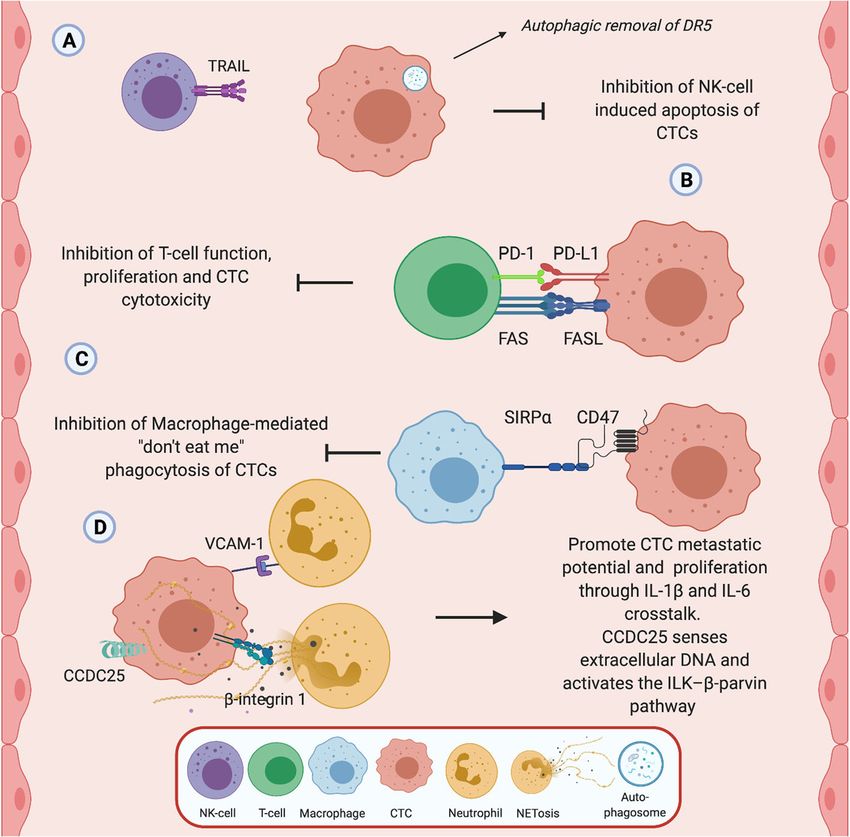
Ward et al. Molecular Cancer   (2021) 20:59                                                                         Page 2 of 17

Introduction                                                       allowing them to leave the primary tumour, journey
Metastatic progression is the most significant cause of cancer     through the body to a distal organ, and successfully es-
associated morbidity and mortality, causing over 8 million         tablish a metastatic niche is critical to the understanding
cancer deaths each year [1, 2]. While metastasis is typically      of metastatic disease progression [8, 17, 18]. Cancer cells
viewed as a process that is indicative of advanced stage can-      within the circulation are known as circulating tumour
cers, recent research suggests that dissemination of tumour        cells (CTCs) [19]. CTCs have unique phenotypic and
cells from the primary malignancy may be an early event in         genotypic characteristics, which allow them to survive
cancer progression [3, 4]. In a clinical setting, there has been   within the circulation and subsequently extravasate to
limited success in reversing metastatic progression using spe-     form a secondary tumour [20]. CTCs have been used as
cific targeting molecules, with the primary barrier being the      a non-invasive source of cancer cells for the analysis of
biological heterogeneity of the cancer cells in the primary        tumour phenotypes and genotypes (using blood as a so-
and metastatic tumour microenvironment [5, 6]. The process         called liquid biopsy), but their detailed characterisation
of metastasis is highly complex and understanding the mo-          also holds the key to understanding the biology of
lecular and cellular components involved is critical to our        blood-borne transition and therefore metastasis. Indeed,
ability to effectively treat cancer, but has proven extremely      the number of CTCs that can be detected at any one
difficult to define [6, 7].                                        time in a patient appears to be relative to the number of
   Metastasis is known to involve several sequential steps         “successful” metastatic events. However, effective isola-
referred to as the “metastatic cascade”. This is commonly          tion and accurate enumeration of these cells while they
regarded as the intricate journey a cancer cell must take          are in the circulation has proven difficult due to the con-
through different conditions in order to find a suitable dis-      straints associated with selectively analysing these rela-
tant environment to invade and establish [8, 9]. The ‘seed         tively rare cells. Although cells that disseminate from the
and soil’ hypothesis put forward in 1889 describes cancer          primary tumour are usually of epithelial origin, tumour cells
cells as “seeds” which must seek out the appropriate organ         can undergo a process known as epithelial-mesenchymal
microenvironment or ‘soil’ that will support their sus-            transition (EMT). During EMT cells lose polarity - adhe-
tained growth if they are to thrive [10, 11]. This hypothesis      rens and tight junctions are dissolved - resulting in a loose
still remains a strong argument for the reasoning behind           epithelial cell that has been dissociated from the epithelial
why certain tumour types have a tendency to metastasise            cell sheet [21, 22]. This independent cell differentiates to
to specific organs [5]. Several studies have shown that the        exhibit several mesenchymal attributes and a more motile
distal site acquired for metastatic progression can be de-         and invasive phenotype [21, 23–25]. Within this review
termined by specific gene patterns or signatures within            we will discuss how platelets promote the initial EMT
the primary tumour, which relate to specific organ sites           changes that enables the tumour cells to enter the blood-
[12, 13]. The complexity of the tumour microenvironment            stream. We will also discuss the sequential interactions
and cancer cell heterogeneity is further compounded by             between CTCs in the metastatic cascade and cells of the
exposure to the blood circulation system and its physical          blood circulation, highlighting the complex biology sur-
and cellular components [14–16]. Identifying the molecu-           rounding these putative biomarkers (Fig. 1).
lar mechanisms involved in the initiation of haematogen-              Given that most tumour cells are of epithelial origin,
ous metastasis and the interactions with platelets, the            many methods of detection and isolation of CTCs from
coagulation cascade and immune cells could help us bet-            patient blood samples involve the use of epithelial
ter understand specific outcomes in patients with meta-            markers such as pan cytokeratin (panCK) and epithelial
static disease. Thus, the overall aims of this review are to:      cell adhesion molecule (EpCAM) [20, 26–28]. Standard
                                                                   identification and the FDA-approved method of CTC
  1. Outline the contribution of platelets, the                    isolation and enumeration from patient blood uses
     coagulation cascade and immune cells on                       EpCAM positivity and leukocyte common antigen nega-
     circulating tumour cells (CTCs) in metastasis.                tivity (CD45-) for CTC classification [29]. However,
  2. Discuss the influence these haematological factors            studies have shown that these isolation methods do not
     have on CTC biology and the impact on their                   fully represent the vast array of CTCs in the circulation.
     clinical utility.                                             Consequently up to one third of patients with advanced
  3. Define the impact of these heterotypical cell-                colorectal, breast or prostate cancer do not possess
     interactions and discuss potential avenues of target-         CTCs that meet the standard criteria for cell enumer-
     ing CTCs in metastatic disease.                               ation [30]. As previously mentioned, the major use for
                                                                   CTCs in the clinic has been in the form of prognostic
Circulating tumour cells (CTCs)                                    biomarkers, and their use as biomarkers in many cancers
The identification and subsequent characterisation of              such as metastatic breast cancer and ovarian cancer have
tumour cells that possess distinguishing features,                 been studied extensively [31, 32]. However, despite the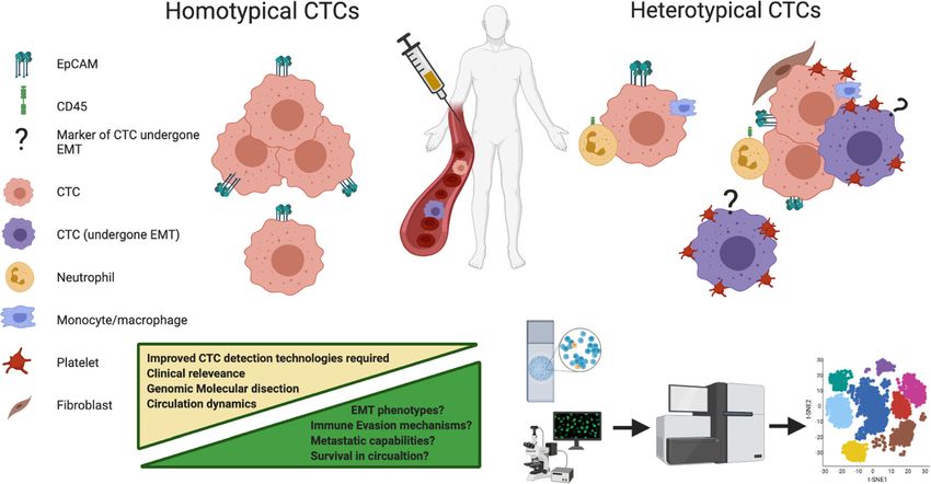
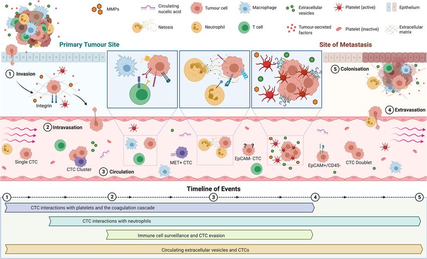
Ward et al. Molecular Cancer        (2021) 20:59                                                                                               Page 3 of 17

 Fig. 1 Overview and timeline of CTC-blood interactions during haematogenous dissemination. 1 Invasion: Tumour cells detach from the primary
 tumour and invade the surrounding tissue. Within the primary tumour, detached CTCs come into contact with platelets and neutrophils within
 minutes and hours of their dissemination. 2 Intravasation: Degradation of the extracellular matrix and the process of epithelial-to-mesenchymal
 transition (EMT) resulting from platelet interactions enables the tumour cells to move through the surrounding tissue and finally enter the blood
 circulation.3 Circulation: CTCs travel though the circulation. Here, they can exist as single cells, doublets or clusters of CTCs and have been shown
 to express heterotypical surface receptors, making them difficult to isolate using current technologies. CTCs are constantly interacting with
 circulating immune cells and other factors in the blood (platelets, circulating nucleic acids, EVs). 4 Extravasation: following the arrival to the site of
 distal metastasis, mesenchymal-to-epithelial transition (MET) occurs. Platelets aid in the recruitment of neutrophils to metastatic niche. Also,
 disseminated neutrophil-associated CTCs that arrive have enhanced extravasation capabilities. 5 Colonisation: CTC colonises a secondary site,
 aided and protected by immune cell-rich microthrombi and host EVs. Here, CTCs and CTC clusters can multiply and eventually develop into a
 metastatic tumour

prognostic impact of CTC counts that are seen mainly                            [25, 32, 38]. The metastatic potential of a tumour can also
using EpCAM-based capturing methods, this method-                               be based on the presence of a low number of stem cell-
ology is not capable of detecting the entire, highly                            like tumour cells found in the tumour tissue [25, 39].
heterogenous population of CTCs in patient blood sam-                           However, no guidelines currently exist for defining a
ples. This is due partly to varying EpCAM antigen dens-                         CTC-CSC phenotype, although a common classification
ities on CTCs as well as potential loss of EpCAM                                of CSCs of high CD44 and low CD24 expression (CD44+/
expression following EMT [26, 29, 33]. The process of                           CD24−/low), is frequently utilised in breast cancer, and
tumour cell dissemination seen in EMT is often accom-                           breast tumours with this expression tend to exhibit en-
panied by a loss or reduction of EpCAM expression on                            hanced invasion and metastasis [40]. The variable bio-
the surface of CTCs, rendering EpCAM-based detection                            marker expression in CTCs, the capacity to transition
methods unable to capture cells with weak or no                                 between different cellular phenotypes, the detection of sin-
EpCAM expression [34–36]. With more recent observa-                             gle CTCs and clusters of cancer cells, and the detection of
tions, many have come to the conclusion that a subset                           CTC and immune cell clusters raises the important ques-
of CTCs possess the unique tumour-initiating capabil-                           tion of which hound in the night is the dangerous one?
ities or stem cell-like properties that enable them to give                        The classic problems encountered for cancer detection
rise to a metastatic tumour [17, 20, 25, 37]. Multiple                          and prognosis apply directly to CTC biology with the
studies have shown that cancer stem cell (CSC) markers                          search for a “common” biomarker to facilitate efficient
are often expressed by CTCs in patient blood samples                            isolation, and more specific biomarkers to enable
Ward et al. Molecular Cancer (2021) 20:59 Page 4 of 17
accurate prognosis. Interpatient variability has also been CTCs survive more than 24 h in the bloodstream, with a
shown to play an important role in the expression of CTC's half life estimated to be around 1 h, impacting on
growth factor receptors, adhesion molecules, major the cells ability to metastasise [47, 48]. The platelet rich
histocompatibility complex antigens and proteases [37, thrombi that is thought to surround CTCs during their
41]. For example, several studies have indicated that the initial introduction to the circulatory system offers them
HER2 proto-oncogene defines a particularly aggressive physical protection from fluid sheer stresses (FSS) [49].
subset of CTCs, that when expressed by the CTCs of Platelet cloaking has been shown to protect ovarian can-
breast cancer patients, can indicate a poor prognosis [42, cer cells from FSS in vitro and to increase the produc-
43]. Similarly, the detection of CK-19 mRNA-positive tion of lactate dehydrogenase, conferring protection to
CTCs in the blood of patients with early stage breast these cells against shear induced damage [50]. At low
cancer after adjuvant chemotherapy has been shown to levels of FSS, thrombin-activated platelets have the cap-
be an independent risk factor for chemotherapy- acity to produce a 5-fold increase in endothelial adher-
resistant disease [44]. However, the problem associated ence in cervical cancer cells [51]. FSS can also stimulate
with CTC surface markers is the heterogeneity of the intravascular survival by upregulation of hexokinase 2
primary tumours. There are currently no known markers (HK-2) mediated glycolysis in CTCs [52]. The addition
that are universally expressed by all CTCs from a par- of a platelet cloak under these conditions may provide
ticular tumour type [45]. Despite attempts to standardise CTCs with a metabolic advantage in circulation, prefer-
criteria, there is conflict regarding the characterisation of entially shunting CTC metabolism to glycolysis. The dis-
CTCs and even more so for immunohistochemical tech- ruption of platelet-cancer cell interactions could
niques, where reproducibility across various laboratories potentially increase shear stress induced destruction of
has been poor [29, 30]. This raises another important cancer cells, limiting the metabolic advantage and meta-
point; the CTC field relies heavily on high quality immu- static potential of CTCs in circulation.
nochemical reagents which together with robust meth- Platelets contain within their α-granules, various
odology are essential to assess CTC variability. For growth factors that are secreted during platelet activa-
example, the non-classical CTCs, such as those lacking tion, such as platelet-derived growth factor (PDGF), vas-
EpCAM expression, or possessing the leucocyte marker cular endothelial growth factor (VEGF) and
CD45 are not well understood and defining this bio- transforming growth factor beta (TGF-β) [53]. These
logical variability is critical. The full extent of CTC util- growth factors can be utilised by CTCs to aid cell
ity as potential clinical biomarkers is currently unknown growth [54, 55] and to evade apoptosis when exposed to
due to our imperfect methods of isolation and enumer- chemotherapy [56]. Platelets have been found to increase
ation, as well as our overall lack of understanding of bio- the metastatic potential of solid tumours by inducing
logical differences between homotypic CTCs and EMT through TGF-β signalling [57, 58]. Platelet-derived
heterotypic CTCs. To complicate matters even further, TGF-β and direct platelet-tumour cell interactions
we will now discuss the consequences of CTC interac- have been shown to harmoniously activate the TGF-
tions with cells from the blood circulation. We will dis- β/SMAD and NF-κB pathways in cancer cells, result-
cuss how these interactions influence a CTC’s ability to ing in their transition to an invasive MET phenotype
survive within the circulation and the biological chal- with enhanced metastasis. Inhibition of either TGF-β
lenges arising from their reciprocal actions. from platelets or the NF-κB pathway in cancer cells
prevented metastasis in vitro [58]. Cancer cell adhe-
Platelets and CTCs sion to, or degranulation of, platelets has also been
Cancer cell–platelet interactions are a crucial part of found to induce pro-survival and pro-angiogenic sig-
cancer metastasis and there may be a physical as well as nalling within cancer cells [59]. The role of platelets
a biochemical basis for this important biological inter- in tumour cell proliferation is somewhat more contro-
action. However, even though the link has been docu- versial, with platelets manifesting both pro- and anti-
mented since the late nineteenth century, the interaction proliferative phenotypes [54, 60]. While inhibiting cell
and role of platelets in haematogenous metastasis re- proliferation in colorectal cancer cells, platelets pro-
mains largely unknown. Thrombocytosis (excess plate- moted metastasis through the release of extracellular
lets in the blood) has been linked with a poor prognosis vesicles (EVs) that induce EMT and endothelial cell
in cancer patients [46]. Platelets are among the first cells activation [60]. Platelet cargo and release of tumour
of the circulation that CTCs encounter on their journey promoting circulating EVs is an exciting hypothesis
to metastasise (Fig. 1). CTCs, once disseminated, spend that CTCs may utilise within the circulation. Future
a short time within the circulation before being either studies are warranted as to the impact of circulating
trapped within the capillaries they encounter or cleared EVs derived from both platelets and other sources in
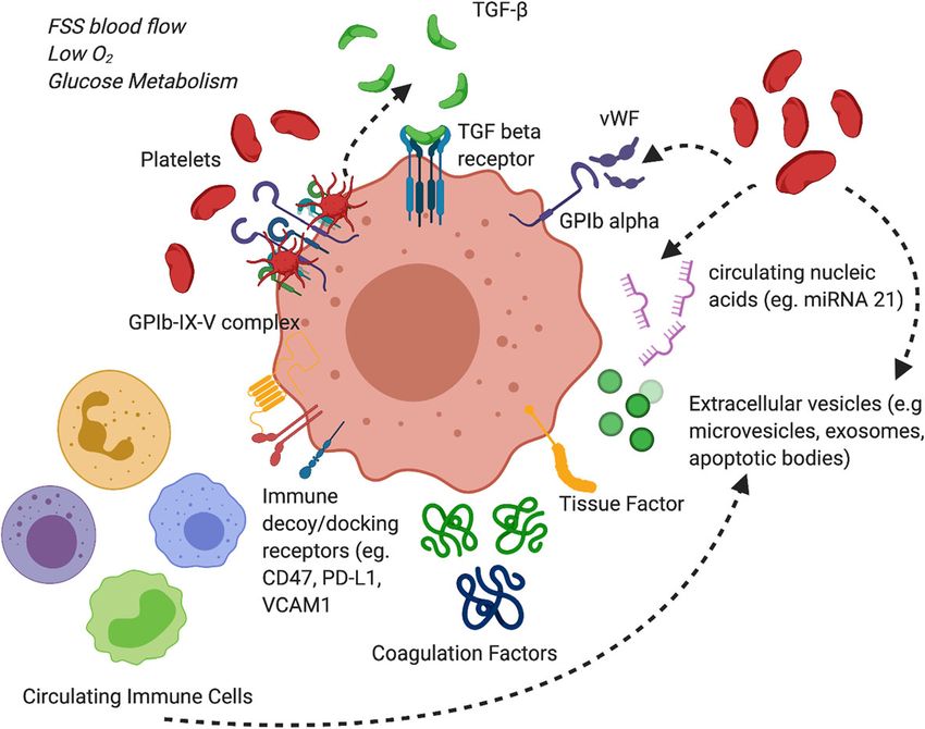
by patrolling immune cells. Only about 0.1% of single aiding CTC survival.Ward et al. Molecular Cancer (2021) 20:59 Page 5 of 17 The subsequent down-regulation of epithelial marker markers of platelets to be overexpressed in their CTC surface expression through the induction of EMT by datasets [65, 66]. ITGA2B (integrin alpha-IIb;CD41) and platelets may also hide platelet cloaked CTCs from clas- ITGB3 (integrin alpha-V beta 3;CD61) can be expressed sical antibody based detection [61, 62]. The presence of by CTCs and are critical for the platelet-cancer cell inter- platelet-derived TGF-β1 in situ in the bloodstream is also action, as inhibition of ITGB3 prevents the platelet- crucial for metastasis, as pre-treating tumour cells with tumour cell interactions [67]. These mRNA signatures platelets from WT mice fails to enhance metastasis forma- may result from platelet vesicle transfer as a consequence tion in mice lacking TGF-β1 in their platelets [58]. Conse- of the platelet cloak and could be a putative marker of ag- quently, it is postulated that the platelet cloak provides gressive CTCs. CTC established cell lines too have been CTCs with a source of TGF-β1 within the circulation, giv- found to express high levels of CXCL5, further elucidating ing them a more invasive, mesenchymal-like phenotype the importance of platelets in fuelling the metastatic po- and extravasation capabilities. Platelets themselves have tential of CTCs [68]. been found to dictate the formation of early metastatic Platelet-derived autotaxin (ATX), a secreted enzyme niches, promoting the recruitment of granulocytes inde- important for generating the lipid signalling molecule pendent of tumour signals through the release of CXCL5/ lysophosphatidic acid (LPA), interacts with tumour in- 7 chemokines [63]. Blockade of the CXCL5/7 receptor tegrin αVβ3 to promote metastasis of breast cancer cells CXCR2, or depletion of either platelets or granulocytes to bone [69]. As ATX is physiologically present in blood, has been shown to prevent the formation of early metasta- platelet cloaked CTCs may utilise this mechanism for sis in mice. This study also reported that the formation of metastasis to the bone in circulation. Interestingly, plate- the platelet induced early metastatic niche occurs within let TGF-β1 and MMP-1 regulate bone metastasis forma- 2 h of tumour cell arrival in the lung vasculature empha- tion, with platelet uptake of tumour-derived proteins sising the near immediate role of platelets in CTC colon- aiding in distal metastasis [70]. Platelets can also pro- isation events. The importance of the CXCL5 axis in CTC mote tumour angiogenesis by secreting numerous angio- metastasis is further exemplified by studies investigating genic regulators and VEGF [71, 72]. There is a need to its role in mediating breast cancer metastasis to the bone better understand the physical and biochemical basis of [64]. Interestingly, RNA sequencing studies for both single CTC platelet interactions as they are central to cancer cell CTCs and CTC clusters have found gene expression cell survival and metastatic potential (Fig. 2). Fig. 2 CTCs interactions with constituents of the blood circulation. CTCs are exposed to a number of influencing factors while in circulation including fluid sheer stress (FSS), hypoxia, nutrient starvation/glucose metabolism. Platelets, coagulation proteins and immune cells provide either direct or indirect contacts with CTCs to aid in their survival. Platelets are a rich source of TGF-β which promotes EMT. Platelets and coagulation proteins also protect CTCs from FSS through the creation of a rich microthrombi surrounding CTCs. CTCs evade immune detection through the expression of immune decoy receptors such as CD47 and PD-L1. These cells, proteins and circulating nucleic acids/extracellular vesicles can influence not only the phenotype of the CTC in circulation but also its molecular make up and cellular fate within the peripheral blood circulation
Ward et al. Molecular Cancer   (2021) 20:59                                                                    Page 6 of 17

Coagulation cascade and CTCs                                  recruitment of immune cells such as neutrophils which
Like platelets, proteins of the coagulation cascade are       activate endothelial cells and promote CTC extravasa-
thought to contribute to the protective thrombi formed        tion from the circulation [63]. It is postulated that co-
around CTCs following intravasation to the circulation        agulation proteins promote a hypercoagulable milieu
system. Patients with metastatic cancer have global           that CTCs utilise on the road to haematogenous metas-
platelet hyperactivity, which could contribute to the risk    tasis. However, mechanistic studies are required to fully
of thrombosis [73]. Indeed, venous thromboembolisms           investigate whether expression of these coagulation pro-
(VTEs) are frequent complications in patients with can-       teins by CTCs is critical for their dissemination, circula-
cer, with the incidence being high in pancreatic, brain,      tion and colonisation.
and gynaecological malignancies [74–76]. This hyperco-           As previously mentioned, patients with advanced stage
agulable state is due to multiple factors including sys-      cancers have a high risk of developing a VTE which is
temic inflammation and altered expression of circulating      thought to be both tumour and treatment related. The
blood coagulation proteins such as fibrinogen, Tissue         presence of CTCs is associated with an increased risk of
Factor (TF), Factor V (FV), FVII, FVIII, FIX and FX [77,      VTE in breast cancer, with CTC positive patients being
78]. CTCs themselves have been found to express TF,           5 times more likely to develop VTE compared to those
the known receptor for coagulation factors VIIa and X,        who are CTC negative [95, 96]. CTC positivity is also
which acts as the principle initiator of coagulation [79].    linked to plasma D-dimer levels in patients with meta-
TF plays a key role in aiding thrombin-mediated prote-        static breast cancer, further linking CTCs with an in-
olysis and the formation of tumour cell-associated            creased risk of VTE and hypercoagulation in metastatic
microthrombi [80, 81]. TF binding with factor VIIa has        cancer patients [97]. However, although increased CTC
the potential to facilitate CTC adhesion to endothelial       counts are associated with an increased risk of VTE in
cells as well as the stimulate activation of several inter-   cancer, the lethal subpopulation of pro-thrombotic
cellular signalling pathways (MAPK, PI3K, AKT,                CTCs has yet to be identified, with further experiments
mTOR), extracellular matrix remodelling and cell prolif-      and animal model studies being required. While the
eration [82, 83]. TF overexpression in tumour cells has       thrombotic potential and the pathogenesis of coagula-
been found to be directly related to the overexpression       tion proteins in mesenchymal and CSC biology remains
of mutant oncogenes such as K-RAS and EGFR as well            to be elucidated, it is clear that multiple aspects of plate-
as the loss of tumour suppressor genes p53 and PTEN           let biology and coagulation proteins are directly involved
[84, 85]. TF expression and signalling is implicated in       in CTC survival and metastatic potential.
the formation of the metastatic niche and can be upreg-
ulated on cancer stem cells [86, 87]. TF can stimulate        Immune cells and CTC interactions
tumour thrombin production as well as blood coagula-          While the immune response to cancer is vast and intri-
tion serine proteases such as PARs (protease-activated        cate, involving a range of cell types and molecular mech-
receptors). The proteolytic activation of these receptors     anisms [98–100], we focus in this review on the specific
in tumour cells triggers signalling pathways that increase    interactions between immune cells and CTCs in periph-
cell migratory/and or invasive abilities through increased    eral blood. We also highlight the mechanisms that CTCs
secretion of MMPs, as well as the activation and release      use to circumvent tumour immune responses including
of soluble proangiogenic factors VEGF and IL-8 [88, 89].      their ability to hijack specific immune cells (Fig. 3).
Indeed, FVIIa itself has been found to have a role in            Neutrophils are among the first immune cells that dis-
tumour pathogenesis, with overexpression increasing the       seminated CTCs encounter following entry to the blood
migratory and invasive potential of breast cancer cells       circulation. Neutrophils are a subset of mature poly-
through PAR2 activation and the upregulation of β-            morphonuclear myeloid cells that are first responders to
catenin [90]. Members of the activated protein C path-        a site of inflammation, and represent one of the body’s
way, a key anticoagulant pathway, are expressed in gy-        first line of defence against pathogens and “foreign” cells
naecological tumours and also play a role in cancer           [101]. Neutrophils were previously regarded as passive
pathogenesis [91]. These non-coagulation functions of         players in the inflammatory response in cancer. How-
the coagulation cascade postulates a role for these pro-      ever, studies have shown these cells can exhibit both
teins in CTC mediated survival and metastatic potential       pro-tumour and anti-tumour functions, as a direct result
in the circulation. Procoagulant circulating EVs too may      of manipulation from various signals emanating from
influence CTCs in circulation by facilitating the transfer    cancer cells [102, 103]. In metastatic breast cancer, the
of proteins and nucleic acids, for instance pro-oncogenic     number of CTCs have been found to correlate with neu-
miRNAs to CTCS, including miR-21 and abundant                 trophil to lymphocyte ratios (NLRs), with patients with
platelet miRNAs [92–94]. The procoagulant nature of           CTCs and a NLR < 3 having an 8 times greater risk of
the microthrombi utilised by CTCs promotes the                disease recurrence [104]. The secretion of granulocyte-
Ward et al. Molecular Cancer       (2021) 20:59                                                                                           Page 7 of 17

 Fig. 3 Interactions between CTCs and immune cells. CTC interaction with immune cells in the circulation is central to both their survival and ability
 to form metastatic niches. a NK cells, b T-cells, c macrophages, and d neutrophils in the blood circulation have all be found to interact with CTCs.
 CTCs have shown the ability to resist TRAIL-induced apoptosis via autophagic removal of death receptor 5 (DR5) in vitro, thus circumventing
 cytokine-mediated immune surveillance. CTCs also have been found to express PD-L1 receptor and interact with T-cell PD-1 to reduce anti-CTC
 T-cell function. Expression of CTC PD-L1 may prevent T-cell mediated cell destruction and offer a potential therapeutic target towards CTCs.
 Expression of CD47 on CTCs may stimulate “don’t eat me” signals, evading macrophage-mediated phagocytosis and promoting intercellular
 adhesion and migration of CTCs. Neutrophils, using both direct cell contact and through the production of extracellular traps can promote the
 metastatic potential of CTCs through increased cellular proliferation. VCAM-1 and β-Integrin1 interactions between CTCs and neutrophils
 promotes an inflammatory milieu that is conducive for CTC extravasation and formation of the metastatic niche. CTCs too use CCDC25 to sense
 neutrophil extracellular DNA produced by NETs deposits in organs acting as a chemotactic factor to attract CTCs for distal metastasis

colony stimulating factor (G-CSF) by tumours recruits                        survive, develop resistance to H2O2-mediated cell death.
circulating neutrophils to the site of a primary tumour                      Indeed, exome sequencing of breast cancer patients
[105]. CTCs identified within CTC-neutrophil clusters                        positive for CTC-neutrophil clusters revealed a mutation
were also found to express G-CSF and other cytokines                         within the TLE1 gene, which may offer some explan-
involved in neutrophil stimulation, suggesting that re-                      ation as to why these cells have the ability to overcome
cruitment of neutrophils to CTCs may occur rapidly in                        neutrophil-mediated killing [106]. Loss of TLE1 results
the circulation or indeed at the primary tumour itself                       in excessive activation of NF-κB–mediated inflammation
[106]. Tumour killing neutrophils have been found to                         in cells and has been shown to aid in cancer progression
eliminate cancer cells via the production of hydrogen                        [110]. Primary tumours with TLE1 mutations too, were
peroxide (H2O2) [107]. However, the enhanced presence                        also found to have increased levels of neutrophil infiltra-
of neutrophils at the site of a primary tumour has been                      tion and shed significantly more CTC-neutrophil clus-
associated with an overall poor prognosis in a number of                     ters into the bloodstream [106]. The inflammatory
cancers [108, 109]. In the context of CTCs, it is specu-                     milieu created by this mutation creates the perfect op-
lated that they too are cleared within the circulation                       portunity for CTCs to utilise neutrophils for colonisa-
using this mechanism but that those cells that do                            tion. The CTC-neutrophil cluster interaction is also
Ward et al. Molecular Cancer   (2021) 20:59                                                                   Page 8 of 17

mediated through VCAM-1-dependent intercellular               phenotypes [116]. As immature patrolling cells, mono-
junctions based on mouse xenograft experiments. It is         cytes can be adapted for clearing cellular debris and to
also conciliated by a cytokine-receptor crosstalk involv-     aid in inflammation. Monocytes can also be recruited to
ing IL-1β and IL-6 [106]. This inflammatory CTC-              the pre-metastatic niche, interacting with tumour cells
neutrophil crosstalk also leads to increased proliferation    to prevent their attachment and facilitate their elimin-
of CTCs within the circulation. Thus, this CTC-               ation by Natural killer (NK) cells through the secretion
neutrophil “piggy back” aids in dissemination, survival       of chemokines such as CCL3, CCL4 and CCL5 [117,
and CTC colonisation improving the metastatic capacity        118]. CTC counts from metastatic breast, colorectal and
of CTCs in the blood of patients.                             prostate cancer were found to inversely correlate with
   Neutrophils also have the ability to form an antibacter-   the expression of TLR2 and TLR4 on peripheral mono-
ial trap in the extracellular space to combat bacterial ac-   cytes suggesting that these cells play a role in CTC cir-
tivity which is also of significant importance for CTCs       culation [119]. Macrophages are mature monocytes with
[111]. These neutrophil extracellular traps (NETs) are        phagocytotic potential that can migrate from the blood-
extrusions of plasma membrane and nuclear material            stream into tissue. They can be responsible for clearing
composed of granule components and histones that can          pathogens and aged/damaged cells like CTCs from the
bind to and kill pathogens in circulation or within resi-     blood stream by phagocytosis [120]. Macrophages detect
dent tissues [111, 112]. Cell death that occurs as a result   and destroy altered cells in the blood that are not “self”
of these NETs is NADPH oxidase dependent and is re-           or which can be regarded as foreign; distinguishing cells
ferred to as NETosis [112], with aged neutrophils poten-      that are “self” from those that are not, like CTCs, by
tially having a higher potential for releasing NETs           identifying “‘don’t eat me” signals on the cell surface of
compared to non-aged neutrophils. Neutrophils maybe           “self-cells”. One of these “‘don’t eat me” signals is CD47,
capable of using these NETs to bind to CTCs while in          a cell surface glycoprotein utilised by red blood cells,
the blood. These CTC-NET interactions are a type of           platelets and lymphocytes to protect against their elimin-
“cloak”, similar to the cloak platelets use to promote        ation by macrophages [121, 122]. CTCs have been found
CTC adhesion to capillaries and subsequent extravasa-         to express CD47 and utilise it to avoid phagocytosis
tion at sites of colonisation [113]. NET deposition has       within the circulation [123]. CD47 binds to the inhibi-
been found to increase tumour cell adhesion to the hep-       tory immunoreceptor signal regulatory protein alpha
atic and pulmonary microvasculature in vivo, and              (SIRPα) on phagocytotic cells, including macrophages, to
in vitro resulting in increased tumour cell migration and     induce a signal cascade within the cell that inhibits
invasion [114]. Moreover, NET formation in the absence        phagocytosis [123, 124]. As a result, CD47 has been im-
of systemic inflammation has also found to be sufficient      plicated in aiding tumour cell evasion from immune sys-
to increase tumour adhesion in vivo. In a murine model,       tem signals [123]. CD47+ CTCs have also been found to
the interaction between CTCs and NETs was found to            be responsible for tumour relapse and metastasis in pa-
be mediated through CTC and neutrophil β1-integrin            tients with breast cancer [125], suggesting that the CTCs
[115]. NETs contain granule components and histones           may also use this surface marker for colonisation.
that have been found to act as chemotactic factors to at-        While the tumour microenvironment is inherently im-
tract cancer cells, rather than just merely acting as a       munosuppressive [126], cancer progression within the
‘trap’ for them. The presences of NET deposits in the         blood circulation is paradoxically associated with an in-
liver or lungs have been found to attract metastasising       tense immune response. Tumour-infiltrating immune
CTCs [114]. The transmembrane protein CCDC25,                 cells, particularly CD8+ T-cells and NK cells, have the
which is found on cancer cells, can act as a NET-DNA          potential to restrict tumour outgrowth or reject meta-
receptor and senses extracellular DNA to subsequently         static tumour cells [127]. Indeed, in most primary tu-
activate the ILK–β-parvin pathway, enhancing cell motil-      mours, a strong Th1/cytotoxic T-cell infiltration is
ity. Why CTCs were primarily attracted to the liver or        correlated with increased patient survival [9, 127]. In
lungs by NETs remains an unanswered question, but it          comparison, the peripheral circulation system is an im-
is speculated that this maybe in part due to favourable       munomodulatory complex that is highly reactive to for-
inflammatory conditions created by these organs. Future       eign cells such as CTCs. CTCs have been found to
studies are required as to the potential role of neutro-      express ‘immune decoy receptors’ which aid in CTC T-
phils and NETosis in aiding colonisation of other distal      cell immune evasion within the circulation [128, 129].
organs by CTCs, such as the brain.                            Programmed death 1 (PD-1) receptor and its ligand
   Circulating monocytes are also myeloid derived im-         (PD-L1) are important checkpoint proteins for the regu-
mune cells like neutrophils, which can be differentiated      lation of the anti-tumour immune response [130]. By
into different subpopulations of cells in response to vari-   binding to PD-1, CTC’s expressing PD-L1 have been
ous stimuli, resulting in juxtaposing inflammatory            shown to limit T-cell function and proliferation to
Ward et al. Molecular Cancer   (2021) 20:59                                                                        Page 9 of 17

facilitate immune tolerance [130, 131]. However, few               homotypic and heterotypic phenotypes depending on
studies to date have looked at the exact role of PD-L1 in          their interactions with other cells in the circulation. Al-
CTC biology. NK cells are major circulating immune                 though the molecular mechanisms involved in their for-
cells that protect the body from a plethora of potentially         mation and biology remain largely unknown, CTC-CTC
damaging agents such as virus-infected cells and malig-            interactions are also of the utmost importance. Clusters
nant transformation [132, 133]. NK cells are responsible           of several individual CTCs have been reported in numer-
for marking cells that lack the Major Histocompatibility           ous cancers and are indicative of a poor clinical outcome
Class (MHC) 1 marker, which is present on all nucleated            in a number of cancer types [65, 150, 151]. CTC clusters
cells, to facilitate their destruction by apoptosis [132]. In      tend to express more mesenchymal markers than epithe-
the early stages of tumorigenesis, NK cells can also de-           lial markers, and given that the process of invasion re-
tect and eliminate tumour cells [134], but patients where          quires a mesenchymal phenotype, this expression profile
CTCs have been detected, have been found to have re-               may confer a selective survival advantage on these cell
duced NK cell numbers as well as decreased NK cell ac-             clusters when in circulation [151, 152]. While it may be
tivity [119, 132]. The exact killing mechanism NK cells            obvious, cooperation between similar cell types shielding
use for CTC elimination in the peripheral circulation is           one another from shear forces, environmental or oxida-
not clear, but direct cell-cell contact is thought to be re-       tive stress, and or immune assault in the circulation is
quired [135]. NK cells induce tumour cell lysis in the circula-    advantageous to CTC clusters (Fig. 4). In this context,
tion via the secretion of tumour necrosis factor-related           heterotypic clusters containing more durable stromal or
apoptosis-inducing ligand (TRAIL), a transmembrane protein         immune cells aggregated with CTCs may provide add-
of the tumour necrosis factor family [136, 137]. TRAIL             itional benefit, which in the most simple terms, protect
expressed on the membrane of NK cells can also bind to a           the core of the CTC cluster. Following on, it has been
series of death receptors expressed on the cancer cell surface     proposed that CTC clusters are less likely to undergo
to induce apoptosis [138, 139]. While FSS alone can sensitize      anoikis or apoptosis than a single CTC, advocating that
CTCs to TRAIL-induced apoptosis [140], in vitro breast can-        the plakoglobin-dependent intracellular adhesion pro-
cer models have also shown that CTCs can develop a resist-         teins holding the cells together could confer a survival
ance to TRAIL-induced apoptosis via autophagic removal of          advantage on the cluster [153].
death receptor 5 (DR5) [141]. We postulate therefore, that            Understanding single cell CTC and CTC-cluster me-
CTCs could potentially be avoiding TNF cytokine-mediated           tabolism and how these cancer cells interact with other
immunosurveillance by downregulation of DR5, which to-             cells in circulation to influence changes in physical prop-
gether with protective “platelet cloak” may enable the im-         erties, metabolism and metastatic potential is of the ut-
munosuppressive nature of CTCs. While the cloaked CTCs             most importance. While in the circulation, oxygen
are shielded from TNF-α and NK cell cell-mediated cytotox-         deprivation may be even more severely restricted than
icity [142, 143], the biochemical basis for this protection may    that of the primary tumour such that only the toughest
extend beyond the physical barrier provided by the cloak.          cells survive, it may nevertheless provide access to nutri-
Platelets can down-regulate NK cell-surface receptor NKG2D         ents to support the CTCs that promote or require alter-
via paracrine signalling [144, 145] and actively supress NK cell   nate metabolism. Understanding the influence of
degranulation and inflammatory cytokine (interferon-γ) pro-        circulatory cells on single CTCs, and CTC homotypic
duction [146], with CTCs potentially benefiting from this          and heterotypic cell clusters in the rewiring of the cancer
mechanism. Platelet-derived VEGF can also supress antigen          cell metabolic network and phenotypic potential is
presentation in mature dendritic cells which in turn limits        therefore paramount. As cells such as platelets during
their immune surveillance capabilities [147]. CTCs can also        cloaking are known to alter EMT which itself impacts
interact with CD4+ Treg cells and have been associated with        the expression of genes involved in metabolic pathways,
defects in T-cell adaptive immunity [148, 149], suggesting that    the EMT phenotype of CTC clusters needs to be investi-
CTCs may actively suppress immune function. Additional             gated [154–156] given the strong relationship between
functional studies are needed to fully dissect the interaction     cancer cell growth and migratory potential, and their
between CTCs and T-cells to reveal the influence T-cells play      contribution to the metastatic process. While these path-
in CTC mediated metastasis and to understand the molecular         ways have been shown to be relevant in single CTCs,
basis of NK cell immune suppression.                               differences in cancer cell metabolism associated to CTCs
                                                                   with mesenchymal characteristics or CTC clusters in-
CTC singlets, clusters and survival strategies;                    cluding platelets and /or other immune cells remains to
safety with friends or potential therapeutic                       be elucidated. The influence of coagulation factors on
targets?                                                           CTC metabolism also remains unclear. However, as glu-
CTCs in the circulation have been shown to exist both              cose metabolism has been found to influence coagula-
as individual cells and as clusters of cells that display          tion factor expression and is associated with an increase
Ward et al. Molecular Cancer (2021) 20:59 Page 10 of 17 Fig. 4 Homotypical and Heterotypical CTCs and their clinical relevance in malignancies. For the use of liquid biopsies to reach their full capabilities, CTC isolation technologies are needed to capture the full array of homotypical and heterotypical CTCs that exist in the circulation. Molecular dissection using single cell genomics, transcriptomics and proteomics integration too will reveal the molecular mechanisms that allow for CTCs interactions. This will allow for greater clinical utilisation by identifying the clinically relevant cells and also reveal potential targets for CTC therapeutic interventions. Overcoming these factors must be considered for future CTC enumeration and molecular taxonomy studies risk in VTEs [157], it is postulated that the metabolism Na+/K+ ATPase inhibitor, and if shown efficacious, of the procoagulant microthrombi cluster of CTCs is al- could be given in combination with standard chemother- tered and influenced by the expression of coagulation apy for metastatic breast cancer. Further observational proteins such as TF in the circulation. CTCs may tap clinical trials are also under way to interrogate key sig- into or directly modulate the metabolic function of sur- nalling networks that are active in CTCs clusters rounding cells and this could be a primary weapon in (NCT04520672. Data from these trials will better inform disarming immune functions that are highly glucose us as of potential drug candidates to target CTC clusters. dependent. The delineation of potential mechanisms of The targeting of heterotypical clusters may prove some- the metabolic “champion” profiles of metastatic CTCs, what more difficult due to the interactions of platelets and its influence on the colonisation potential of CTC and immune cells. clusters may lead to the development of new therapeu- Targeting the interactions between platelets/coagula- tics that seek out and interrupt CTC cluster metabolic tion cascade potentially offers an avenue for CTC dis- pathways that are critical to their survival. ruption. Aspirin is a well-established anti-platelet agent Further molecular dissection of the cells within the and has shown the ability to disrupt the platelet-cancer CTC heterotypic cluster and the development of stan- cell interaction. Aspirin has the ability to inhibit the re- dardised markers to detect CTCs that have undergone lease of MMPs from platelets thereby preventing the EMT are necessary to fully dissect the biology of CTC degradation of the ECM and reducing the invasive po- clusters and elucidate any further potential weakness of tential of CTCs [158]. Previous studies have shown that these lethal CTCs. (Fig. 4). Interestingly, next generation ovarian cell line models induce high levels of tumour cell sequencing of homotypic CTC clusters illuminated a po- induced platelet aggregates (TCIPAs) and therefore may tential weaknesses; in a mouse model, Na+/K+ ATPase provide a useful model to investigate if aspirin can in- inhibitors enabled the dissociation of CTC clusters into hibit ovarian TCIPAs in the initiation of CTC metastasis single cells, leading to DNA methylation remodelling at [158]. Platelet function can also be inhibited by integrin critical sites and metastasis suppression [66]. Currently a subunit αIIb inhibitors, which have been reported to re- clinical trial (NCT03928210 is underway to investigate duce metastatic cancer progression [159, 160]. Similarly, the effects of Digitoxin on CTC clusters in breast cancer the experimental blockade or deletion of key platelet re- patients. Digitoxin itself is a well-established and safe ceptors on CTCs, such as Glycoprotein Ib-IX-V and
Ward et al. Molecular Cancer   (2021) 20:59                                                                 Page 11 of 17

Glycoprotein VI, can interfere with the formation of          that of CTC clusters, or whether the cells that constitute
TCIPAs, and significantly decrease their metastatic po-       a heterotypical CTC cluster exhibit increased PD-L1 ex-
tential [161]. The inhibition of platelet aggregation         pression. The evaluation of CTC expression in meta-
in vivo by a highly specific Glycoprotein α-IIb β3            static NSCLC patients that were treated with the PD-1
receptor antagonist or aspirin [162] has the potential        inhibitor Nivolumab [169], showed the detection of PD-
therefore to impact CTC function and downstream               L1 positive CTCs by CellSearch post-treatment corre-
metastasis. Clinical trials such as the “Add-Aspirin”         lated with immunotherapy resistance in 14 of 19 pa-
trial (NCT02804815) are currently underway and de-            tients. In addition to indicating that PD-L1 positive
signed to assess whether regular aspirin use after            CTCs may be a marker of immune escape [169]. This
treatment for an early-stage cancer can prevent recur-        study also highlights the potential for CTCs as a tool to
rence and enhance survival rates in patients [163]. It        monitor changes in PD-L1 expression in tumour cells
would be very interesting if CTC monitoring could be          during radiation therapy, which can potentially be prog-
included in such a trial, as the use of aspirin may po-       nostic for response to treatment [131]. Further studies
tentially diminish the platelet-rich thrombi associated       are needed to illustrate whether PD-L1 expression in
with the initial stages of metastasis and prevent the         CTCs is acquired at the primary tumour or only while in
onset of metastatic disease. The use of direct oral an-       the circulation, and to elucidate the possible reasoning
ticoagulants (DOACs) may also offer a potential av-           behind differing PD-L1 expression profiles of CTCs and
enue for targeting CTCs. However, current data is             primary and secondary tumours. Interestingly, combin-
inconclusive as to their effectiveness at reducing            ation therapies targeting markers of CTC expression
metastatic disease in patients and in vitro studies           such as HER2/3 or EpCAM in conjunction with im-
have so far been disappointing [164].                         munotherapies may offer an alternative approach to
   Targeting CTCs ability to evade immune detection is        single-agent immunotherapy [170, 171]. The combin-
an exciting avenue in immuno-oncology. The utilisation        ation of EpAb2–6 (anti-EpCAM monoclonal antibody)
of PD-L1 by CTCs as a means of immune evasion is a            with atezolizumab (anti-PD-L1 antibody) has been
potential therapeutic target for metastatic disease, but is   shown to almost completely eliminate tumours in an
still somewhat in its infancy for solid tumours that          orthotopic model of human colorectal cancer. Also, duel
undergo haematogenous metastasis. PD-1/PD-L1 inhibi-          targeting of the HER3 receptor using an antibody-drug
tors that are currently in use in the clinic and undergo-     conjugate, U3–1402, has been found to charge the anti-
ing clinical trials use CTCs to monitor treatment             tumour effects of PD-1 inhibition alone in mice [172].
response [165, 166]. However, to date no trial data exists    These combination therapies significantly enhanced
for the direct targeting of CTCs by immunotherapies. In       anti-tumour immunity and may offer an avenue to
advanced lung cancer and gastrointestinal cancer there        prime otherwise immunotherapeutic “cold” tumours.
is an association between PD-L1 expression in CTCs            Further studies are warranted to investigate whether
and poor prognosis in patients [167, 168]. This associ-       dual-targeting of these markers results in altered CTC
ation is hypothesised to be a result of PD-L1 positive        expression in patients. However, these studies do high-
CTCs that reflect an immunosuppressive tumour micro-          light potential new combination strategies for cancer im-
environment, which can promote tumour relapse, and            munotherapy in patients with EpCAM+ or HER3+
PD-L1 positive CTCs with a higher metastatic potential        CTCs and opens the avenue for further combination im-
owing to their increased immune evasion capacity [131].       munotherapies targeting both CTC surface and immune
This heterogeneity between primary and metastatic tu-         evasion markers. Finally, with the ever expanding field of
mours indicates that a single core biopsy could be ineffi-    CAR-T cell immunotherapies, therapies designed to dir-
cient at gauging the level of PD-L1 expression at all         ectly target an individual patient’s CTCs maybe soon be
tumour sites, and CTC expression levels could also be         on the horizon.
flawed in this way. This comes back to a basic problem
in the CTC field where there are limitations with CTC         Conclusion
isolation and their characterisation, which is com-           Circulating tumour cells are critical components of the
pounded by the need for alternate biomarkers that can         metastatic cascade that can be isolated from patients by
effectively report on the complex biology of CTCs, as         a simple liquid biopsy, and can provide valuable infor-
discussed in this review. It is possible that some meta-      mation about the patient’s tumour profile and prognosis.
static or circulatory event could confer a change of PD-      The clinical utility of CTCs is yet to be fully recognised
L1 expression on CTCs. Further research is need investi-      or appreciated, mainly because we only have a limited
gating the altering of PD-L1 profile between the meta-        understanding of CTC biology. The difficulties associ-
static tumour and CTCs. It is also not known whether          ated with the isolation and enumeration of CTCs have
PD-L1 expression is altered between single CTCs and           hindered our ability to progress the technology for CTC
Ward et al. Molecular Cancer          (2021) 20:59                                                                                                 Page 12 of 17

characterisation and this has generated conflict within                          involved in funding acquisition. The authors read and approved the final
the field regarding appropriate characterisation, and con-                       manuscript.

sequently, variable data sets and conclusions. While
                                                                                 Authors’ information
many methods of CTC detection exist, the invention of                            Not applicable.
a single standardised method to capture the full plethora
of CTCs will allow for a more uniform body of research                           Funding
                                                                                 The study was supported by an Innovation Partnership Fund from Enterprise
that would greatly aid future studies and clinical prac-                         Ireland, which is co-funded by the European Regional Development Fund
tice. However, this may reveal the inherent adaptivity of                        (ERDF) under Ireland’s European Structural and Investment Funds Pro-
CTCs and a greater appreciation for the environment                              grammes 2014–2020.
that CTCs inhabit and importance of the cells that con-
                                                                                 Availability of data and materials
stitute this environment. Understanding aspects of CTC                           Data sharing not applicable to this article as no datasets were generated or
cluster biology will be important to better comprehend                           analysed during the current study. All data and information in this review
the dynamics of homotypic and heterotypic CTC clus-                              can be found in the reference list.
ters as well as how this is influenced by the mutational
                                                                                 Declarations
heterogeneity and metabolic adaptability at a critical
point in the metastatic cascade. Targeting the cells of                          Ethics approval and consent to participate
the blood circulation to either prevent the formation of                         Not applicable.

CTC clusters or their interactions with single CTCs in                           Consent for publication
the circulation may lead to a significant delay in meta-                         Not applicable.
static potential and thus increased patient survival. Fu-
ture CTC studies must take into consideration the                                Competing interests
                                                                                 The authors declare no conflicts of interest.
influence that the cells of the circulation have on CTC
imitation, militance and adaptability during the progres-                        Author details
                                                                                 1
sion to advanced metastatic disease.                                              Department of Histopathology and Morbid Anatomy, Trinity College Dublin,
                                                                                 Dublin 8, Ireland. 2Emer Casey Molecular Pathology Research Laboratory,
Abbreviations                                                                    Coombe Women and Infants University Hospital, Dublin 8, Ireland. 3Trinity St.
ATX: Autotaxin; CCL: C-C Motif Chemokine Ligand; CD24: Cluster of                James’s Cancer Institute, St James’s Hospital, Dublin 8, Ireland. 4Department
differentiation 24; CD44: CD44 molecule (Indian blood group);                    of Obstetrics and Gynaecology, Trinity College Dublin, Dublin 8, Ireland.
                                                                                 5
CD45: Leukocyte common antigen; CD47: Integrin associated protein;                Cancer Research Institute, University of South Australia, 5001 Adelaide,
CSC: Cancer stem cell; CTC: Circulating tumour tell; DNA: Deoxyribonucleic       Australia. 6School of Health and Biomedical Sciences, RMIT University, Victoria
acid; ECM: Extracellular matrix; EGFR: Epidermal growth factor receptor;         3083 Bundoora, Australia. 7BD Research Centre Ireland, Limerick, Ireland. 8BD
EMT: Epithelial-mesenchymal transition; EpCAM: Epithelial cell adhesion          Technologies and Innovation, Research Triangle Park, NC, USA.
molecule; FSS: Fluid shear stress; FV: Coagulation Factor V; FVII: Coagulation
Factor VII; FX: Coagulation Factor X; G-CSF: Granulocyte-colony stimulating      Received: 11 November 2020 Accepted: 15 March 2021
factor; HER2: Human epidermal growth factor receptor 2; HER3: Human
epidermal growth factor receptor 3; HK-2: Hexokinase 2; IL: Interleukin;
iNOS: Nitric oxide synthase; ITGA2B: Integrin alpha-IIb; CD41; ITGB3: Integrin   References
alpha-V beta 3; CD61; K-RAS: Kirsten ras oncogene; LPA: Lysophosphatidic         1. Bray F, Ferlay J, Soerjomataram I, Siegel RL, Torre LA, Jemal A. Global cancer
acid; MAPK: Mitogen-activated protein kinase; MDSC: Myeloid-derived                  statistics 2018: GLOBOCAN estimates of incidence and mortality worldwide
suppressor cell; MET: Mesenchymal-epithelial transition; MHC: Major                  for 36 cancers in 185 countries. CA Cancer J Clin. 2018;68(6):394–424.
Histocompatibility Class; miRNA: microRNA; MMP: Matrix metalloproteinase;            https://doi.org/10.3322/caac.21492.
mTOR: Mammalian target of rapamycin; NET: Neutrophil extracellular trap; NK      2. Gupta GP, Massagué J. Cancer metastasis: building a framework. Cell. 2006;
cells: Natural killer cells; NKG2D: Natural killer group 2D; NLR: Neutrophil         127(4):679–95. https://doi.org/10.1016/j.cell.2006.11.001.
leucocyte ratio; NSCLC: Non-small cell lung cancer; p53: Tumour protein p53;     3. Hüsemann Y, Geigl JB, Schubert F, Musiani P, Meyer M, Burghart E, Forni G,
PAI-1: Plasminogen activator inhibitor-1; panCK: pan Cytokeratin;                    Eils R, Fehm T, Riethmüller G. Systemic spread is an early step in breast
PARs: Protease-activated receptors; PD-1: Programmed death 1; PD-                    cancer. Cancer Cell. 2008;13(1):58–68. https://doi.org/10.1016/j.ccr.2007.12.
L1: Programmed death ligand 1; PDGF: Platelet-derived growth factor;                 003.
PI3K: Phosphoinositide 3-kinases; PMN: Polymorphonuclear;                        4. Pantel K, Brakenhoff RH. Dissecting the metastatic cascade. Nat Rev Cancer.
PTEN: Phosphatase and tensin homolog; SMAD: Small mothers against                    2004;4(6):448–56. https://doi.org/10.1038/nrc1370.
decapentaplegic protein 1; T reg: Regulatory T-cells; TCIPA: Tumour cell-        5. Fidler IJ. The pathogenesis of cancer metastasis: the ‘seed and soil’
induced platelet aggregates; TF: Tissue Factor; TGF-β: Transforming growth           hypothesis revisited. Nat Rev Cancer. 2003;3(6):453–8. https://doi.org/10.103
factor beta; TLR: Toll-like receptor; TNF: Tumour necrosis factor; TRAI              8/nrc1098.
L: Tumour necrosis factor-related apoptosis-inducing ligand; VEGF: Vascular      6. Valastyan S, Weinberg RA. Tumor metastasis: molecular insights and
endothelial growth factor; VTE: Venous thromboembolism                               evolving paradigms. Cell. 2011;147(2):275–92. https://doi.org/10.1016/j.cell.2
                                                                                     011.09.024.
Acknowledgements                                                                 7. Parcesepe P, Giordano G, Laudanna C, Febbraro A, Pancione M. Cancer-
Figures 1, 2, 3 and 4 in this review were created using BioRender.com                associated immune resistance and evasion of immune surveillance in
(https://biorender.com).                                                             colorectal cancer. Gastroenterol Res Pract. 2016;2016:1–8. https://doi.org/1
                                                                                     0.1155/2016/6261721.
Authors’ contributions                                                           8. Sethi N, Kang Y. Unravelling the complexity of metastasis—molecular
MW, SOT and JOL contributed to conception and manuscript design. MW,                 understanding and targeted therapies. Nat Rev Cancer. 2011;11(10):735–48.
LK, SOT and JOL wrote the manuscript. LN, BM, TK, MB, AC, NB, CM, DB, RB,            https://doi.org/10.1038/nrc3125.
SS SH, and ED were all involved in manuscript revision and critique of the       9. Kitamura T, Qian B-Z, Pollard JW. Immune cell promotion of metastasis. Nat
manuscript for important intellectual content. CM, SOT and JOL were                  Rev Immunol. 2015;15(2):73–86. https://doi.org/10.1038/nri3789.
You can also read