Nanoengineered photoactive theranostic agents for cancer
←
→
Page content transcription
If your browser does not render page correctly, please read the page content below
Nanophotonics 2021; aop

Review

Nishant K. Jain, Bavya M. Chathoth, Vinil S. Bhaskar, Himanshu Meena, Rajendra Prasad*
and Rohit Srivastava*

Nanoengineered photoactive theranostic agents
for cancer
https://doi.org/10.1515/nanoph-2021-0205                                 nanotheranostics. Moreover, the challenges involved in
Received April 30, 2021; accepted June 29, 2021;                         clinical translation of photoactive materials along with their
published online July 13, 2021
                                                                         application in vivid areas of cancer nanomedicine and
                                                                         elucidate the future implications on photoactive therapy have
Abstract: Cancer has gained much attention because of
slow development of advanced diagnostics and therapeutic                 been addressed here.
strategies. So far, conventional procedures like surgery, ra-            Keywords: cancer therapy; nanotheranostics; photoactive
diation therapy and chemotherapy are only available options              materials; photodynamic probes; photothermal agents.
for cancer treatment which have various limitations. To
overcome the limitations of conventional procedures, nano-
diagnostics, and therapeutics are emerging approaches for                1 Introduction
localized diagnosis and treatment of cancer nowadays. So far,
various bio-mimicking and stimuli active cancer theranostic              Cancer stills remains as one of the leading causes of death
platforms have been established but they are limited only for            worldwide and accounts for about 10 million deaths per year
animal studies and their clinical translational progress is              [1, 2]. Approximately 70% of deaths from cancer occur in low-
slow. Among various cancer theranostics platforms, photo-                and middle-income countries mainly due to delayed diag-
responsive systems have shown promising outcomes for                     nosis and lack of access to diagnosis or treatment facilities [3].
cancer theranostics applications due to their specific physi-            Further, the monetary expenses involved in treating cancer
cochemical properties, biocompatibility, multifunctionality              are quite high and results in economic burden. Conventional
etc. Moreover, these photothermal agents in combination                  treatment strategies such as surgery, radiotherapy, and
with diagnostics probes and surface functional targeting                 chemotherapy are still widely used in spite of several draw-
moieties demonstrate their synergistic response for site se-             backs that include nonspecificity, toxicity, and poor thera-
lective imaging and ablating cancer cells/tumor. Photoactive             peutic efficacy [4]. Hence, various researchers across the
principles are rife and with increasing access to light irradi-          globe have been trying to explore novel strategies that can
ation setups, more the discovery of photoactive products,                overcome these limitations. Nanotechnology has emerged as
more would be the success reaped in cancer battle. This re-              an innovative technology to cater the unmet needs in the field
view highlights recent developments in cancer nano-                      of cancer therapy [5–7]. Several materials i.e., organic as well
theranostics with a special focus on photoactive functional              as inorganic in origin have been designed to exploit their
                                                                         unique properties at nanoscale dimension. These nanoplat-
Nishant K. Jain and Bavya M. Chathoth have contributed equally to        forms exhibit diverse physicochemical properties and
this work.                                                               demonstrate significant therapeutic activity in treating cancer
                                                                         by active or passive targeting mechanism. Furthermore, these
*Corresponding authors: Rohit Srivastava and Rajendra Prasad,
                                                                         nanosystems can be surface functionalized with targeting
Department of Biosciences and Bioengineering, Indian Institute of
Technology Bombay, Powai, Mumbai, 400076, India,                         ligands and can also be made bioresponsive resulting in
E-mail: rsrivasta@iitb.ac.in (R. Srivastava), rpmeena@iitb.ac.in         localized action at the tumor site yielding better therapeutic
(R. Prasad). https://orcid.org/0000-0002-3937-5139 (R. Srivastava).      response with less side effects [5–11]. Photothermal therapy
https://orcid.org/0000-0001-9851-8630 (R. Prasad)                        (PTT) has emerged as one of the potential modality in treating
Nishant K. Jain, Bavya M. Chathoth and Himanshu Meena,
                                                                         cancer offering several advantages that includes non-
Department of Biosciences and Bioengineering, Indian Institute of
Technology Bombay, Powai, Mumbai, 400076, India
                                                                         invasiveness, low toxicity, and localized action [12, 13]. It
Vinil S. Bhaskar, Department of Anaesthesia, Saifee Hospital,            makes use of photoactive agents that generate heat upon
Girgaon, Mumbai, 400004, India                                           irradiation with specific wavelength of light leading to
  Open Access. © 2021 Nishant K. Jain et al., published by De Gruyter.     This work is licensed under the Creative Commons Attribution 4.0
International License.
2        N.K. Jain et al.: Nanoengineered photoactive theranostic agents for cancer

denaturation of proteins, DNA damage and cellular mem-                2 Approaches in cancer diagnosis
brane destruction [14, 15]. This results in ablation of cancer
cell with subsequent reduction in size of tumor tissue.                 and therapy
Further, the most widely used light for PTT is near infrared
(NIR-I) (650–950 nm) often mentioned as first biological               2.1 Traditional treatment approaches
window because the specified wavelength reduces NIR
                                                                      Treatment strategies for cancer have undergone huge
absorption of biological tissues mainly blood and water
                                                                      progress in bringing back patients to live quality life
[16, 17]. While, some studies have indicated 1000–1700 nm
                                                                      overshadowing the holy grail. According to expert opinion,
as second wavelength (NIR-II) or second biological window
                                                                      the advancement in screening and treatment has largely
for which bioimaging of deep rooted tumors show immense               improved or doubled the survival rates in decades after
progress with improved signal to noise ratio [17]. Recently,          diagnosis. Cancer diagnosis has broadly been divided into
several materials of organic and inorganic origin are                 traditional and modern screening, with a focus to under-
designed with an aim to improve the photothermal con-                 stand the cell morphology at microscopic level. Traditional
version efficiency with high biocompatibility and enhanced             procedure includes biopsy, scans (X-ray, computerized
localization of nanomedicine at target site for better in vivo        tomography, ultrasonography, and magnetic resonance
PTT performance [18, 19]. Additionally, PTT mediated                  imaging), and endoscopy [28]. Among the above, biopsy is
nanomaterials are used in combination with other strategies           regarded as the gold standard confirmatory test for cancer,
like chemotherapy, photodynamic therapy, and immuno-                  for which the abnormal tissue is removed and further
therapy to achieve synergistic effect for better tumor                delved to understand the cellular pathology. Once the
regression [16, 17, 20]. Moreover, these hybrid systems can           disease is confirmed treatment proceeds based on the
                                                                      severity/degree of the ailment. The treatment options used
be designed as stimuli responsive nanocarriers and
                                                                      in the past and still relied to date are radiation, chemo-
functionalized with ligands for on-demand release of
                                                                      therapy, immunotherapy or a combinatorial approach of
chemotherapeutic drug and site-specific localization of the
                                                                      chemo-immunotherapy, and surgery [29].
nanomedicine, respectively [21, 22]. The heat generated
during the PTT activates the antitumor immunity
through immune-stimulatory molecules and release of                   2.2 Modernized techniques for cancer
antigens from the ablating tumor cells [23–25]. Further, it               treatment
also alters the enzymatic activity and gene expression
of living cells that regulates the biological events [26, 27].        2.2.1 Photodynamic therapy (PDT)
     In this review, we have highlighted the recent de-
velopments in the field of photo-activated nanomaterials              PDT is a procedure by which photosensitizing agent is
and their applications as cancer nanotheranostics. Design of          targeted onto tumor cells which upon stimulation with
various advanced NIR active hybrid materials for localized            visible light of specific wavelength produces reactive
imaging and synergistic PTT have been discussed. Further,             singlet oxygen species (Figure 1a) [30]. The procedure re-
the principle of heat generation from photoactive materials           quires vigilant selection of photoactive agents capable of
upon irradiation with NIR light and mechanism of cancer               tumor localization and metabolic synthesis resulting in
cell death from generated heat has been deciphered. The               irremediable cytolysis of cancer cells [31]. Additionally,
                                                                      tumor vasculature adds to protumorigenic and immuno-
role of these nanohybrids as an efficient multimode-imaging
                                                                      suppressive microenvironment creating physical barrier to
agent for visualization of tumor area has also been reflected.
                                                                      T cell infiltration favoring PDT [32]. The major challenge in
Besides thermal effect, the synergistic effect achieved with
                                                                      this therapy is the photobleaching of sensitizers. Therefore,
nanohybrids through multimodal therapies with delivery of             loading of these photosensitizers in nanocarriers could
drug, gene or enzyme, and photodynamic therapy (PDT) has              overcome this issue [33]. Commonly employed sensitizers
been elaborated. Further, we have summarized the                      being hydrophobic, use of nanocarriers has shown to
challenges in clinical translation and future perspectives of         improve the bioavailability [29]. Mainly noble metals are
these photoactive nanotheranostic agents in the field of              suitable for PDT owing to their absorption capacity
cancer.                                                               compared to that of photoabsorbing dyes [34].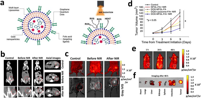
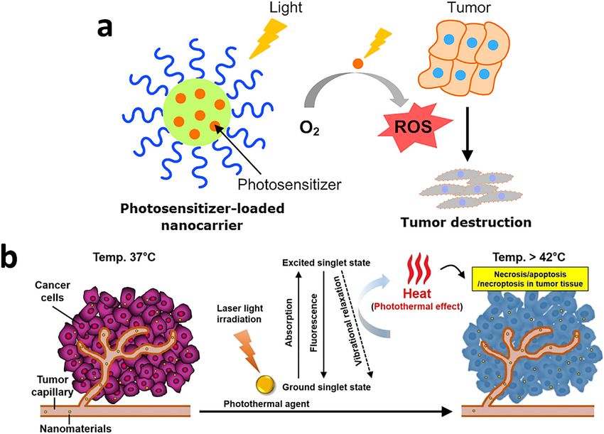
N.K. Jain et al.: Nanoengineered photoactive theranostic agents for cancer     3

Figure 1: Mechanism of action for photoactive nanotheranostic agent resulting in tumor regression.
(a) Photodynamic therapy (PDT) involving generation of reactive oxygen species (ROS) resulting in cytolysis of tumor cells [78]. (b)
Photothermal therapy (PTT) involving generation of heat upon irradiation resulting in ablation of tumor cells [79].

2.2.1.1 Commonly used agents for PDT                                     concentration [42]. Owing to the amphiphilic nature, ICG
The commonly used photosensitizers in PDT mainly belong                  has tendency to adsorb lipids [43]. Therefore, in certain
to tetrapyrrole family. Among them the most used are the                 cases ICG may interact with normal tissues and may lead to
ones found in nature; e.g.: phthalocyanines and porphy-                  chaos during surgical intervention. Notably, ICG in contact
rins [35]. Besides, many other compounds also serve as                   with living tissues bind and exhibits in vivo lipoprotein
photosensitizers (PS). The suitable commonly used dyes                   dynamics. Therefore, it is hard to predict the material
include phenothiazines, phenalenones, squaraines, and                    movement completely using ICG [44–46]. Studies have
indocyanine green (ICG). ICG is negatively charged poly-                 described that ICG enabled deep light tissue permeability
methine dye which serves in eradicating multiple cancers                 improvising PDT [38, 39]. Apart natural products are also
on irradiation with NIR laser at a wavelength range of 800–              useful as PS [33]. Further, with advancing technology
810 nm and has reduced toxicity to nonsubject host tissue                inorganic nanoparticles has contributed enormously
[36, 37]. Studies suggest ICG successfully absorbs light                 which is described in the later part.
above 800 nm, and the photochemical nature of the com-
pound makes it a suitable candidate for PDT [29, 38, 39].                2.2.2 Photothermal therapy (PTT)
However, ICG has certain reported drawbacks as well.
Stability of ICG differs in different solvents when used for             PTT aims to sentence cancer cell death via engineering heat
clinical applications, which could be accounted due to its               exposure (electromagnetic radiation) to near infrared light
physiochemical nature [40]. Presence of sulfonyl groups                  (Figure 1b) [47, 48]. Instilling contrast agents or dyes with
promotes water solubility of ICG, while hydrophobic                      weaker emission converts photo energy to thermal energy
polycyclic groups make it lipophilic [41]. While ICG810 is               and results in tumor cell necrosis or apoptosis [39].
seen to aggregate or degrade in water which may further                  Recently, tremendous progress is witnessed in cancer
result in selfquenching and fluorescent reduction, while                  therapy using PTT. NIR absorbents for efficient heat pro-
the optical properties strictly depend on the dye                        duction and incorporating drug delivery system (DDS) for
4        N.K. Jain et al.: Nanoengineered photoactive theranostic agents for cancer

enhanced heat exposure to abnormal cells has helped PTT               penetration. The second criteria for selection; the materials
to attain success. The notable merit of DDS in PTT is                 should not interact with cells and should be able to trigger
improvising the efficacy and ensuring safety to healthy                immune responses without external stimuli. Further, for
cells from photothermal damage. The only concern before               targeted therapies the agents should be capable of
starting the therapy is the optimization of fluence rate and           clearing from blood and normal tissues for assured
irradiation time. Later, DDS NIR absorbent and simulta-               phototherapy agent. To brief all these characteristics
neous PTT is performed irradiating tumor for specified time            with good photostability make the material apt for
followed by absorbent administration [49]. However, hur-              photoactive therapies [60].
dles exist in determining tumor degree, size, and hetero-                  The principle involved in the generation of heat by the
geneity for which maximum therapeutic effect is hard to               photoactive materials upon irradiation with NIR light in-
exert. At 50 °C or above the risk of thermal damage in                cludes absorption of photon resulting in excitement of elec-
healthy tissue raises concern as it might denature protein            tron. The excited electron shifts from low excited singlet state
and may likely cause healthy cell necrosis [50]. Generally,           (S0) to highly unstable excited singlet state (S1) that subse-
the temperature threshold for PTT therapy is chosen from              quently comes back to stable S0 state with release of energy
40 to 60 °C above which will lead to cell death by coagu-             during the relaxation process [61–65]. The energy released
lation necrosis [51, 52]. Above this temperature, it may              can be in the form of fluorescence emission or nonradiative
affect the integrity of cell membrane and induce the release          vibrational emission in the form of photothermal energy.
of cellular contents [51, 53, 54]. When a comparison of PTT           Various inorganic structures made from gold, silver, iron
is made over PDT, it could be concluded that PTT is more              oxide, graphene, carbon nanotubes, and upconversion
preferred than PDT due to its longer wavelength of light              nanoparticles have been explored for photothermal therapy
used and is well tolerated by normal cells; additionally the          [12, 13, 17, 65, 66], whereas, organic materials like dyes and
oxygen requirement for the procedure is very minimal [55].            polymers are also used in photothermal based treatment of
Well, the specificity of photosensitizers restricts extension          cancer. All these nanostructures upon irradiation with NIR
of PDT [56].                                                          light generate heat that ablates the cancer cells. Further,
                                                                      these materials due to their diverse physicochemical prop-
2.2.2.1 Commonly used agents for PTT                                  erties also aid in the multimodal imaging of the tumor area
Photothermal agents are selected based on their size,                 as well image guided tumor regression. The optical,
shape, and ability to transfer energy from one form to                electronic, and magnetic properties of these materials
another [33]. Mostly used agents comes in the category of             play a crucial role in contributing to this application
inorganic (metallic, carbon nanostructures, and quantum               [67, 68]. Such nanomaterials which are used for therapy
dots) or organic–inorganic nanohybrids. Inorganic metal-              as well as diagnosis of a disease are known as nano-
based photoactive agents are selected due to the desired              theranostic agents [69]. The field of nanotheranostics is
shape and size favoring heat and optical properties [57].             rapidly emerging in cancer with different novel materials
Whereas, various forms of carbon based nanostructures                 being explored by various researchers cross the world.
demonstrate ability to transfer energy from one form to               Their application in cancer has revolutionized the
another upon light irradiation [58]. Importantly, quan-               treatment regime as they offer several advantages over
tum confinement make quantum dots suitable optically                   the conventional treatment like biocompatibility,
active probes for theranostic applications [59]. Inorganic            biodegradability, minimal dose requirement with less
nanoparticles are not widely used because of the chal-                frequency, and low systemic toxicity [70].
lenges in biodegradability/clearance issue, bioavail-                      The most widely used light source for PTT is NIR due to
ability, and toxicity caused for long term [60]. However,             high penetration and less toxicity. The first window of NIR
hybrids derived from combination of organic and inor-                 (NIR-I, 650–950 nm) demonstrates lesser penetration when
ganic has demonstrated good biocompatibility, high                    compared to second NIR window (NIR-II, 1000–1700 nm)
renal clearance, less toxicity, and high photothermal                 through biological tissue [68, 71]. Additionally, the maximal
performance.                                                          permissible exposure (MPE) for skin is higher with NIR-II
                                                                      (1 W/cm2 for 1064 nm) when compared to NIR-I (0.33 W/cm2
2.2.2.2 Principle of photoactive agents                               for 808 nm) [72]. Hence, materials with photoexcitation in
Photoactive agent is the basic element for contrasting                NIR-II window have recently attracted wide attention in PTT
phototherapy and need to be selected judiciously. There-              based cancer nanomedicine. This includes inorganic as well
fore, these agents basically absorb light of particular               organic nanomaterials that are tuned to exhibit distinctive
wavelength, precisely in near infrared for improved light             photoexcitation in the NIR window. These photoactive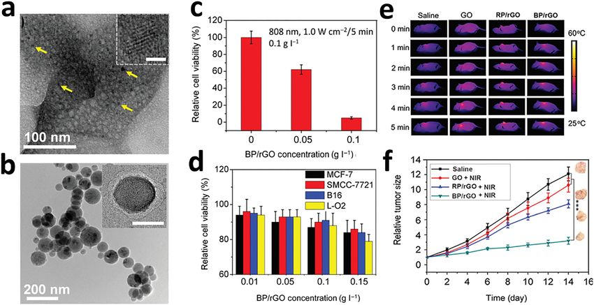
N.K. Jain et al.: Nanoengineered photoactive theranostic agents for cancer 5
carriers can be elements or derived as the products of (PS), which further needs wavelength lasers for activation
chemical bond with temperature sensitive material. The of two PS [75]. While with the advancement in studies it is
generated heat from these photoactive materials not only recognized that some PS is able to function both in PDT and
kills the malignant cells but also alters the enzyme activity PTT, e.g.; carbon dots [76]. Besides, ICG loaded liposomes
and gene expression. Further, these NIR photoactivable were also equipped in PDT and PTT assisted therapies [77].
nanosystems when used in combination with chemothera- An overview of different photoactive agents used in cancer
peutic drugs, enzymes or gene exhibit synergistic effect with for PDT, PTT and their combinatorial therapy has been
better therapeutic action [73]. Additionally, some of these provided in Table 1.
materials exhibit fluorescence or contrast due to their
unique physicochemical properties that aids in imaging of 2.2.4 PDT versus PTT: curbs in promoting to clinics
the tumor tissue which are recognized as nanotheranostic
agents. Recently, such photoactive nanotheranostic agents PDT has been introduced to clinics in the last 40 years and
are gaining attention and are actively reported for diverse holds propitious treatment options for cancers of head,
applications in cancer therapy [74]. neck, breast, bladder, bile duct, pancreas and other vital
organs [103]. The therapy is affirmed with the production of
reactive oxygen species (ROS). Irradiation triggers the
2.2.3 Combinatorial agents for PDT and PTT photosensitizer to absorb photons and to get excited to
higher electronic states. The singlet state photosensitizer
The efficacy could be pronounced with the use of combi- tries to achieve triplet state and further emits energy in the
natorial therapy of PDT and PTT. The dual therapy is form of fluorescence, heat or light. In this state, the ROS is
generally made use of two different phosphatidylserine produced via two mechanisms. The triplet molecular
Table : Different photoactive materials used in cancer.
Chemical family of photosensitizer Cancer type Wavelength (nm) References
used in PDT
Pyrolipid T murine breast tumor []
Murine colorectal cancer (MC , CT ) [, ]
Porphyrin Skin, bladder, esophagus, breast, lung, and brain [–]
Basal cell White light []
Chlorin Superficial, skin, breast, and colon [–]
Nanomaterials for cancer treatment
via PTT
Carbon nanotubes RKO (rectal carcinoma cell line), HCT (human colon cancer cells) []
AuNPs CC (colon carcinoma cells) []
Graphene oxide NPs HCT (human colon cancer cells), human dermal fibroblast []
PLGA-ICG-R T breast cancer cell line, CT colon rectal cancer cell line [, ]
Gold nanostars MB bladder tumor []
Combination therapies for cancer
PTT + CT HeLa cells (human cervix cancer cell line) LTH () + drug []
Polydopamine/rGo/MSN release
PTT + CT Breast cancer LTH () + drug []
CD AuNP release
PTT + CT HeLa cells (human cervix cancer cell line) LTH () + drug []
Polypyrrole/MIL/DOX release
PTT + PDT T LTH + ROS () []
Te ND Breast cancer cell line
GNcHyNA MDA-MB- LTH () + ROS []
Breast cancer cell line ()
UCNP-NGO/ZnPC KB cells (human nasopharyngeal epidermal carcinoma), HeLa cells LTH () + ROS []
(human cervix cancer cell line) ()
CP-TPP/Au/PEG nanospheres HeLa cells (human cervix cancer cell line) LTH () + ROS []
()
6 N.K. Jain et al.: Nanoengineered photoactive theranostic agents for cancer
oxygen in ground state stimulates to produce singlet oxy- 1350 nm. Skin tumors are treated using light emitting
gen species [104–106]. An ideal photosensitizer is equip- diode (LED) with regulated exposure to sunlight [114].
ped with ample biocompatibility and absorbs high light While, most of the lasers are available commercially
levels. PS can be classified into several categories like (Table 2).
metals, organic, inorganic, polymer based and others [107].
Apart from PS selection, it is often a cumbersome task to
decide drug to device match and the mode to which the 2.3 Role of nanoparticles in cancer cell
light needs to be applied, or in brief PDT dosimetry (irra-
screening and therapy
diation geometry with parameters which includes fluence
rate, wavelength, and total fluence), light source and
Nanoparticles (NPs) have growing interest in cancer ther-
exposure, PS dose and intervals for drug to light adminis-
apies. Certain nanoparticles are capable of absorbing light
tration is a topic which needs to be worked up diligently
and get heated up parallelly with their plasmon band. The
[108, 109].
optical transmission in tissue is optimal due to possible
PTT is a technique for treating tumors in local arena.
manipulation of plasmonic band in NPs from visible to
Here, the tissue temperature is raised above 60 °C and re-
infrared favoring deep tissue treatment [121]. Therefore,
sults in protein denaturation followed by plasma mem-
NPs need to be well orchestrated for; heat generation after
brane destruction. The mechanism is that photosensitizers
absorption of innocuous light in the near infrared range.
when activated by light of particular wavelength absorbs
The efficacy of PTT is largely dependent upon the accu-
photons and excites from singlet ground state to excited
mulation of light responsive NPs, excitation time with po-
singlet state. A cascade of event takes place, the electronic
wer density of light, and effectiveness of light energy to
excitation energy undergoes vibrational relaxation, a
heat conversion [47].
nonradiative decay and comes back to ground state which
Recent studies have demonstrated the use of nano-
is mediated by collisions from photothermal agents and
shells and nanocages of metallic and noble metals in
other molecules. During this event, tissue temperature el-
organic polymeric shells to combat cancer [49]. Study
evates as a result of kinetic energy, were heat shock pro-
conducted by Hirsch et al., revealed that gold–silica
teins and gene expression changes occurs to overcome the
nanoshell by thermal ablative therapy on tumor cells
thermal damage [110]. Further, as the temperature goes
demonstrated effective outcomes in breast carcinoma cells.
higher it causes microvascular thrombosis and ischemia
The study was performed tailoring the nanoshells to absorb
[111]. Both PDT and PTT are superior compared to other
near infrared where optical transmission was optimally
treatments due to selectivity of PS. In addition, the tech-
controlled. Photothermal morbidity (with 820 nm, 35 W/
nique provides less scar formation and can be utilized for
cm2) was further confirmed with viable fluorescent stain-
treating multiple times [112, 113].
ing. Simultaneously the in vivo studies conducted sub-
stantiated the irreversible thermal damage precisely to the
2.2.5 Laser and suitability tumor arena, using magnetic resonance imaging (MRI) for
concordant planning and temperature monitoring by
Light absorption for PTT and PDT in visible light ranges phase sensitive gradient-echo MRI. Further, magnetic
from 400 to 700 nm while for near infra-red from 700 to resonance temperature imaging (MRTI) evidence was
supported with histopathological findings, proving that
nanoshells can be used as potential candidate for thermal
Table : List of commercially available lasers along with their
ablative tumor destruction [122]. Recent study conducted
wavelength range [–].
by Norton et al. on PTT effects of plasmonic metal nano-
Laser Wavelength range
particles summarized that it is apt to consider NP whose
(nm) plasmon resonance is near to therapeutic window for
maximum possible light exposure onto soft tissues.
Green laser
Alexandrite laser – Though it is possible to tune the plasmon resonance of
Dye laser – nanoshells the absorption is narrow. Therefore,
Diode laser – maximum efficacy is obtained when optical wavelength
Neodymium doped yttrium aluminium garnet gets along with each other. The study has given more
laser
insights to which the NP diffusion can be confined to the
LED (red light)
tumor region [123].N.K. Jain et al.: Nanoengineered photoactive theranostic agents for cancer 7
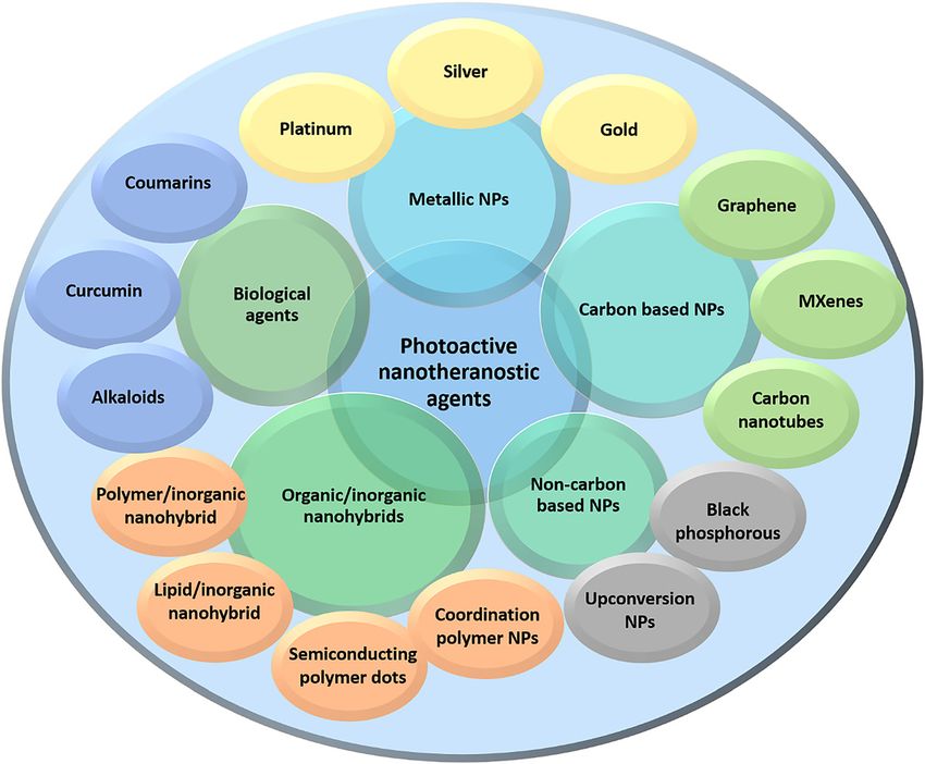
3 Nanotheranostic agents in phosphorous), organic/inorganic nanohybrids (lipid/inor-
ganic, polymer/inorganic, semiconductor polymeric dots,
cancer and coordination polymeric nanoparticles), and biologically
derived photoactive agents etc. (Figure 2) [126–128].
Conventional treatment of cancer involves chemotherapy,
radiation, and surgery. Based on the stage of cancer and
extent of tumor area, combination of these strategies is also
3.1 Gold nanoparticles
used for better therapeutic action. The widely used cancer
chemotherapy suffers from the drawbacks of non-
Gold nanoparticles (AuNPs) are the most commonly
specificity of drug action leading to poor drug concentra-
used metallic nanoparticles for PTT applications with
tion levels in tumor area, toxicity of nontargeted tissue,
vivid morphological structures like nanoshells, nano-
and multiple dose requirements [124, 125]. However, the
rods, nanospheres, and nanostars [60]. Of the ones
emergence of nanotechnology has added new dimension
mentioned, nanorods find use in numerous applications
to cancer treatment approach with minimal side effects.
due to their unique physiochemical properties and their
They exhibit unique physicochemical properties due to
high efficiency to converts light to heat [129]. Moreover,
their nanoscale dimension that plays a crucial role in
the ease of surface functionalization and biocompati-
cancer nanomedicine. Various nanotheranostics agents
bility has extended their applications in imaging, di-
derived from inorganic as well as organic origin have been
agnostics and cancer therapies [130]. Au nanoshells were
designed for cancer applications. However, photoactive
the first to get into clinical translation in the year 2008
materials have gained much attention due to their prom-
[131]. In vivo studies had confirmed the enhanced
ising potential to overcome the drawbacks of conventional permeability and retention (EPR) upon intravenous
chemotherapy. In addition, the delivery of therapeutic administration of Au nanoshells in mice, further ther-
molecules like drugs, enzymes or genes along with these mally ablated using 808 NIR laser [122]. Further pre-
nanohybrids results in synergistic therapy at minimal dose clinical assessment also confirmed that there was no
with reduced side effects. Different materials developed for obvious toxicity although the nanoshells accumulated in
PTT includes metallic nanoparticles (NPs) (gold NPs, silver liver and spleen. Therefore, full-fledged clinical trials on
NPs, and platinum NPs), carbon based NPs (carbon nano- Au nanoshells are in progress, for which lung cancer
tubes and nanodots, graphene based NPs, and Mxene), (primary/metastasis) patients are given Au nanoshell
noncarbon based NPs (upconversion NPs and black intravenously and temperature is raised via exposure to
Figure 2: Different types of photoactive
nanotheranostic agents used in cancer.8 N.K. Jain et al.: Nanoengineered photoactive theranostic agents for cancer radiation through bronchoscopy. Investigations are still and PDT employing Au nanoechinus both in in vivo and on the adult stage for treatment tumors of head and neck on HeLa cells. using Au nanoshells [47]. Mooney et al., were successful Further, Wang et al., proved Ce-6 Aptamer conjugated in showing the distribution of Au nanorods throughout Au nanorods for cancer therapy. Aptamer (Sgc8) targeted the tumors when transported by neural stem cells in leukemia T cells and the conjugation through Au-thiol breast cancer xenografts in mice in vivo. In doing so, the covalent bond. Fluorescence quenching was observed due gold nanorods were able to improve the tumor ablation to the proximity of Ce-6 to Au surface. Upon aptamer with tremendous reduction in tumor recurrence. The binding to cancer cells, Ce-6 released due to DNA structure work also compared the results from free Au nanorods. and purposed as agent for PDT irradiating with NIR The authors concluded that a combination of nano- (812 nm). The study highlighted PDT/PTT to serve as technology with cell therapy would benefit for cancer treatment for targeted multimodal therapy [134]. Yet curation [132]. Vijayaraghavan et al. [133], showed the another study by Lin et al., successfully demonstrated the complete elimination of deep tumors by the combinato- synergism of PDT/PTT in ruling out cancer (Figure 3). The rial treatment with gene silencing (ultralow NIR light) photosensitizer utilized was Ce-6 loaded plasmonic gold Figure 3: Morphological characterization of plasmonic gold vesicles (GV) and in vivo trimodality imaging using GV loaded with photosensitizer Ce6 (GV-Ce6). (a) Scanning electron microscopic (SEM) image and (b–d) Transmission electron microscopic (TEM) images of plasmonic gold nanovesicles. (e) NIR fluorescence image at preinjection and postinjection of GV-Ce6 in MDA-MB-435 tumor-bearing mice. (f) Thermal images at postinjection of GV-Ce6 in tumor-bearing mice upon 6 min irradiation with laser of 671 nm (2.0 W/cm2). Red circles indicate the tumor location. (g) Heating curves of tumors upon laser irradiation as a function of irradiation time. (h) In vivo photoacoustic (PA) images. Yellow circles indicate the injected location and (i) Average PA intensity of tumor tissues at preinjection and postinjection of GV-Ce6 [135].
N.K. Jain et al.: Nanoengineered photoactive theranostic agents for cancer   9

vesicles. The technology used was trimodality assisted          successful to prove that the nanocomposite system pur-
fluorescence/thermal/acoustic image guided combination           poses to be a promising PTT agent for treating cancer [137].
with PDT/PTT. The study reported that the absorbance by
vesicles in NIR region was strong, and the NIR irradiation
(671 nm) was successful in exciting Ce-6 along with Au          3.3 Platinum nanoparticles
vesicles producing singlet oxygen species together with
heat and eradicated cancer cells. When investigated in          Platinum (Pt) is a metal that absorbs light in biological
vivo, the results were fruitful and were visualized via         range and generates DNA strand breaks [140]. Though Pt
fluorescence/thermal/acoustic signals. There the study           NPs act as antioxidants at higher concentration, the
clearly descries the importance of combinatorial approach       toxicity reported is also high in literature [141]. While
with PDT/PTT in cancer cell destruction [135].                  cancer therapies have also evidenced synergistic effect
                                                                with the use of these NP’s further reducing the side effects,
                                                                which is a factor of longer time period patient prognosis
                                                                [142]. To date Pt NPs when used in reduced concentration
3.2 Silver nanoparticles
                                                                are apt in biological stability and tolerance [143]. Study
                                                                conducted by Depciuch et al., was on assessment of size of
Silver nanoparticles (Ag NPs) are popular due to their
                                                                Pt NPs for PTT therapy. The study concluded that the
antibacterial and tumor destructing properties. Besides,
                                                                antitumor activity of Pt NPs like DNA damage and enzyme
their plasmon tunability in the NIR biological window
                                                                activity, apart the smaller size could serve as best PS agent
range (650–1200 nm) have paved for increased applica-
                                                                [144]. Another study conducted by Song et al., demon-
tions of Ag in medical field [136]. More interesting is its
                                                                strated Au–Pt NPs uptake with cell targeting folic acid and
antitumor properties. However, Ag is shown to exhibit
                                                                triphenylphosphine targeting mitochondria in tumor cells.
cancer killing properties only at higher concentrations
                                                                Mitochondria targeting was achieved via loading PS (Ce6)
[137]. Ag in photoactive therapy seems to aid as NPs for
                                                                onto Au–Pt NPs. The authors were successful in designing
effective drug loading or photosensitizers to improve
                                                                multifunctional theranostic approach with ability to
antitumor therapy [138]. Ag NPs are suitable to load heat
                                                                target mitochondria using combinatorial PDT and PTT
labile or water soluble drugs because of the synthesis
                                                                [145]. While the study conducted by Phan et al., revealed
method (reduction of Ag ions which is devoid of organic         that the prepared Iron–Platinum NPs (Fe–Pt NPs) with
solvent or heat) [34]. Studies conducted by Bose et al.,        polypyrrole coating with evidence of high biocompati-
demonstrated folate receptor targeted plasmonic Ag NPs          bility, NIR absorbance, and photothermal stability can
intended for breast cancer where, Ag NPs to purport as          excellently serve as multifunctional system for photo
efficient nanocarrier system for delivery of quercetin           diagnostic application like PDT and photo acoustic im-
thereby inducing PTT. The quercetin folate receptor Ag NPs      aging (PAI) [141].
were synthesized by one pot method and later the
hydrogen bond between stabilizer and reductant were
tuned according to the need. The outcome of the study was       3.4 Graphene
that PTT induced Ag NPs complemented to the antitumor
efficiency by hyperthermia induction resulting in selective      Graphene based materials are of major importance in
lysis of abnormal cells with a fate of apoptosis. Further,      biomedical applications due to their uniqueness in prog-
quercetin incorporated Ag NPs showed double antitumor           nosis, diagnosis, and therapeutic agents in cancer thera-
effect in lab and animal studies. The study successfully        pies [146]. This is due to their easiness in surface
proved that quercetin loaded Ag NPs are more effective          modification, for being rich in carboxyl and hydroxyl
than free quercetin and can be beneficial for breast cancers     groups [147]. Graphene lays superior position in cancer
[139]. While recent study conducted by Park et al., tried to    treatment due to its toxic effects in tumor cells [148]. Hence,
delve effectiveness and applications of indocyanine green       graphene is used actively in battling cancer in PTT, PDT,
loaded Ag NPs. The study was successful in high loading         and imaging [149]. On the other hand, there needs an un-
capacity of indocyanine with good stability against light       met research to address its biocompatibility and biode-
induced degradation and hepatic clearance. The composite        gradability [150]. Wang et al., demonstrated in his studies
delivery system addressed significant tumor accumulation         by covalent grafting of nanographene oxide as core shell
combining local irradiation of laser at tumor site resulting    and upconversion through bifunctional polyethylene gly-
in tremendous inhibition of tumor growth. The author was        col, further, loading phthalocyanines on graphene oxide
10 N.K. Jain et al.: Nanoengineered photoactive theranostic agents for cancer
surface. The study reports the greater efficacy of the pre- upto 1 mg/mL, and was shown to exhibit photoactive
pared nanocomposite system with good biocompatibility. ability upon green, blue light activation [161]. Zheng et al.,
The study leaves a remark that the system can be a great in his studies a solution for hypoxia condition caused in
success to be used as upconversion luminescence probe of solid tumors. He designed a multifunctional nano-
cells and to image whole body with greater contrast [101]. composite comprising of carbon dots decorated with car-
Recently, a notable study conducted by Thapa et al., bon nitride NPs against hypoxic tumor water splitting. The
revealed that graphene was suitable for prostate cancer results were so promising that improved intracellular ox-
treatment, where the authors intratumorally-injected ygen concentration with reactive oxygen species under
palladium nanoparticle decorated with graphene oxide. hypoxia and normoxia with light irradiation. While in vivo
The outcome was that the graphene oxide decorated studies confirm that the multifunctional nanosystem sur-
nanoparticulate system showed enhanced local distribu- passed tumor hypoxic condition. The author suggests us-
tion, photothermal ablation, and inhibited cancer cells in ing water-splitting materials which has the capability of
PC3 xenograft mouse models, with reduced organ toxicity enhancing oxygen level and reversing hypoxia induced
[151]. Besides, these a study conducted in 2016 by Zhang PDT resistance and metastasis [162]. A recent study by
et al., proved that BaGdF5 nanoparticles were seemed to get Sundaram et al., in 2020, was successful in coating hyal-
attached intact on graphene oxide surface nanosheets to uronic acid and Ce6 onto carbon nanotubes which pur-
form the GO/BaGdF5/PEG nanocomposites. This nano- ported as photosensitizer. This further on when tested on
composite system exhibited low toxicity, efficient magnetic CaCo-2 colorectal cancer cells via PDT approach (660 nm)
contrast. The author with evidence suggested that the inhibited apoptotic cell death, the study confers the
system could serve as dual imaging (MR and X-ray) models nanocomposite system to be an efficient vehicle for
for in vivo tumor models [152]. Study conducted by Nur- photosensitizer localization in Colorectal cancer cells [163].
unnabi et al. [153], was yet a further confirmation that
graphene nanoparticles being photoluminescent was
suitable candidate for PTT and imaging. Notably Nafiujja- 3.6 Mxene
man et al. [154], through his work elucidated the use of
graphene quantum dots in targeted cancer therapy (PDT) MXenes are two-dimensional (2D) novel structures derived
and imaging. In short, graphene will be a promising com- from transition metal carbides and nitrides [164]. They are
pound in future for treating and imaging cancer, except the characterized by the presence of layered arrays of transi-
challenges faced for its biocompatibility. tion metal atoms that are interconnected by a carbon or
nitrogen atom. The general formula for MXenes is
Mn + 1XnTx (n = 1–3), where M stands for early transition
3.5 Carbon nanostructures metal carbides (Ti, Hf, Zr, Nb, Ta, V, Mo Sc etc.), X denotes
carbon or nitrogen and Tx represent functional groups
Carbon dots are basically diamond or graphitic core, which (–Cl, –F, –OH, and O) terminating the M surface [165–167].
is sp2 or sp3 hybridized. Low dimensional carbon is clas- They have attracted increased attention as they exhibit
sified as carbon nanoparticle, carbon nanodot or carbon excellent electrical, optical, magnetic, and mechanical
quantum dot, and fullerenes [155–157]. The ease of surface properties [168, 169]. These properties can be tuned by
functionalization of carbon dots with amine (NH2), car- modulating the transition metal atom and surface func-
boxylic acid (–COOH–), alcohol (OH), aldehyde (CHO) and tional groups. Further, they demonstrate thermal stability
production tuning their size together with the ability to and extreme biocompatibility that makes them suitable for
deliver photoluminescence has made them popular in catalysis, energy storage and biomedical applications [164,
cancer therapeutics and imaging [158]. Besides, they have 166, 170, 171]. They have structural similarity with gra-
gained much attention in biomedical applications owing to phene and their morphology as well as size can be altered
their optical absorption, photostability, high penetration, as per the application requirement. Recently, MXenes have
good electrical conductivity, biocompatibility, high emerged as a promising photothermal agent for cancer
chemical stability, and low toxicity [159, 160]. Study con- therapy due to their high surface area, hydrophilic nature,
ducted by Serda et al., in 2018 has affirmed the use of broad absorption band in UV/NIR region and high photo-
fullerene in PDT applications. The authors have developed thermal conversion efficiency (PCE) [172, 173]. The hydro-
glycoconjugated C60 derivative for adenocarcinoma. As a philic nature of MXenes can attributed to the presence of
result, it was seen that fullerenes completely accumulated hydroxyl (OH), oxygen (O), and fluorine (F). Additionally,
in nucleus of stellate pancreatic cells, which is not toxic their surface can be functionalized to achieve activeN.K. Jain et al.: Nanoengineered photoactive theranostic agents for cancer 11
targeting as well as combinatorial therapy. Moreover, the ultraviolet/visible. Large stokes shift, photo-blinking and
planar structure of MXenes can acts as a carrier for cargo stable energy levels in micro/milli seconds makes UCNPs
delivery [172]. Highly photostable MXene QDs with good outstanding compared to other techniques [177]. Besides,
quantum yield and tunable wavelength demonstrating minimal scattering and absorption levels improved pene-
luminescent properties have been studied extensively for tration depth favors UCNP for in vivo biological applica-
PTT. Therefore, they can be used for imaging guided syn- tions [178, 179]. The host materials selected for UCNP
ergistic PTT integrated with chemotherapy/photodynamic should preferably be of low lattice energy, to lessen non-
therapy in cancer. The potential of MXene as theranostics radiative loss and deliver maximum radiative emission.
agents in cancer has been explored by researchers across Nonradiative energy loss needs the presence of phonons in
the globe. host lattice [180]. UCNP when excited to long wavelengths
For instance, the surface of Ti3C2 MXenes were been (e.g.: 980 or 808 nm) the upconversion is to shorter
coated with superparamagnetic iron oxide nanoparticles wavelength ranging from deep ultraviolet to near infrared.
(IONPs) for cancer theranostic applications [174]. The In turn NIR light excitation results in low auto fluorescence,
formed composite of Ti3C2-IONPs demonstrated good reducing photo damage and enhancing the penetration
biocompatibility and high PCE of 48.6% revealed through depth, and benefitting cell label and imaging in live or-
systemic in vitro and in vivo studies. Further, the large ganisms [181, 182].
surface area provided by the nanosheets of MXene acts as a Upconversion mechanisms are mainly classified into
carrier for loading of therapeutic molecules or nano- five types namely; energy transfer, photon avalanche,
particles. Herein, the functionalization of MXene with migration mediated energy upconversion, excited state
superparamagnetic IONPs resulted in imaging-guided PTT conversion, and cooperative energy transfer upconversion.
against cancer through contrast-enhanced T2-weighted Although there is no gold standard procedure for upcon-
MRI and efficient photothermal ablation of cancer cells. version mechanisms these are purely based on host matrix
Hence, such unique design of safe and efficient photo- type and concentration of doped activator [181, 183].
thermal based composite system with high therapeutic Lanthanide doped UCNPs will be able to get excited with
efficacy holds promising potential for clinical translation. negligible background providing future possibilities in the
Additionally, recent research has been widely focused field of bioimaging. Surface modification of UCNP opens a
on photoactive active materials that are triggered in NIR-II good possibility for better biocompatibility (silica coating).
(1000–1350 nm) biowindow as it shows high tissue pene- In addition, efforts to remove the capping ligand on hy-
tration with less adverse effects. Lin et al. reported the drophobic UCNPs were also successful [184]. Though UCNP
design of biodegradable Nb2C nanosheets (150 nm) that has high possibility to excel in bio-applications, immense
demonstrated absorption in both NIR-I (808 nm, PCE effort is needed to explore and tune the biological and
36.4%) and NIR-II (1064 nm, PCE 45.65%) biowindow [175]. physiochemical properties especially in cellular applica-
Modification of these Nb2C nanosheets with poly- tions [121, 177, 185].
vinylpyrrolidone (PVP) resulted in composite (Nb2C-PVP)
with increased biocompatibility and less toxicity. Results
of both in vitro as well as in vivo experiments confirmed the 3.8 Black phosphorous quantum dots
non-toxic nature and excellent photothermal performance (BPQDs)
of composite in the NIR-I and NIR-II biological windows.
Effective photothermal conversion with considerable tu- Black phosphorous (BP) is two-dimensional (2D) material
mor regression composite in the NIR-I and NIR-II biological with high surface area and serves as a carrier for drug
windows was seen in vivo and such systems can be highly loading [186, 187]. It also acts as a photosensitizer due to its
beneficial for deep tissue PTT applications. electronic properties and generates singlet oxygen for PDT
[188, 189]. The bulk properties of BP widely differ when
they are brought down to single layered structure. Addi-
3.7 Upconversion nanomaterials tionally, the nanoparticles (NPs) and quantum dots (QDs)
of BP exhibits wide absorption spectrum that can be used
Upconversion nanomaterials abbreviated as UCNPs makes for the near infrared (NIR) light triggered PTT [190]. Hence,
it unique due to the ability of generating shorter wave- BP shows huge potential in the field of biomedicine and
length emissions in longer wavelength excitations [176]. photo-electronics due to its diverse properties [186, 190–
This requires two or more than two low energy NIR photons 192]. Black phosphorous quantum dots (BPQDs) are
for high-energy photon generation that ranges from NIR to nonmetallic, optically active and semiconductor based12 N.K. Jain et al.: Nanoengineered photoactive theranostic agents for cancer
material with tunable band gap. They demonstrate absor- visualized synergistic therapy involving photoacoustic and
bance in the NIR region of spectrum. They exhibit diag- photothermal imaging. Also, the system demonstrated effec-
nostic as well as therapeutic properties for cancer tive therapeutic response using photodynamic, photothermal,
applications. Their surface can be functionalized and can and chemotherapy. Cellular toxicity of targeted system of
serve as carrier for chemotherapeutic drug. Hence, they FA-PEG@BPQD@DOX was approximately 10 times
can be used to achieve image guided synergistic photo- more to that of nontargeted PEG@BPQD@DOX under
dynamic–photothermal–chemotherapy [193–196]. Func- same condition of irradiation. Such diverse multifunc-
tionalization of these systems with targeting moieties helps tional targeted systems could serve as an effective
in selective localization in tumor area resulting in precise treatment modality in cancer. In another study, Liang
killing of the cancer cells. Moreover, the BPQDs degrade in et al. [200], described a simple, cost-effective protocol
aqueous medium yielding nontoxic and biocompatible for synthesis of BP and BP reduced graphene oxide
phosphate and phosphonate [197, 198]. (BP/rGO) hybrids. The hybrid system demonstrated high
Wang et al., reported a biocompatible folic acid func- stability attributed to the covalent bond formation be-
tionalized doxorubicin loaded BPQDs (FA-PEG@BPQD@DOX) tween carbon from rGO and phosphorous from BP.
that exhibited selective uptake in the cancer cells over Further, surface modification of the hybrid with PEG
expressing folate receptors [199]. Upon NIR irradiation, the (PEGylated BP/rGO) enhanced the photothermal per-
generated heat resulted in ablation of cancer cells along formance with photothermal conversion efficiency (PCE)
with triggering the drug release at tumor site. In addition, of 57.79% at 808 nm. Results of in vitro as well as in vivo
considerable reduction in tumor size was observed in studies with hybrid revealed promising results with
nude mice animal model without affecting the noncancerous significant antitumor efficacy in cancer biomedicine
cells. Hence, the designed nanotheranostic agent aided in (Figure 4).
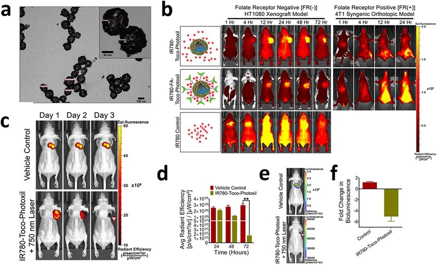
Figure 4: Morphological characterization and evaluation of nanotheranostic property of black phosphorous and reduced graphene oxide
composite (BP/rGO).
(a) TEM image of BP/rGO composite. Inset shows a typical HRTEM image of dark, small particles; scale bar, 2 nm. (b) TEM image of PEG-coated
BP/rGO. Inset shows an enlarged image of a typical PEG-coated BP/rGO particle; scale bar, 100 nm). (c) In vitro relative cell viability of
SMCC-7721 cells after photothermal ablation using NIR laser irradiation (808 nm, 1.0 W/cm2) for 5 min. (d) In vitro relative cell viability of
MCF-7, SMCC-7721, B16, and L-O2 cells after co-incubation with different concentrations (0.01, 0.05, 0.1, and 0.15 g L−1 ) of PEGylated BP/rGO
for 24 h. Error bars represent the standard deviations (SDs) calculated by three parallel samples. (e) In vivo real-time IR thermal photos of
SMCC-7721 tumor-bearing nude mice exposed to a NIR-laser after intratumor injection of PEGylated BP/rGO (10 μg per mouse). (f) Typical
photographs and corresponding growth curves of tumors collected from the mice treated with saline solution, PEGylated GO with NIR laser
irradiation, PEGylated RP/rGO with NIR laser irradiation, and PEGylated BP/rGO with NIR laser irradiation. All the relative tumor volumes were
normalized to the initial sizes. Data were shown as mean ± SD, ***p < 0.001, n = 5, Dunnett’s multiple comparison test [200].N.K. Jain et al.: Nanoengineered photoactive theranostic agents for cancer 13 3.9 Organic/inorganic nanohybrids 3.9.1 Polymer/inorganic Recently, organic/inorganic nanohybrids have attracted Chauhan et al., designed a novel biodegradable and wide attention due to their desirable properties that extend biocompatible plasmonic system for imaging guided pho- their applications in cancer nanomedicine. They demon- tothermal therapy named as Toco-Photoxil (Figure 5) [203]. strate the properties of inorganic materials (such as elec- The nanotheranostic system consisted of vitamin E modi- trical, optical, and magnetic properties) and at the same fied gold-coated poly (lactic-co-glycolic acid) nanoshells time, the organic component helps to improve the loaded with Pgp inhibitor D-α-tocopheryl polyethylene biocompatibility, biodegradability, and clearance glycol 1000 succinate (TPGS). Toco-Photoxil was tuned to [201, 202]. Organic materials like polymer, lipids etc., are exhibit absorption at 750 nm for effective therapeutic effi- used to design versatile nanohybrid for biomedical appli- cacy. Systemic in vivo studies demonstrated that Toco- cations. The functional groups present in the organic Photoxil passively accumulates in the solid tumor and component also help to functionalize the nanohybrid sys- disintegrates upon NIR light irradiation resulting in gen- tem with targeting molecules resulting in site-specific eration of heat that ablates the cancer cell. The system localization of nanohybrid. Such nanohybrids can be upon disintegration is easily cleared from the body mini- extensively used for less-invasive imaging as well as mizing the toxicity issues that can be caused due to accu- imaging-guided PTT. mulation of material. Functionalization of Toco-Photoxil Figure 5: Morphological characterization and in vivo nanotheranostic evaluation of Toco-Photoxil. (a) FEG-TEM image of Toco-Photoxil. (b) Representative Near Infra-Red Fluorescence (NIRF) imaging at different time points in HT1080 FR(−) xenograft and 4T1 FR(+) orthotopic tumor-bearing mice after systemic delivery of IR780-Toco-Photoxil (top), IR780-FA-Toco-Photoxil (middle), and IR780 dye control (bottom). (c) Qualitative representation of TurboFP fluorescence images of mice bearing HT1080-fluc2-turboFP tumors during the course of photothermal treatment (arrow head indicates the treated tumor region). (d) Quantitative assessment of changes in light output of the TurboFP fluorescent protein (p < 0.01). (e) Representative follow up bioluminescence images of mice at day 10 (arrow head indicates the treated tumor region). (f) Fold change in bioluminescence light output between the vehicle control treated mice and mice treated with a combination of Toco-Photoxil and 750 nm laser [203].
14          N.K. Jain et al.: Nanoengineered photoactive theranostic agents for cancer

with folic acid (FA) and IR780 reduced the stability leading                    Similarly, they have also reported NIR light-triggered
to aggregation and decreased the photothermal trans-                       thermoresponsive nanoshell for plasmonic PTT based
duction potential due to disturbance in plasmon reso-                      cancer theranostic application [205]. The hybrid nano-
nance. Computed tomography (CT) imaging studies with                       shell (Au PNVCL NS) was formed by ascorbic acid-driven
Toco-Photoxil revealed comparable contrast to that of                      in situ gold coating over thermoresponsive chitosan-
iodine based contrast agent Omnipaque at five times lesser                  grafted poly(N-vinyl caprolactam) nanoparticles. The
concentration due to high X-ray attenuation power. Hence,                  plasmonic absorption peak was tuned in NIR region
Toco-Photoxil with high photothermal conversion and                        (750 nm) for its application in PTT and loading of drug in
localized accumulation can serve as an effective and safe                  the polymeric core results in controlled release due to
material that can be clinically translated for cancer nano-                hyperthermia triggered shrinkage of polymer. Grafting of
theranostic applications in future.                                        chitosan was found to increase the biocompatibility,
     Chauhan et al. [204], has also reported a facile and                  biodegradability, and elevates the lower critical solution
green synthesis of gold deposited zein nanoshells (AuZNS)                  temperature (LCST) to desired value. Au PNVCL NS
for image guided plasmonic photothermal therapy. The                       demonstrated superior contrast over Omnipaque in X-ray
zein nanoparticles were surface functionalized with                        imaging. In vitro studies in mouse normal fibroblast L929
cationic glycol chitosan that helps in stabilizing the system              cells confirmed the biocompatible nature of the nano-
and ex-situ coating of gold was done over the zein nano-                   hybrid and studies in breast cancer cells MCF-7 revealed
particles. The designed system demonstrated high                           the therapeutic potential of Au PNVCL NS. Based on the
biocompatibility and effectively killed cancer cells under                 results, Au PNVCL NS could be considered as a promising
NIR light (808 nm) irradiation. Further, it also assisted in               multifunctional theranostic agent for image-guided PTT
diagnosis of tumor through CT imaging and hence the                        and can be explored for in future for combinatorial
system functions as a nanotheranostic agent in cancer.                     chemo-photothermal therapy.

Figure 6: Liposomal nanotheranostics (NFGL–FA) for multimode-targeted bioimaging and phototriggered cancer therapy.
(a) Schematic showing folic acid targeting ligand decorated self-assembled liposomal nanohybrid loaded with multimode imaging probes,
viz., gold nanoparticles (AuNPs) and graphene quantum dots (GQDs). (b) Localized tumor diagnosis and specific biodistribution measure-
ments after 48 h of time before and after NIR light exposure (750 nm, 1 W/cm2 for 10 min) followed by whole body X-ray computed tomography
scans with coronal and axial CT slices of mice body (c) Visualization of targeted deep tumor localization in mice body before and after NIR light
exposure using the in vivo imaging system (IVIS). (d) Measurements of tumor reduction by tumor volume (mm3, *p < 0.05) analysis (n = 3 mice
per group) during various therapeutic conditions using different formulations of NFGL–FA nanotheranostics with and without NIR light
exposure (e) Whole body in vivo imaging for site-selective 4T1 tumor diagnosis at various time points (1, 24, and 48 h) of intravenously injected
NFGL–FA. (f) Ex vivo imaging of collected major organs and 4T1 tumor after 48 h from intravenously nanotheranostics injected animals [207].
You can also read