Molecular Modelling Guided Modulation of Molecular Shape and Charge for Design of Smart Self-Assembled Polymeric Drug Transporters - MDPI
←
→
Page content transcription
If your browser does not render page correctly, please read the page content below
pharmaceutics

Review
Molecular Modelling Guided Modulation of Molecular Shape
and Charge for Design of Smart Self-Assembled Polymeric
Drug Transporters
Sousa Javan Nikkhah *                 and Damien Thompson

                                          Department of Physics, Bernal Institute, University of Limerick, V94 T9PX Limerick, Ireland;
                                          damien.thompson@ul.ie
                                          * Correspondence: Sousa.Javannikkhah@ul.ie

                                          Abstract: Nanomedicine employs molecular materials for prevention and treatment of disease.
                                          Recently, smart nanoparticle (NP)-based drug delivery systems were developed for the advanced
                                          transport of drug molecules. Rationally engineered organic and inorganic NP platforms hold the
                                          promise of improving drug targeting, solubility, prolonged circulation, and tissue penetration. How-
                                          ever, despite great progress in the synthesis of NP building blocks, more interdisciplinary research
                                          is needed to understand their self-assembly and optimize their performance as smart nanocarriers.
                                          Multi-scale modeling and simulations provide a valuable ally to experiment by mapping the potential
                                          energy landscape of self-assembly, translocation, and delivery of smart drug-loaded NPs. Here,
                                          we highlight key recent advances to illustrate the concepts, methods, and applications of smart
                                          polymer-based NP drug delivery. We summarize the key design principles emerging for advanced
                                          multifunctional polymer topologies, illustrating how the unusual architecture and chemistry of

                                   dendritic polymers, self-assembling polyelectrolytes and cyclic polymers can provide exceptional
Citation: Javan Nikkhah, S.;              drug delivery platforms. We provide a roadmap outlining the opportunities and challenges for the
Thompson, D. Molecular Modelling          effective use of predictive multiscale molecular modeling techniques to accelerate the development
Guided Modulation of Molecular            of smart polymer-based drug delivery systems.
Shape and Charge for Design of
Smart Self-Assembled Polymeric            Keywords: dendritic polymers; polyelectrolytes; cyclic polymers; self-assembly; smart drug nanocar-
Drug Transporters. Pharmaceutics          riers; molecular modeling
2021, 13, 141. https://doi.org/
10.3390/pharmaceutics13020141

Academic Editor: Manuela Curcio
                                          1. Introduction
Received: 22 December 2020
Accepted: 18 January 2021
                                                The efficacy of Active Pharmaceutical Ingredients (API) is often hampered by low
Published: 22 January 2021
                                          aqueous solubility and short residence times in the body [1]. Conventional drug delivery
                                          formulations such as tablets, capsules, pills, solutions, powders, suspensions, injectables,
Publisher’s Note: MDPI stays neutral
                                          lotions, creams, pastes, and, etc., have contributed greatly to the treatment of disease.
with regard to jurisdictional claims in
                                          The impetus for smart delivery methods has escalated due to several factors. These include
published maps and institutional affil-   low efficacy, the difficulty in keeping the drug levels within a desired range, minimising
iations.                                  side effects and toxicity, and the emergence of specific biological therapeutics [2,3]. Research
                                          into newer smart drug delivery systems is being carried out using liposomes, nanoparticles,
                                          niosomes, transdermal drug delivery, implants, microencapsulation, polymers, aerosols,
                                          etc. These studies show their potential for higher bioavailability, less side effects, less total
                                          amount of required drug, more patient compliance, less tissue damage and lower price,
Copyright: © 2021 by the authors.
Licensee MDPI, Basel, Switzerland.
                                          compared to the conventional methods [4].
This article is an open access article
                                                One promising approach to improve the physicochemical properties of APIs is to
distributed under the terms and           engineer smart nanoparticle (NP)-based drug delivery systems [5–7]. The ability of the NPs
conditions of the Creative Commons        to enter the cell is determined by both physicochemical parameters and biological barriers.
Attribution (CC BY) license (https://     Due to the high surface area to volume ratio (small size), they are able to penetrate the cell
creativecommons.org/licenses/by/          membrane and deliver the drug inside the cell [8]. NP drug carriers can improve drug
4.0/).                                    solubility, biocompatibility and half-life, and can reduce dosage frequency and side effects

Pharmaceutics 2021, 13, 141. https://doi.org/10.3390/pharmaceutics13020141                             https://www.mdpi.com/journal/pharmaceutics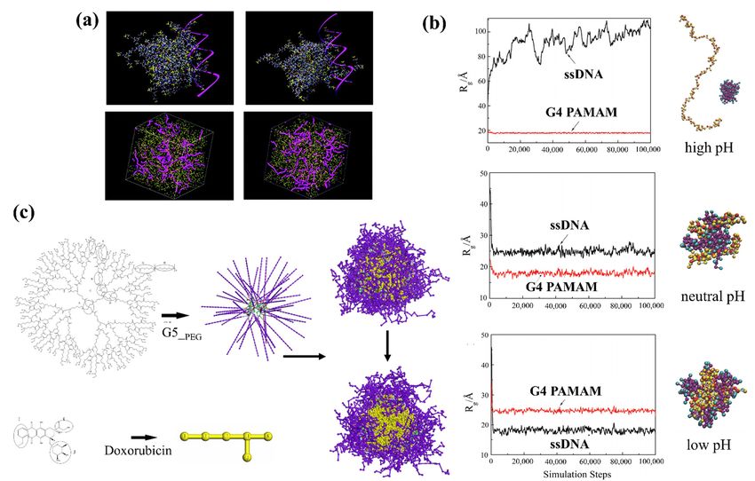
Pharmaceutics 2021, 13, 141                                                                                            2 of 28

                              by transporting drugs to specific and targeted sites. This novel class of multifunctional
                              drug carrier platform can potentially combine diagnostic agents and targeted multidrug
                              therapies together in a single system [9–13].
                                    Smart nanostructured systems can be categorized into two main groups of organic
                              and inorganic nanocarriers, the physiochemical characteristics of which can be tuned by
                              changing their compositions, shape, dimension, and surface properties [14,15]. The organic
                              group includes polymer micelles [16], liposomes [17–20], dendrimers [21–23], glycopep-
                              tides [24,25] and protein assemblies [26,27], while carbon-based nanomaterials [28,29],
                              gold nanoparticles [30,31], silver nanoparticles [32,33], porous silica-based nanomateri-
                              als [34–36], and various metal-based quantum dots [37,38] belong to the inorganic group.
                                    The promise and potential of NPs has been discussed in numerous review articles
                              covering their history, advances, advantages, potentials and limitations [39–50]. Research
                              articles have reported valuable knowledge on their intracellular transport [51,52], targeted
                              drug delivery to tumors [53], internalization and cellular uptake mechanism (localization
                              of intracellular nanoparticles) [54,55], interfacial biophysicochemical interactions [56],
                              mechanisms of nanoparticle endocytosis [51,57], cellular excretion and degradation of
                              nanoparticles [58] and toxicity (cytotoxicity and immunogenicity) [12,59]. The amount of
                              often-conflicting data makes it difficult to draw general conclusions about how to produce
                              particles for optimal drug delivery. There is still a lot to learn about NP-based drug delivery
                              mechanisms in order to interpret data from in vitro studies and to improve the in vivo
                              use of the particles. Thus, understanding the whole cellular process induced by NPs
                              would provide a rational basis for materials design, tuning their interaction with the cell
                              membrane, and improving their uptake by cells [60,61].
                                    The in vivo availability and efficacy of drug delivery systems are mainly determined
                              by their pharmacokinetics. The pharmacokinetic parameters of drug delivery systems
                              are size, shape, composition, administration route, and surface modification. All these
                              parameters also influence pharmacodynamics. While the main advantage of the smart
                              NPs drug delivery systems as compared to the conventional ones is having controllable
                              pharmacokinetic parameters [8], it can be very difficult to predict a priori the material
                              performance given the broad range and interdependence of the parameters.
                                    Understanding how the smart NPs form and perform becomes crucial for the future
                              development of efficient drug delivery. However, there is still a lack of knowledge about
                              the interactions and processes in mediated drug transport due to the short time and
                              length scales at which nanocarriers operate, which can rarely be detected by experiments
                              alone. Appropriately benchmarked and parameterized computer simulation methods can
                              supply the necessary molecular details to build a deeper understanding of how the API is
                              encapsulated and transported [13,62].
                                    Different molecular modeling approaches scaling from ab initio quantum mechanics
                              (QM) to classical molecular mechanics (MM), molecular dynamics (MD), Monte Carlo (MC)
                              methods and out to mesoscale (MS) techniques cover the broad range of both length and
                              time scales to design a complete drug delivery system [63–67] (see Table 1 for more details
                              of the simulation methods). QM methods can provide exceptional accuracy by obtaining
                              the electron distribution of any molecular system but with practical size limitations of few
                              hundred atoms due to sharply rising computational cost. The MD level can capture all non-
                              covalent interactions at atomic resolution for systems of, typically, a few hundred thousand
                              atoms with microsecond sampling times [68]. However, many critical problems in this
                              field still require time and length scales far beyond atomistic MD, which can be modelled
                              by mesoscale simulations that have been appropriately parametrized using atomistic
                              simulations of the component building blocks and interfaces. Mesoscopic simulations are
                              performed using a coarse-grained molecular model formed by particles which are related
                              to a group of atoms in the corresponding atomistic structure [13]. Coarse-grained molecular
                              dynamics (GCMD) [69], MARTINI [70], and dissipative particle dynamics (DPD) [71–73]
                              are some of the most popular mesoscopic simulation techniques that have been applied to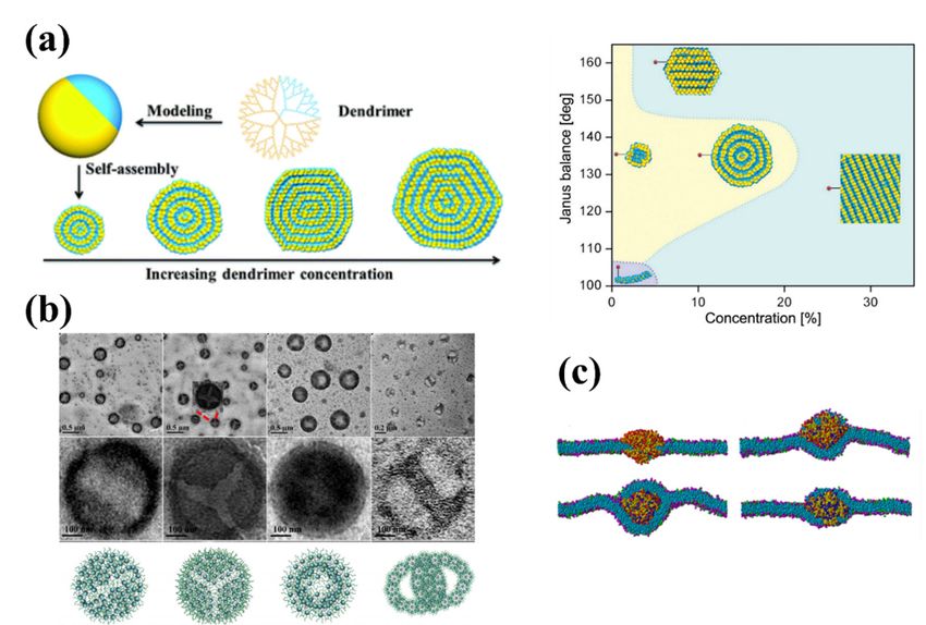
Pharmaceutics 2021, 13, 141                                                                                                          3 of 28

                                  study the self-assembly of NPs from polymeric building blocks, providing valuable insights
                                  and design principles for the rational engineering of novel drug delivery systems [13].

                  Table 1. General descriptions of the common simulation methods applied for drug-delivery studies.

       Scales          Length and Time Scales [74]             Descriptions                               Formulations
                                                                                               2
                                                     •    The nuclei and electrons are    − 8πh2 m ∇2 φ(r )k + U (r )φ(r )k = Ek φ(r )k ,
                                                          the particles of interest at    φ(r )k : Wave equation
                                                          this scale and quantum          Ek : energy eigenstate
 Quantum scale         ~10−10 m and ~10−12 s              mechanics (QM) methods          U (r ) : Potential
                                                          are used to model their state   h: Planck constant
                                                          by solving the Schrödinger      r: coordinates vector
                                                          wave equation [74].             m: mass
                                                                                          A new configuration can be produced by
                                                                                          arbitrarily or systematically moving one
                                                                                          atom from position i → j and can be
                                                                                          accepted if ∆H = H ( j) − H (i ) < 0
                                                                                          If ∆H > 0 the move is accepted only
                                                     •    The Monte Carlo (MC)
                                                                                          with a certain probability pi→ j which is
                                                          technique is a stochastic
                                                          method that uses random         given by pi→ j ∝ exp − k∆HB T   .
                                                          numbers to generate a           According to Metropolis et al. [76], one
                                                          sample population of the        can determine the new configuration
                                                          system from which one can       according to the
                                                                                                           following rule:
                                                          calculate the properties of
                                                                                          ξ ≤ exp − k∆H   BT
                                                                                                              , the move is accepted;
                                                          interest [74,75].
                                                                                          ξ >
                                                                                          exp − k∆H BT
                                                                                                         , the move is not accepted.
                                                                                          H: Hamiltonian
                                                                                          kB : the Boltzmann constant
 Atomistic scale      ~10−9 m, ~10−9 –10−6 s                                              ξ: a random number between 0 and 1
                                                                                          The simulation of a many-body system
                                                                                          would require the formulation and
                                                                                          solution of equations of motion of all
                                                                                          constituting particles, which for a particle
                                                     •    The Molecular dynamics          i is 2
                                                          (MD) simulation technique       mi ddtr2i = fi ,
                                                          allows one to predict the       mi : the particle mass
                                                          time evolution of a system of   ri : the particle position vector.
                                                          interacting particles (e.g.,    fi : the force acting on the ith particle
                                                          atoms, molecules, granules,     The interaction potentials describe in
                                                          etc.) and estimate the          detail how the particles in a system
                                                          relevant physical               interact with each other, i.e., how the
                                                          properties [75].                potential energy of a system depends on
                                                                                          the particle coordinates. Some of the
                                                                                          most common simulations use AMBER
                                                                                          [77], GROMOS [78] CHARMM [79] and
                                                                                          OPLS [80].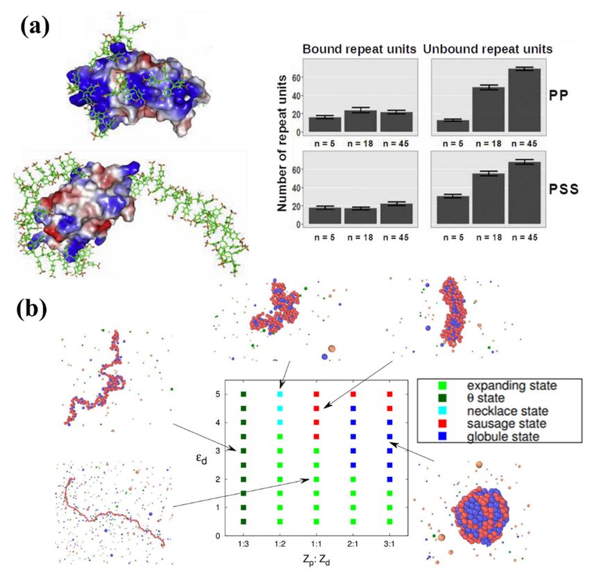
Pharmaceutics 2021, 13, 141                                                                                                        4 of 28

                                                          Table 1. Cont.

       Scales          Length and Time Scales [74]            Descriptions                               Formulations

                                                     •   Molecular mechanics (MM)
                                                         is a simulation technique to
                                                         minimize large molecular
                                                         structures such as DNA,
                                                         RNA, proteins and their
                                                         complexes, in which atoms
                                                         are treated as masses, and       Similar to MD simulation, MM is based
                                                         bonds as springs with            on Newton’s equation of motion. The
                                                         appropriate force constants.     interactions between the particles in the
                                                         For minimizations                system can be described via the
                                                         calculations, the positions of   force-field potentials applied in MD
                                                         the atoms within a molecule      simulations [82].
                                                         must be systematically or
                                                         randomly moved and the
                                                         energy recalculated with the
                                                         goal of finding a lower
                                                         energy and hence more
                                                         stable molecule [81,82].

                                                     •   Coarse-grained molecular         Commonly used forcefields in CGMD
                                                         dynamics (CGMD) methods          are:
                                                         overcome length and time         •    Weeks–Chandler–Andersen
                                                         scale limitations of atomistic        potential, COS potential and Finite
                                                         simulations though                    Extensible Elastic (FENE) bond
                                                         coarse-graining large                 potential [83].
                                                         molecules by several             •    MARTINI forcefileds [70].
                                                         connected beads [13].

                                                     •   The Dissipative particle
 Mesoscopic scale ~10−6 m, ~10−6 –10−3 s                 dynamics (DPD) method is
                                                         also a mesoscopic simulation
                                                         technique which can              Beads i and j interact through simple
                                                         correctly account for the        pairwise force consisting of a
                                                         hydrodynamic interactions        conservative force (FC i j ), a dissipative
                                                         by considering water             force (FD i j ), and a random force (FR i j ).
                                                         molecules explicitly. In DPD     The total force applied on each bead i due
                                                         simulations, a cluster of        to bead j is given as a sum of these three
                                                         atoms are represented by         terms [71]
                                                         one bead and its dynamics is     F = FijC + FijD + FijR
                                                         governed by Newton’s
                                                         equation of
                                                         motion [13,74,75].

                                       The present review aims to highlight the key common features of recent successful
                                  modeling-led investigations into the use of organic polymer-based NPs in drug delivery.
                                  We discuss the advanced topologies and chemistries of functional polymers including
                                  dendrimers, hyperbranched polymers, cyclic polymer and polyelectrolyte-based micelles
                                  (Figure 1) that can self-assemble into multifunctional smart drug carriers.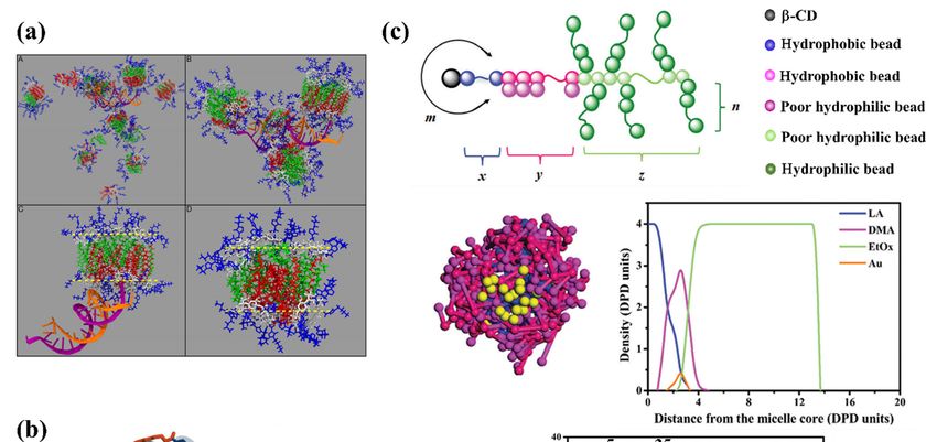
of motion[13,74,75].
The present review aims to highlight the key common features of recent successful
modeling-led investigations into the use of organic polymer-based NPs in drug delivery.
Pharmaceutics 2021, 13, 141 We discuss the advanced topologies and chemistries of functional polymers including5 of 28
dendrimers, hyperbranched polymers, cyclic polymer and polyelectrolyte-based micelles
(Figure 1) that can self-assemble into multifunctional smart drug carriers.
Schematic structure of
Figure1.1. Schematic
Figure of (a)
(a)“star
“starburst”
burst”dendrimer
dendrimer(of(of
generation 4, G4),
generation (b) (b)
4, G4), hyperbranched
hyperbranched polymer, (c) dendrimer
polymer, (c) den-
drimer multi-arm
multi-arm copolymer,
copolymer, (d) polyelectrolyte
(d) polyelectrolyte (polycation
(polycation and polyanion)
and polyanion) chains,chains,
and (e)and (e)polymer.
cyclic cyclic polymer.
2.2.Polymer-Based
Polymer-BasedSmart SmartNanocarriers
Nanocarriers
Organic
Organic nanocarriersare
nanocarriers aregenerally
generallycharacterized
characterizedby bytheir
theirtunable
tunablemorphology,
morphology,col-
col-
loidal
loidal stability, relatively large size, high biocompatibility and improveddrug
stability, relatively large size, high biocompatibility and improved drugloading
loading
capacity,
capacity, which make them
which make themsuitable
suitableforfor transporting
transporting a wide
a wide variety
variety of drugs
of drugs [84–92].
[84–92]. How-
However, it is very
ever, it is very important
important to make
to make surethe
sure that that the polymeric
polymeric drug nanocarriers
drug nanocarriers are safeare safe
and do
and
not do not trigger
trigger cytotoxicity
cytotoxicity at thelevel.
at the tissue tissueSafe,
level. Safe,NPs
smart smart NPs formulations
formulations for drugfor drug
delivery
must be biocompatible and have low immunogenicity; thus, they should be carefully
designed and evaluated [93].
They can be divided into two main categories: (1) nanostructures that form through
the self-assembly of short polymer chains and (2) synthesized large polymers (such as
the dendrimers, hyperbranched polymers, chemical nanogels). The latest generation of
smart supramolecular nanocarrier often is made by combining the two types [94–96]. How-
ever, the common point between these smart NPs categories is that they form through
self-assembly of smart polymers. These so-called smart polymers have been defined as poly-
mers that can undergo physical and structural conformational changes/rearrangements
in response to mild changes in their surrounding environment, categorized as thermo-,
pH-, electro- and magneto-responsive polymers [97]. In the following, we discuss the main
characteristics of dendritic, cyclic polymers and polyelectrolytes, and critically assess recent
efforts to predict their NP drug carrier potential via modelling.
2.1. Dendritic Polymers
Dendritic polymers are branched polymers with useful encapsulation properties
including high degree of branching, high density of terminal functional groups, and
nanometric size [98]. There are two main groups of dendritic polymers: dendrimers
and hyperbranched polymers (see Figure 1a,b). Dendrimers are monodisperse polymers
with perfectly branched architectures that are known for their well-organized structures,
versatility in drug delivery and high functionality. Their potential abilities to physically
entrap or conjugate high molecular weight molecules have been proven. Dendrimers could
also be decorated to make them smart enough to carry the drug to the desired locus and
release it in a controlled manner [99–103].
The introduction of stimuli responsive functionality on dendrimers allows the release
of drugs in response to a specific trigger only. These triggers described below could be
endogenous in nature (acid, enzyme, and redox potentials) or could be applied exter-
nally (light and temperature) [104]. pH-responsive dendrimers: Presence of ionizable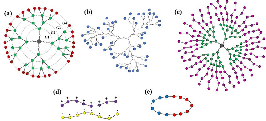
Pharmaceutics 2021, 13, 141                                                                                           6 of 28

                              functional groups such as amine and carboxylic acid on the surface or in the core of
                              the dendrimer exhibit a pH-dependent release due to change of amphiphilicity of the
                              system [105]. Redox-responsive dendrimers: The frequently used redox-responsive link-
                              ers for dendrimer–drug conjugate, such as disulfide bonds, diselenide or ditellurium
                              bonds [106]. Enzyme-responsive dendrimers: Incorporation of drug molecules as the tail
                              units and an enzyme substrate as the trigger in dendrimers, generating a prodrug unit that
                              is triggered upon a single enzymatic cleavage. The enzymatic trigger commonly utilized
                              is 38C2 antibody, penicillin-G-amidase or β-galactosidase [107]. Temperature-sensitive
                              dendrimers: Modification of dendrimer surfaces with oligo- and poly(ethylene oxide)-
                              based groups endows them with temperature-sensitive characteristics. There is an inverse
                              relationship between aqueous solubility and temperature for temperature sensitivity func-
                              tionalities. As temperature is increased, the degree of hydrogen bonding between the
                              temperature sensitive moieties and water decreases, and this leads to phase separation.
                              Lower critical solution temperature (LCST) is the phrase used to describe such phase
                              transition [108,109]. Light-responsive dendrimers: The principle governing the release
                              of drug from dendrimers using light as a stimulus is based on (i) the absorption of light
                              by photosensitive ligands that would trigger configurational changes (e.g., trans-cis iso-
                              merization) and cause the release of the encapsulated drugs and (ii) the absorption of
                              light by photosensitive ligands causing irreversible cleavage reactions. The most common
                              photosensitive ligands for (i) are azobenzene derivatives and for (ii) are o-nitrobenzyl ether
                              derivatives grafted on the surface of dendrimers [110].
                                    Dendritic polymers have tuneable surface charge and chemical composition obtained
                              by surface modification with charged moieties (such as amine-, carboxyl-, and acetyl-) [111]
                              and by special functional side groups (such as isobutyramide and poly(ethylene gly-
                              col)) [112,113], which also makes them controllable in size (reported between 1 nm and
                              100 nm) and physiochemical properties [114]. Owing to these features, dendrimers have
                              been used in the development of drug nanocarriers and many therapeutic and biomedical
                              applications [115–117]. In Table 2, some clinical studies that have been done on dendritic
                              polymer NPs as drug delivery systems are presented.
                                    A complete dendrimer structure (Figure 1a) consists of an exterior multivalent (multi-
                              site) surface with a multitude of potentially active or passive sites and the interior layers
                              surround the core [118,119]. Higher-generation dendrimers have more branches. Depend-
                              ing on their hydrophobicity/hydrophilicity, drug molecules can occupy the vacant spaces
                              (voids) within the interior layers, or else bind to dendrimer surface functional groups either
                              physically or covalently [120–122].
                                    Although the properties of dendrimers render them suitable as pharmaceutical excipi-
                              ents, they also present less advantageous properties, which may hinder their use, namely,
                              their cytotoxicity, the limitation of incorporation of the drug into the dendrimer cavities,
                              and the inability to control the rate of drug release. Furthermore, this type of polymer
                              also presents high manufacturing costs and the need for a specialized workforce [123].
                              The new developments have allowed for increased industrial manufacturing efficiency
                              and lowered production costs [124]. In order to reduce the cytotoxicity and to increase
                              the space on the dendrimer cavity, numerous modifications have been proposed to the
                              chemical structure of the dendrimers. These alterations make dendrimers more suitable for
                              use as pharmaceutical excipients. Additionally, PEGylation of dendrimers increases their
                              blood circulation time. The lack of control of the rate of drug release from the dendrimer
                              can be avoided by covalent conjugation of the drug to the dendrimer surface. Drug release
                              is then dependent on the cleavage of the dendrimer–drug linkage [114].
                                    On the other hand, randomly branched polymers, also known as hyperbranched
                              polymers, have advantages due to their low intrinsic viscosity, low tendency to chain entan-
                              glements, good solubility and high degree of branching (see Figure 1b), which have been
                              exploited for development of smart nanocarriers for drug delivery [125,126]. In addition,
                              the composition of the branching, linear, and terminal units of hyperbranched polymers
                              can be engineered to be responsive to one or multiple stimuli, which leads to a signifi-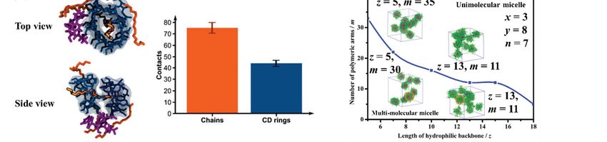
Pharmaceutics 2021, 13, 141 7 of 28
cant degree of freedom in the molecular design of smart hyperbranched polymer-based
nanocarriers for drug delivery [127].
Table 2. Clinical studies on the reviewed smart nanoparticles (NPs).
Delivery System Platform Nanoparticles Clinical Study
• DEP™-Docetaxel DTX-SPL8783 [128] (DEP
dendrimer with docetaxel and PEG terminal • Advanced or metastatic cancer
Dendritic polymers blocks)
• VivaGel® SPL7013 [128] (an active ingredient is
a generation 4 lysine dendrimer, ended by a
• Bacterial vaginosis
2-[(3,6-disulfo-1-naphthalenyl)oxy] acetic acid
disodium salt)
• Hydroxyl-terminated PAMAM • Neuroinflammation in a
dendrimers [129] large animal
• Approved by the FDA for clinical
applications, including insulin
Polyelectrolytes • Protamines [130]
delivery and reverting
heparin-induced anticoagulation.
• An anticancer agent CALAA01, a targeted,
• In phase I clinical trials for the
self-assembling nanoparticles system based on
treatment of solid tumours
Cyclic polymers CD complexed siRNA [131]
• Anti-inflammation
• Piroxicam-beta-Cyclodextrin [132]
• Anti-inflammation
• Nimesulide-beta-Cyclodextrin [132]
• Anti-inflammation
• Aceclofenac-beta-Cyclodextrin [132]
Dendritic polymers have also shown great potential as building units for self-assembled
NPs. Known generally as dendrimer multi-arm copolymers or hyperbranched multi-arm
copolymers, the dendritic polymers used in self-assembly (Figure 1c) exhibit an amphiphilic
structure with a hydrophobic (or hydrophilic) core and many hydrophilic (or hydrophobic)
linear arms [57]. Many supramolecular aggregates with diverse structures and morpholo-
gies have been formed through self-assembly of amphiphilic dendritic polymers, including
macroscopic tubes [133], physical gels [134], vesicles [135–137], spherical micelles [138–141],
and honeycomb films [142], bridging atomic to macro space-time scales. Below, we select
recent research work that illustrates how molecular modeling of dendritic polymers can
guide the engineering of useful drug carriers by controlling supramolecular interactions
and self-assembly.
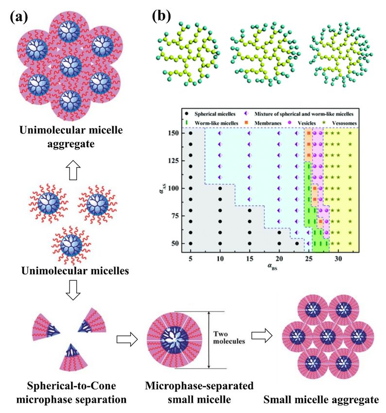
Liu et al. [143] compared the interaction of triethanolamine (TEA) core and NH3 core
polyamidoamine (PAMAM) dendrimers with DNA using atomistic molecular simulations
in explicit solvent, at physiological ionic strength (0.15 M) and pH = 7.4. The TEA-core
PAMAM showed open flexible conformations with voids localized within its interior shells,
while the NH3 -core structure showed more rigid conformation, with a more homogeneous
distribution of the monomer units and voids throughout the entire molecule. The TEA-core
dendrimers then showed a preference for binding the charged phosphate backbone of
DNA to their outer branches during complex formation. The simulations showed that the
more flexible dendrimer architecture could achieve conformational rearrangement of its
amine to optimize induced-fit with DNA (see Figure 2a).
Su et al. [144] employed DPD simulations to study the complexation of the PAMAM
dendrimer and short ssDNA molecules. They built the coarse-grained model of ssDNA
model according to the wormlike chain (WLC) potential [145] and quantified the effects
of pH, dendrimer generation, salt concentration, and dendrimer/ssDNA charge ratio onPharmaceutics 2021, 13, 141 8 of 28
the structure of the ssDNA–PAMAM complexes. They found that the ssDNA molecules
were significantly compacted by PAMAM dendrimers at neutral or low pH, with the most
stable ssDNA–PAMAM complex observed at low pH (see Figure 2b). They suggested that
the release of ssDNA from dendrimer could be modified by using different generations
of dendrimer. They also pointed out that the charge ratio between PAMAM dendrimer
and ssDNA can be used to tune the size and morphology of the self-assembled aggregates,
which could increase the transfection efficiency of ssDNA molecules in dendrimer-based
gene vectors.
PEG–polyester dendrimers are one of the most attractive dendrimers for in vivo drug
delivery due to their biodegradability and solubility achieved by PEGylation, their low
cytotoxicity, and long half-life in the circulation system [146]. The potential of polyester-
PEG dendrimers for in vivo delivery of anti-cancer drug doxorubicin (DOX) was studied
by Wen et al. [147] using DPD simulations to investigate the loading/release mechanism of
DOX in generation 5 polyester-PEG (G5-PEG polyester) dendrimers. In each dendrimer
Pharmaceutics 2021, 13, molecule, units G1 to G4 consisted of aliphatic polyester blocks and the outermost G5 8layer
of 28
contained 56 PEG blocks (see Figure 2c).
Figure 2. (a) Triethanolamine (TEA)-core
(a) Triethanolamine (TEA)-core dendrimer
dendrimer (top left), NH
(top left), NH3-core dendrimer (top right) in complex with a small
Figure 2. 3 -core dendrimer (top right) in complex with a small
fragment of double-helix DNA (highlighted as a magenta ribbon). Mesoscale morphologies of the self-assembled systems
fragment of double-helix DNA (highlighted as a magenta ribbon). Mesoscale morphologies of the self-assembled systems
between TEA-core dendrimers and DNA (bottom left) and NH3-core and DNA (bottom right) [143]. (b) Evolution of the
between TEA-core dendrimers and DNA (bottom left) and NH3 -core and DNA (bottom right) [143]. (b) Evolution of
radius of gyration (Rg) of ssDNA and G4 PAMAM dendrimer at various pH values [144]. (c) Chemical structures of den-
the radius
drimer andofdrug
gyration (Rg ) ofinssDNA
molecules and G4 PAMAM
DPD simulation dendrimer
(left), their at various
corresponding pH values
simplified [144].
beads in (c)
theChemical structures
coarse-gained modelsof
dendrimer and drug molecules in DPD simulation (left), their
(middle), and the computed G5-PEG/DOX microspheres (right) [147]. corresponding simplified beads in the coarse-gained models
(middle), and the computed G5-PEG/DOX microspheres (right) [147].
Modelling studies have provided useful insights to guide design of polymer-based
They found four sequential transient stages during DOX encapsulation: (1) initial
NPs structures and morphologies through self-assembly of copolymers with different ar-
random distribution of all the components in the simulation box, (2) dispersion of DOX
chitectures, in order to create novel multifunctional platforms with promising application
molecules in the G5-PEG dendritic microsphere, (3) core-shell microsphere growth by coa-
in drug delivery. One excellent example came from Wang et al. [148] in a systematic DPD
simulation study of micelle formation from amphiphilic dendritic multi-arm copolymers
in dilute solution. The authors simulated three models for dendritic multi-arm copoly-
mers with different lengths of the arms and one model for hyperbranched multi-arm co-
polymers. Two kinds of mechanisms, namely the unimolecular micelle aggregate mecha-
nism and the small micelle aggregate mechanism, were found to support the formation ofPharmaceutics 2021, 13, 141 9 of 28
lescence of the small core–shell dendritic microspheres, and (4) stabilization (see Figure 2c).
According to their results, in their system that contained 12.5 and 2.5% (mass fraction) of
G5-PEG and DOX, 16.7% of DOX molecules were loaded to the core–shell dendritic micro-
spheres with a loading efficiency of 100%, which is close to the experiment results [113].
The simulation also confirmed that no DOX molecule was released from G5-PEG/DOX at
pH 7.4 in the simulation temperature range, from 25 to 37 ◦ C, which means temperature
was not the major driver for drug release at pH 7.4. Furthermore, at pH 5, the formation of
some pores on the surface of G5-PEG/DOX microspheres increased the exposure of DOX
molecules to water but did not trigger release. The authors concluded that the protonation
of G5-PEG may facilitate the drug release process but it is not the major factor governing
rapid release.
Modelling studies have provided useful insights to guide design of polymer-based
NPs structures and morphologies through self-assembly of copolymers with different
architectures, in order to create novel multifunctional platforms with promising application
in drug delivery. One excellent example came from Wang et al. [148] in a systematic DPD
simulation study of micelle formation from amphiphilic dendritic multi-arm copolymers
in dilute solution. The authors simulated three models for dendritic multi-arm copolymers
with different lengths of the arms and one model for hyperbranched multi-arm copolymers.
Two kinds of mechanisms, namely the unimolecular micelle aggregate mechanism and
the small micelle aggregate mechanism, were found to support the formation of large
multimolecular micelles from the dendritic multi-arm copolymers (see Figure 3a). For the
unimolecular micelle aggregate mechanism, the dendritic multi-arm copolymers first form
the unimolecular micelles, and then the unimolecular micelles further aggregate into large
micelles without microphase separations. For the small micelle aggregate mechanism,
the dendritic multi-arm copolymers first self-assemble into microphase-separated small
micelles, and then the small micelles further aggregate into large ones. These simulation
results supported the experiments very well and extended general understanding of the
micellization processes of dendritic multi-arm copolymers.
Amphiphilic hyperbranched multi-arm copolymers were also studied by Tan et al. [149]
using DPD simulations. Their comprehensive study of the self-assembly of amphiphilic hy-
perbranched multi-arm copolymers with different hydrophilic fractions in various solvents
resulted in three morphological phase diagrams. A variety of morphologies, ranging from
spherical micelles and worm-like micelles to membranes and vesicles, were obtained (see
Figure 3b). They also discovered several novel structures, such as aggregates of spherical
and worm-like micelles, vesosomes and helical micelles, generated during self-assembly
of amphiphilic hyperbranched multi-arm copolymers. Their results extend knowledge of
the self-assembly of amphiphilic hyperbranched multi-arm copolymers, especially on the
control of supramolecular interactions for the realization of novel self-assemblies.
Hu et al. [150] investigated the formation mechanism of dendrimersomes through
MD simulations of the formation of synthetic vesicles from amphiphilic Janus (two-sided)
dendrimers. They created the spherical single-site Janus particle using an anisotropic
potential to mimic the two distinct surfaces, one hydrophobic side and another hydrophilic
side (see Figure 4a). This simple model allowed them to model both the concentration-
dependent growth of structures, both the enthalpy-mediated formation process of onion-
like dendrimersomes and the alternative entropy-mediated self-assembly of amphiphilic
flexible chains. Linear micelles, lamellar structures and vesicles were observed in the
simulations (see Figure 4a). They also found that the size of dendrimersomes will not
increase through mutual fusion once the well-defined onion-like structure is formed,
unlike numerous lipidsomes and polymersomes that can spontaneously coalesce. Future
work combining their hard chain model with the previous reports using flexible chain
models [144,147–149,151,152] could more fully characterize and predict the potential of
dendrimersomes for applications in drug and gene delivery.gies, ranging from spherical micelles and worm-like micelles to membranes and vesicles,
were obtained (see Figure 3b). They also discovered several novel structures, such as ag-
gregates of spherical and worm-like micelles, vesosomes and helical micelles, generated
during self-assembly of amphiphilic hyperbranched multi-arm copolymers. Their results
extend knowledge of the self-assembly of amphiphilic hyperbranched multi-arm copoly-
Pharmaceutics 2021, 13, 141
mers, especially on the control of supramolecular interactions for the realization of10 of 28
novel
self-assemblies.
Figure 3.3. (a)
Figure (a)Self-assembly
Self-assembly mechanism
mechanism of
of the
the microphase
microphase separated
separated small
small micelle
micelle with
with aa spherical-to-cone
spherical-to-cone microphase
microphase
separation [148] and inset
inset (b)
(b) morphological
morphological phase
phase diagram
diagram of
ofaggregates
aggregatesformed
formedfrom
fromAA4242BB20 in selective
20 in selective solvents
solvents as a
hydrophilic [149].
function of the interaction parameters. A bead is hydrophobic and B bead is hydrophilic [149].
Wang
Hu et al.
et al. [153]
[150] reported a the
investigated combined experiment–simulation
formation co-study detailing
mechanism of dendrimersomes througha
general strategy to construct uniform aggregates by manipulating self-assembly
MD simulations of the formation of synthetic vesicles from amphiphilic Janus (two-sided) of den-
drimers withThey
dendrimers. precisely controlled
created polyhedral
the spherical oligomeric
single-site Janussilsesquioxane (POSS)-embedded
particle using an anisotropic po-
tential to mimic the two distinct surfaces, one hydrophobic side and anotherrigid–flexible
cores. The authors created different types of amphiphilic dendrimers with hydrophilic
coupling POSS-embedded cores, different PEG chain lengths, and various geometries.
They found that the rigid POSS molecules could grow in ordered arrangements while
the flexible alkyl chains could rearrange to minimize the free energy of the assembly.
PEG chains with extended conformations influenced the configuration of the inner core
by occupying the excluded volume of the hydrophobic regions. DPD simulations sub-
stantiated their experimental findings, allowing better understanding of the underlying
self-assembly processes. As an example, they studied the self-assembly of amphiphilic
C3-POSS3-(PEG550)x polymers with different hydrophobic/hydrophilic ratios created by
simply changing the PEG length x. With increasing hydrophobicity, spherical micelles, rodPharmaceutics 2021, 13, 141 11 of 28
micelles, and vesicles were obtained from DPD simulations in good agreement with the
images obtained from electron microscopy (see Figure 4b). The results obtained in this
aceutics 2021, 13, work provide a methodology to broaden the variety of rigid–flexible11 core
of 28dendrimers to
fabricate responsive hierarchical self-assemblies for biomaterial science and biomimetic
nanotechnology [154,155].
Figure 4. (a)Figure
Schematic4. (a)of
Schematic of self-assembly
self-assembly of nanoarchitectures
of nanoarchitectures from Janusfrom Janus dendrimers
dendrimers and the corre-structure–
and the corresponding
sponding structure–concentration phase diagram for amphiphilic dendrimer aqueous solutions
concentration phase diagram for amphiphilic dendrimer aqueous solutions [150]. (b) Self-assembled morphologies by
[150]. (b) Self-assembled morphologies by various Ca-POSSb-PEGc polymers observed by micros-
various Ca -POSSb -PEGc polymers observed by microscopy (top, with zoom-in on nanoscopic feature in the middle
copy (top, with zoom-in on nanoscopic feature in the middle panel) and corresponding DPD mod-
panel) and corresponding
elling images of DPD modelling images
self-assemblies of self-assemblies
by the amphiphilic polymersby the amphiphilic
at the polymers
initial polymer at the initial polymer
concentration
concentration ofof 1 mg/mL
1 mg/mL in aqueous
in aqueous solutions
solutions (bottom)
(bottom) [153].
[153]. (c) (c) Typical
Typical calculated
calculated states
states of the
of the interactions between a
interactions
lipid bilayer between
membrane and abilayer
a lipid PAMAM dendrimer
membrane conjugated
and a PAMAM with ligands. The
dendrimer receptorwith
conjugated density of theThe
ligands. membrane
recep- is fixed at
fR = 0.33. Thetor density
surface of theofmembrane
tension the membrane,is fixed at fRthe
σ, and = 0.33. Thedensity
ligand surfaceoftension of the membrane,
the dendrimer, fL , are (topσ,left) = 2.09 kB T/rc 2
andσthe
and fL = 0.1,ligand density
σ =of0.10 2
the kdendrimer, 2
B T/rc andfLf,Lare (top(bottom
left) σ =left)
2.09 σkB=T/r andkfBLT/r
= 0.1, (top fright) σ =and
0.10(bottom right)
2
(top right) = 0.4, −c0.65 c and L = 0.6,
kBcT/r
σ = 1.10 kB T/r c2 and
2 and fL 0.8.
fL = = 0.4, (bottom
Color left)lipid
scheme: σ = −0.65
head kbeads
BT/rc2(pink),
and fL =lipid
0.6, tail
andbeads
(bottom right)
(cyan), σ = 1.10
receptor kBT/r
beads c2
(green), charged
and fL = 0.8. Color scheme: lipid head beads (pink), lipid tail beads (cyan), receptor beads (green),
beads of dendrimer (red), uncharged beads of dendrimer (yellow), and ligand beads (blue) [156].
charged beads of dendrimer (red), uncharged beads of dendrimer (yellow), and ligand beads
(blue) [156]. Combining DPD simulation and analytic models, Yan et al. [156,157] studied the
molecular mechanism and kinetic behavior of the transmembrane transport of PAMAM
2.2. Polyelectrolytes
dendrimer-like soft NPs conjugated with ligands. They identified three different states
Polymersduring
carryingtheainteraction
net negative or positive
between charge (seeand
the dendrimer Figure 1d) membrane:
the cell at or near neutral
penetration, partial
pH are calledwrapping,
polyelectrolytes
and full(PEs). Their (see
wrapping solubility
Figurein4c).
water
Bothisthe
driven by electrostatic
membrane tension and the ligand
interactions between
density water
on theand
NPsthe charged all
influenced monomer [158]. Examples of such polymers
three stages.
include DNA, protein and some derivatives of cellulose polymers. PEs are classified ac-
cording to four 2.2. Polyelectrolytes
main categories: natural/synthetic, homopolymers/copolymers, lin-
Polymersand
ear/branched/cross-linked carrying a net negative or positive charge (see[159].
polyanions/polycations/polyampholytes Figure 1d) atre-
Many or near neutral
searchers have pH are calledinvestigated
extensively polyelectrolytes (PEs). Their
the properties of solubility in water
the individual is driven by electrostatic
polyelectrolytes
interactions
and the formation between water
of polyelectrolyte and the
complexes charged
(PECs) monomer
[160,161]. [158]. Examples
The structures created of such poly-
by the collapse mers include
of PEs DNA,
by, for proteinmacro-ion
example, and somelike derivatives
proteinsof cellulose
[162], ionic polymers.
surfactantsPEs are classi-
fied according
[163], or oppositely charged to four
PEs main categories:
[164,165] are usuallynatural/synthetic, homopolymers/copolymers,
described as a “polyelectrolytes
complex”, which has wide applications in functional nanomaterials, gene therapy, and [159]. Many
linear/branched/cross-linked and polyanions/polycations/polyampholytes
drug deliveryresearchers
[166–171]. Thehaveformation
extensively investigated
mechanism of the properties of the
a polyelectrolyte individual
complex can bepolyelectrolytes
and the formation of polyelectrolyte complexes (PECs) [160,161].
explained by theories based on the electrostatic forces and Flory–Huggins mixing free en- The structures created by
the collapse of PEs
ergies of the polyelectrolytes by, forWhen
[172,173]. example, macro-ion
the charge like proteins
fraction [162],isionic
of the chains low, surfactants
the [163],
polymer backbone repulsion (Flory–Huggins interaction parameter) is dominant and the
solution separates into two phases, each containing mostly one of the polymers, while at
high charge fraction, the attractive electrostatic interactions between the polymers is the
controlling factor which stimulates precipitation and formation of a complex [174,175].Pharmaceutics 2021, 13, 141 12 of 28
or oppositely charged PEs [164,165] are usually described as a “polyelectrolytes complex”,
which has wide applications in functional nanomaterials, gene therapy, and drug deliv-
ery [166–171]. The formation mechanism of a polyelectrolyte complex can be explained
by theories based on the electrostatic forces and Flory–Huggins mixing free energies of
the polyelectrolytes [172,173]. When the charge fraction of the chains is low, the polymer
backbone repulsion (Flory–Huggins interaction parameter) is dominant and the solution
separates into two phases, each containing mostly one of the polymers, while at high charge
fraction, the attractive electrostatic interactions between the polymers is the controlling
factor which stimulates precipitation and formation of a complex [174,175].
The concept of PECs in the design of drug delivery systems may be useful because they
can alter drug physicochemical properties such as stability and dissolution [171,176–178].
The drug molecules can be encapsulated through incorporation into PECs by being: (1) en-
trapped from the solution during precipitation of the complex, (2) absorbed on the already
formed complex, (3) chemically bonded to at least one partner, or (4) itself a charged partner
for PEC [179]. Polyelectrolyte–drug complexes have an amorphous colloidal structure that
may be suited for delivery of poorly soluble drugs [180–183]. An example of a clinical
study on a polyelectrolyte-based drug delivery system is presented in Table 2. The for-
mation of the PECs is a spontaneous reaction which appears in aqueous environments
at the stage of polymer mixing, yet its complex nature stems from the dual character of
macromolecules and electrolytes. This may, in turn, cause difficulties in providing the
structural homogeneity that may aid their deployment in living systems. A number of
technological factors, including pH alterations, charge density, or polymer concentration,
may determine or modulate the physicochemical and biological properties of the PECs
formed [184]. Upon ionization, polyelectrolytes become pH-responsive polymers that can
undergo various changes in their physical structure, including extension of their coiled
chains as a result of electrostatic repulsion between positive charges generated protona-
tion at low pH [185]. Various physiochemical characteristics of polyelectrolytes including
solubility, chain conformation, self-assembly of polymers/copolymers, aggregation size,
shapes and volume of the individual counterparts can be adjusted by controlling the pH
level [97]. Polyelectrolyte NPs are often polydisperse systems with broad size distributions
ranging between 10 nm and 1 µm [186]. pH-responsive behavior of polyelectrolytes has
been used to induce the controlled release of model compounds such as vitamin B12 [187]
and drugs including ibuprofen [188].
Despite the large amount of research works on the behavior of polyelectrolyte chains,
the size and complexity of the assemblies has limited modelling of the polyelectrolytes-
drug complex, in which the polyelectrolyte chains and opposite charged hydrophobic drug
molecules form a complex that can protect the drugs [189,190]. As an example, Sofronova
et al. [191] studied the influence of degree of polymerization on the structure and properties
of the formed soluble protein–polyelectrolyte complexes. Using MD simulations, they
modelled the structure, dynamics and energetics of complexes of cationic protein lysozyme
with highly charged polyanions poly(styrene sulfonate) and polyphosphate created with
different degrees of polymerization. The computed complexes revealed that the short
charged chains efficiently coated the protein, while the long chains interacted with the
protein mainly through the charged loops and tails (see Figure 5a), keeping the proteins
in their native state and protecting against aggregation. Given this ability to make stable,
specific complexes between PEs and naturally charged macromolecules such as proteins,
one potential application would be the design of platforms for antibody drug delivery
systems that protect against damage during different administration methods. Modelling
results such as those provided by Sofronova et al. can give a general perspective on the
influence of polymer type, length and charge on the mechanism of assembly of the desired
polyelectrolyte-based protein delivery system.Pharmaceutics 2021, 13, 13 of 28
exploring the dynamics aspect of PE–drug complexation, they found that extremely hy-
Pharmaceutics 2021, 13, 141 drophobic drug molecules could trap the complex in a non-equilibrium glass-like13state.
of 28
Their work can provide guidelines to fabricate colloidal PE–drug complexes with “dialed-
in” desirable physical characteristics.
Figure
Figure 5.5. (a)
(a)Computed
Computedprotein–carrier
protein–carrierstructures made
structures madefrom poly(styrene
from poly(styrenesulfonate) (PSS)
sulfonate) andand
(PSS)
polyphosphate (PP), showing PSS5 (top left) and protein-PSS45 (top right) complexes. The
polyphosphate (PP), showing PSS5 (top left) and protein-PSS45 (top right) complexes. The polyanion poly-
anion is shown in sticks, the protein is shown in surface representation and colored according to
is shown in sticks, the protein is shown in surface representation and colored according to electrostatic
electrostatic potential. The number of bound and unbound repeat units of PP (middle) or PSS
potential. The number of bound and unbound repeat units of PP (middle) or PSS (bottom) chains
(bottom) chains (n is a degree of polymerization) is plotted underneath [191]. (b) Morphological
(n is adiagram
phase degree ofofpolymerization) is plotted
the PE–drug complexes underneathusing
characterized [191].drug
(b) Morphological
hydrophobicity phase
εd anddiagram
PE-
of the PE–drug complexes characterized
drug valence ratio Zp:Zd. [192]. using drug hydrophobicity εd and PE-drug valence ratio
Zp :Zd . [192].
2.3. Cyclic Copolymers
Lei et al. [192] combined mean-field theory and extensive molecular simulations to
2.3.1. Overview
study the phase behavior of the PE–drug complex in dilute, salt-free solution. They focused
on theCyclic polymer-based
morphologies of thestructures
complexesare challenging
with to synthesise
varying drug and purify
hydrophobicity andbut show
different
good
PE–drugpotential
valenceas drug
ratios.nanocarriers
They obtaineddue to their special
a phase diagram architecture
in which and
five stimuli
differentrespon-
main
sive behaviors including
morphologies [193–201]. theTheexpanding
relative sizestate,
of the resulting
the copolymer
θ condition assemblies
state (in which the is chain
influ-
enced
behaves byexactly
the conformation
as predictedofby the different
the randomarchitectures.
walk or idealAs the model),
chain core-forming block of
the necklace the
state,
cyclic diblock copolymer assembly is required to loop and cannot stretch
the sausage state and the compact globular state were identified (see Figure 5b). The benefit without re-
striction, the value
of this phase diagram of radius of hydration
is the provided broad (Rinformation
h) for a cyclic ondiblock
PE–drug micellar
complex assembly
response is
expected to be larger thanand
to drug hydrophobicity thatPE–drug
of the equivalent
valence linear
ratios,diblock
which canfor abe
given
usedblock composi-
in both drug
tion (for example,
encapsulation and for cyclic-PEO42-b-PBO8
release processes. They found Rh = that
4.4 nm, while for linear
the complexation is aPEO21-b-PBO8-
first-order-like
phase transition
b-PEO21 Rh = 4.0 controlled
nm) [202]. by the hydrophobic
Zhang attraction
et al. [203] reported thatbetween the drug molecules.
the hydrodynamic diameter
They
of alsopoly(ethylene
cyclic predicted thatglycol)-b-poly(ε-caprolactone)
the stability and morphology of(PEGx-b-PCLy)
the complex was determined
micelles was ap-by
the valence ratio
proximately between
half that the drug
of linear molecule micelles.
PEGx-b-PCLy and PE monomer. Finally, by exploring the
dynamics aspect
Different of PE–drug
cyclic polymercomplexation, they found
materials are designed to that extremely to
be responsive hydrophobic drug
different stimuli
molecules
such could trap
as photonic, the complex
thermal, in aornon-equilibrium
electronic chemical [201]. For glass-like state.
example, Their work
polystyrene can
[204],
provide guidelines to fabricate colloidal PE–drug complexes with “dialed-in” desirable
physical characteristics.Pharmaceutics 2021, 13, 141 14 of 28
2.3. Cyclic Copolymers
2.3.1. Overview
Cyclic polymer-based structures are challenging to synthesise and purify but show
good potential as drug nanocarriers due to their special architecture and stimuli responsive
behaviors [193–201]. The relative size of the resulting copolymer assemblies is influenced
by the conformation of the different architectures. As the core-forming block of the cyclic
diblock copolymer assembly is required to loop and cannot stretch without restriction,
the value of radius of hydration (Rh ) for a cyclic diblock micellar assembly is expected
to be larger than that of the equivalent linear diblock for a given block composition (for
example, for cyclic-PEO42-b-PBO8 Rh = 4.4 nm, while for linear PEO21-b-PBO8-b-PEO21
Rh = 4.0 nm) [202]. Zhang et al. [203] reported that the hydrodynamic diameter of cyclic
poly(ethylene glycol)-b-poly(ε-caprolactone) (PEGx-b-PCLy) micelles was approximately
half that of linear PEGx-b-PCLy micelles.
Different cyclic polymer materials are designed to be responsive to different stimuli
such as photonic, thermal, electronic or chemical [201]. For example, polystyrene [204],
poly(methyl methacrylate) [205] or polythiophene [206] derivatives can make photo re-
sponses in cyclic polymers. Thermo/chemo responses can take place via poly(aldehyde) [207],
and cyclic–linear topological transformation that can be triggered by poly(ethylene oxide)
(or polyethylene glycol) [207]. Additionally, in biomedical fields, the topology effects of
cyclic polymers was exploited to achieve controlled/improved biotransportation as well
as gene/DNA delivery [201]. For the former application, poly(acrylic acid) derivatives
grafted to polyethylene glycol [208] or oligopeptides [209] were incorporated into the cyclic
polymer structure, and for delivery, cationic derivatives of poly(methyl methacrylate) [210]
and polyethylene imine were used [211].
In the following, we discuss some of the most recent and promising predictive
modelling-based research that can guide future experimental efforts to self-assemble func-
tional cyclic polymer-based nanostructures.
Liu et al. [212] combined DPD with all-atom MD simulations based on the ABEEM
(atom-bond electronegativity equalization fluctuating charge force field model) polarizable
force field to study the self-assembly of linear and cyclic polystyrene (PS)-polyisoprene (PI)
di-block copolymers, PS290 -PI110 , in n-heptane. This work was the first try to combine DPD
simulations and all-atom MD simulations based on a polarizable force field, in an effort to
quantify the effect of architecture and blending on the self-assembly properties in solution.
The ABEEM polarizable force field provides a more accurate treatment of the intermolecular
interactions in the system than traditional nonpolarizable force fields [213]. Their results
demonstrate that the combination of DPD and MD with a polarizable force field can
efficiently bridge the gap between atomistic and mesoscopic simulations, and enables the
accessing of larger length scales and longer time scales while preserving atomic scale detail.
By comparing the self-assembly behavior of cyclic di-block copolymers with those of their
analogous linear block copolymers, they found the PS-PI cyclic block copolymers self-
assembled into cylindrical micelles, while spherical micelles with stable structures formed
from the linear PS-PI block copolymer with the same composition. In both structures,
the low-polarity PS blocks were distributed inside the micelle, forming a hydrophobic
core, and the high-polarity PI blocks spread around the surface, forming a protective shell.
According to their results, the self-assembled morphologies could be changed dramatically
by the addition of PS homopolymers; for cyclic copolymer from cylindrical micelles to
vesicle, and for the linear copolymer from spherical to cylindrical micelles
The self-assembly of microstructures from amphiphilic cyclic brush-copolymers in
solution was investigated by Yang [214] using DPD simulations. Cyclic brush copolymers
are innovative materials with a cyclic core hosting radiating polymer brushes producing
myriad polymer topologies. The authors could obtain a series of self-assembled structures,
such as rods, plates, vesicles, large compound vesicles, bilayers, and spheres from the
solutions by changing solvophilic/solvophobic side chain lengths, solvophilic/solvophobic
backbone lengths, and grafting densities. For example, in the case of vesicle structure, theyPharmaceutics 2021, 13, 141 15 of 28
found that increasing the solvophobic side chain length or solvophobic backbone length
decreases the cavity size and increases the membrane thickness, while the whole vesicle
sizes remained near-constant. They also pointed out that self-assembly of a plate structure
with larger thickness and narrower width required increased solvophobic side chain or
backbone lengths. Their most useful finding was that amphiphilic cyclic brush copolymers
with higher backbone asymmetry and larger grafting density can form morphologies with
more curved interfaces. This study can provide valuable guidance to design cyclic brush
copoloymers, as a complex functional material, to form different self-assembly structures
that could be useful in many applications including drug delivery, bioimaging and nano-
or microreactors.
2.3.2. The Informative Representative Example of Cyclodextrin
One of the remarkable cyclic polymers with exceptional properties is cyclodextrin
(CD) [215,216] that has high potential as a promising delivery platform for therapeutic
oligonucleotides [217–219] (see Table 2 to find some clinical studies on the CD based
drug delivery system). Cyclodextrins are natural cyclic oligosaccharides composed of six
(α-CD), seven (β-CD) or eight (γ-CD) glucopyranoside units linked by α-1,4-glycosidic
bonds [220], with hydrophilic primary and secondary faces and a hydrophobic cavity.
The hydroxyl groups on the ring structure provide the opportunity to functionalize and
provide amphiphilic, cationic, anionic, PEGylated and targeted CDs [217,221]. Herein, we
review the recent modeling research works on CDs self-assembly and interactions with
drug molecules.
Zheng et al. [222], investigated the host–guest interaction between α-CD and azobenzene-
containing amphiphile 1-[10-(4-phenylazophenoxy)decyl]pyridinium bromide (AzoC10),
which is a photoresponsive material and can undergo cis–trans photoisomerization in
response to UV and visible light. They used coarse-grained molecular dynamics (CGMD)
simulations with the modified MARTINI force field to investigate the assembly of cis-,
trans-AzoC10, and cis-, trans-AzoC10/α-CD into micelles in water. By analyzing the size
and shape of spontaneously assembled micelles, they realized that the shape of the obtained
aggregate depended on both the molecular structure and the monomer concentration in
the following way: both cis- and trans-AzoC10 aggregated into spherical micelles at low
concentrations, while at high concentrations, cis-AzoC10 showed co-existing disk-like
and spherical micelles but trans-AzoC10 formed co-existing worm-like and spherical mi-
celles. In mapping the dynamics of the aggregation, the authors divided the self-assembly
process into three stages: rapid nucleation; formation and growth of spherical micelles;
and formation of disk-like or worm-like micelles. In the self-assembly of cis-AzoC10/α-CD,
the hydrophobic azobenzene moieties aggregated to form the inner core of worm-like mi-
celles with outward-pointing hydrophilic pyridinium head groups. The worm-like micelles
were surrounded by α-CDs to shield the hydrophobic azobenzene group against water.
However, due to the bulky size of α-CD, some hydrophobic azobenzene groups were still
exposed and loosely packed. According to their results, cis-AzoC10/α-CD aggregated
into worm-like micelles at all concentrations, while in trans-AzoC10/α-CD, the loss of
amphiphilicity caused by axial configuration of the hydrophobic azobenzene moiety in the
α-CD cavity [223] destabilized the micelle structure.
Singh et al. [224] examined the dynamical self-assembly behavior of cation-functionalized
β-cyclodextrin (CD) derivatives around siRNA using classical MD simulations in water
with physiological salt ionic strength. They found the cationic CD molecules spontaneously
formed superstructures in solution, which assembled around siRNA to form a stable host
that stabilized siRNA via electrostatic interactions. They concluded that the superstructures
formed by the cationic CD molecules constitute an ideal platform to encapsulate negatively
charged siRNA molecules, which could provide a promising gene delivery vector (and
in future work, delivery of negatively charged proteins or other macromolecular drugs).
The cation CD lipid-like behavior in solution enabled creation of stable superstructures
(see Figure 6a), providing nanoscale molecular templates with highly controllable size andYou can also read