Development of amphotericin b Based organogels against mucocutaneous fungal infections - SciELO
←
→
Page content transcription
If your browser does not render page correctly, please read the page content below
Brazilian Journal of

                                                                                                                                            Article
                                                                                                               Pharmaceutical Sciences

                                                                                     http://dx.doi.org/10.1590/s2175-97902020000117509

                  Development of amphotericin b Based organogels
                      against mucocutaneous fungal infections

                                                     Kavya Gopalan1, Jobin Jose iD *1
                         Department of Pharmaceutics, N.G.S.M. Institute of Pharmaceutical Sciences, Nitte
                         1

                           Deemed to be University, Paneer, Deralakatte, Mangalore, Karnataka, India

                     Amphotericin B is a broad spectrum antifungal agent used to treat fungal infections. Organogel
                     is a semisolid preparation in which the apolar phase gets immobilized within spaces of the three-
                     dimensional structure. The current study aimed at the formulation and comparative evaluation of
                     sorbitan monostearate organogels and pluronic lecithin organogels (PLO). Different compositions
                     of span 60 based organogels were prepared by varying the concentrations of the span 60 and PLO
                     gels were prepared by varying the concentration of Pluronic F 127. The developed organogels were
                     subjected to various characteristics such as pH, viscosity, spreadability, extrudability, and drug
                     release studies. The optimized formulations were evaluated against Candida albicans and carried out
                     ex vivo release study. The optimized formulation was selected from span 60 based organogels, and
                     pluronic lecithin organogels were S1 and P1, respectively. The optimized formulation (S1) showed
                     effective inhibition against Candida albicans. The skin irritation test was carried out on albino mice
                     for optimized formulations and results showed that no irritation to the skin. Based on the results,
                     organogels prepared by sorbitan monostearate showed better antifungal activity, and also all the
                     formulations were found to be stable and safe throughout the study period.

                     Keywords: Amphotericin B. Organogel. Span 60. Pluronic F127. Lecithin.

INTRODUCTION                                                                 skin texture, skin temperature, age, sex, and disease
                                                                             conditions etc. (Quindós, 2014). Numerous novel topical
     For many decades, topical drug delivery system is                       formulations have evolved in the last decades (Nakhat et
considered as the best and easiest way of administration of                  al., 2005). Gels are the convenient form of topical delivery
therapeutic agents for local effect; however, it also shows                  system which has a lot of application in the pharmaceutical
some systemic effects. Absorption of the drug through                        industry. Compared to other topical semisolid preparations
topical application is easy because of the presence of                       like ointment and cream, the gel has got better application
innumerable blood vessels in the skin. Amongst various                       property and stability. It is also providing controlled drug
topical formulations, the semisolid system holds keen                        delivery. Nowadays, several novel gels are available in the
importance due to its ease of absorption through the skin                    market. Among which organogel, which is a new type of
layers. Topical dosage forms are more effective and less                     gel system became popular in a short time because of its
toxic than conventional dosage forms. There are different                    various advantages. The popularity of organogel increases
factors which can affect the topical absorption of drugs                     because of its penetration power through the skin layers
such as particle size of the drug, chemical nature of the                    without the addition of chemical enhancer, ease of
drug, physiological factors such as moisture of the skin,                    preparation, and it can accommodate both hydrophilic
                                                                             and lipophilic therapeutic agents (Voltan, Fusco-Almeida,
                                                                             Mendes-Giannini, 2014).
*Correspondence: J. Jose, Department of Pharmaceutics, N.G.S.M.                    Cutaneous fungal infection is one of the most
Institute of Pharmaceutical Sciences, Paneer, Deralakatte, Mangalore - 575
018, India. E-mail: jjmattam07@gmail.com                                     common diseases in all over the world. Many fungi

Braz. J. Pharm. Sci. 2020;56: e17509                                                                                            Page 1/17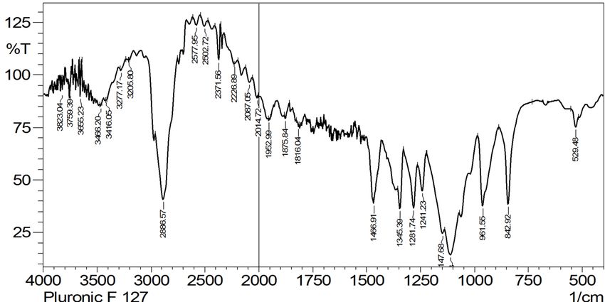
Kavya Gopalan, Jobin Jose

which are occurring in our environment can cause               was procured from Sigma Aldrich, USA. Soya lecithin,
various types of infections in different parts of              isopropyl myristate, isopropyl palmitate, and sorbic acid
the body in both humans and animals. There are                 were obtained from Hi-Media laboratories, Mumbai,
several drugs available to treat these types of fungal         India. All the materials used in this study are of
infections such as ketoconazole, clotrimazole, and             analytical grades.
miconazole etc. Amphotericin B is one of the broad
spectrum antifungal agent which is usually used                METHOD OF PREPARATION OF ORGANOGEL
to treat systemic fungal infection by intravenous
administration. Topical application of amphotericin                For the preparation of organogels two different
B is limited because of its low absorption through             organogelators were selected, like sorbitan monostearate
mucosa or skin. Due to its lipophilic nature, it does          based organogel and lecithin based organogel.
not dissolve in an aqueous medium.
      Various topical formulations are available such          Preparation of sorbitan monostearate organogel
as cream, gel, lotion, but these are undesirable for
many drugs due to its poor absorption and other side                Sorbitan monostearate (span 60) based organogels
effects. To avoid all these drawbacks, it is a new idea to     were prepared by a simple method, in which required the
prepare a formulation of organogel incorporating with          amount of span 60 was dissolved into isopropyl myristate
amphotericin B as the therapeutic agent. Organogel             (10% w/v) in a beaker and 2% w/v of polysorbate was
has better penetration power through the skin layers           added to the above solution. This mixture was heated to
without any chemical catalyst, and it is a good carrier        60°C in a water bath, and a homogenous solution was
for lipophilic drugs. These novel drug delivery systems        obtained. In another beaker, 0.1% w/w of amphotericin
can also produce controlled drug delivery; it can also         B was dissolved in 5 mL of dimethyl sulfoxide (DMSO),
significantly improve its performance in terms of              and this was added to the homogenous solution with
efficacy, safety and stability (Xie et al., 2014).             continuous stirring at room temperature (Chen, Playford,
                                                               Sorrell, 2010). The solution was further cooled, and a
MATERIAL AND METHODS                                           semisolid gel was obtained. Six different formulations
                                                               were prepared by changing the concentration of span
    Amphotericin B was received as a gift sample               60 (Chandrasekar, 2011). The compositions of different
from Cipla Pvt. Ltd. Mumbai, India. Pluronic F127              formulations were summarized in Table I.

TABLE I - Sorbitan monostearate organogel formulations from S1 to S6

                                Drug         Span 60         Tween 20      DMSO
Formulation code                                                                          Isopropyl myristate (w/v)
                               (%w/v)        (% w/v)          (% w/v)      (%w/v)

S1                               0.1            18              2             2                        100

S2                               0.1            20              2             2                        100

S3                               0.1            22              2             2                        100

S4                               0.1            24              2             2                        100

S5                               0.1            26              2             2                        100

S6                               0.1            28              2             2                        100

Page 2/17                                                                                   Braz. J. Pharm. Sci. 2020;56: e17509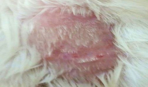
Development of amphotericin b based organogels against mucocutaneous fungal infections

Preparation of pluronic-lecithin                                           recorded on an FTIR spectrometer (Shimadzu FTIR
organogel (lecithin based)                                                 8300 Spectrophotometer). The signals arising out
                                                                           of various intramolecular stretching and bending
     Pluronic lecithin orange consists of aqueous phase                    vibrations have been expressed in wave numbers. The
as well as the oil phase. The aqueous phase was prepared                   spectra obtained for drug, physical mixture and the final
by dissolving the required amount of Pluronic F127 in                      formulation was compared.
cold water with continuous stirring, and this dispersion
was stored in a freezer for 24 h to obtain a clear polymer                 Physical appearance
solution. Potassium sorbate was added as a preservative
(Jose, Charyulu, 2016). Similarly, the oil phase was                             The prepared organogel was visually inspected for
prepared by dissolving the specified quantity of soya                      its colour, homogeneity, and consistency.
lecithin in isopropyl palmitate with continuous stirring.
Sorbic acid was added as a preservative. This solution was                 Measurement of pH
kept at room temperature for 24 h. Finally, the organogel
was prepared by mixing of oil phase with the aqueous                            The pH of all the formulations was determined by
phase. Amphotericin B (0.1%w/w) was dissolved in a                         placing the electrode of electronic pH meter in contact
minimum quantity of DMSO and added to the oil phase.                       with the surface of the prepared gel and allowing
The oil phase containing the drug was added in drop wise                   equilibrating for 1 min (Singh, Pramanik, Pal, 2015).
manner to the aqueous phase with continuous stirring
(Petrikkos, Skiada, 2007). The formulae for preparation                    Gel-sol transition temperature
of pluronic lecithin organogel are given in Table II.
                                                                                The gel-sol transition temperature of all gels
Physicochemical Characterization of Organogel                              was determined by incubating the organogels in a
                                                                           temperature bath at a temperature ranging from 25 ºC
Compatibility studies                                                      – 60 ºC. The temperature of the water bath increased by
                                                                           5°C every 5 min. The temperature was noted at which
     The FTIR spectra of the pure drug (amphotericin                       the gel started to flow when the beaker was inverted
B), pluronic F127, span 60 and the formulations were                       (Mohamed, 2004).

TABLE II - Formulation of pluronic lecithin organogel from P1 to P6

                                                                                               Formulation code
Components                                     Content
                                                                             P1          P2       P3        P4      P5       P6

Drug(% w/v)                                Amphotericin B                    0.1         0.1      0.1       0.1     0.1      0.1

                                             Soya lecithin                   10          10       10        10      10        10
Oil phase
                                          Potassium sorbate                  0.4         0.4      0.4       0.4     0.4      0.4
(% w/v)
                                       Isopropyl palmitate up to            100          100     100        100     100      100

                                            Pluronic F 127                   30          34       38        42      46        50

Aqueous phase (%w/v)                         Sorbic acid                     0.4         0.4      0.4       0.4     0.4      0.4

                                         Distilled water up to              100          100     100        100     100      100

Braz. J. Pharm. Sci. 2020;56: e17509                                                                                       Page 3/17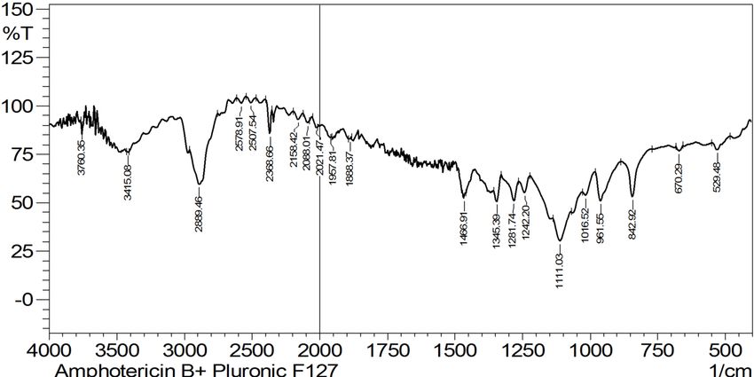
Kavya Gopalan, Jobin Jose

Viscosity                                                    spectrometer. This absorbance was correlated with the
                                                             calibration curve, and drug content was determined
     The viscosity of all the organogel was determined       (Shaikh et al., 2009).
by using Brookfield viscometer (Brookfield DV-II+
Pro). The samples were taken in a beaker and viscosity       In vitro drug release
was measured using spindle no: 96 at room temperature
and increase in angular velocity 5 rpm to 100 rpm and              In vitro, drug release studies were carried out in
constructed rheogram(Gendy, Jun, Kassem, 2002).              modified in vitro permeation apparatus using dialysis
                                                             membrane (MWCO 1000 Daltons). The phosphate
Spreadability                                                buffer of pH 7.4 was used as the dissolution medium.
                                                             The dialysis membrane was soaked in the dissolution
     All the prepared formulations were evaluated for        medium for overnight. Accurately weighed 1 g of
the spreadability by using two glass plates. A circle of 1   organogel and placed in the centre of the dialysis
cm diameter was marked in one of the glass plates, and       membrane and tied to one of the opening ends of the
0.5 g of the formulation was placed on it. The second        specially designed glass cylinder (glass cylinder with an
glass plate placed over the first glass plate, and 1kg of    opening at both ends and a diameter of 3.4 cm called as
weight was kept over it for 5 min. Spreadability of the      donor chamber). The glass cylinder was attached to the
gel was measured by increased diameter after 5 min           metallic shaft and dipped in a beaker containing 50 mL
(Shivhar, Jain, Mathur, 2009).                               of phosphate buffer of pH 7.4, and it is placed in such
                                                             a way that the dialysis membrane just touched to the
Extrudability test                                           receptor dissolution medium surface. The dissolution
                                                             medium was kept at a temperature of 37+ 0.5 ºC and
      Extrudability test of all the prepared formulations    stirred throughout the studies at 50 rpm. In a specified
was done by using Monsanto hardness tester. About 15 g       time interval, aliquots of 3 mL sample from the receptor
of the prepared formulation was placed in the aluminium      medium were withdrawn, and the same volume was
tube, and the plunger of the tester was adjusted to hold     replaced with phosphate buffer of pH 7.4 to maintain
the tube properly. About 1 kg/cm2 pressure was applied       perfect sink conditions. The absorbance of the samples
to it for 30 sec. The amount of formulation extruded out     was measured by UV spectrometer at 409 nm after
was weighed, and the procedure was repeated at three         suitable dilution (Khan et al., 2013).
equidistance places of the tube (Hosadurga et al., 2014).
                                                             Screening of antifungal activity of the
Drug content determination                                   formulation against Candida albicans

     The amphotericin B showed maximum absorbance                 The antifungal activity studies of the optimized
in the UV region at its characteristic wavelength            formulations were carried out by the cup plate method
(409 nm). A calibration curve was constructed at             (Hammer, Carson, Riley,1999). The organism used
different amphotericin B concentrations. Since other         for the study was Candida albicans ATCC-14053. The
ingredients in the formulation give no absorbance at         optimized formulations (S1 and P1) were taken for the
409 nm, the absorbance obtained from the formulation         antifungal activity study. The DMSO was used as the
would be exclusively from amphotericin B. The limits         control. Pure Amphotericin B was used as a reference
of detection (LOD), and the limits of quantification         standard, and it was dissolved in a minimum volume
(LOQ) were found to be 0.24 and 0.72 µg/mL                   of DMSO. The culture medium was prepared by
respectively. From each formulation, weigh accurately        dissolving 6.5 g of sabouraud dextrose agar in 100 mL
1 g of formulation and dissolved in 2 mL DMSO in a           distilled water by heating and sterilized by autoclaving
100 mL volumetric flask. The volume was made up              at 15 lbs/sq inch pressure for 15 min. After that, the
to the mark with phosphate buffer of pH 7.4. Again           medium was poured into three Petri plates and kept
1 mL from this stock solution was taken and diluted          in an incubator at 37±2 ºC. A loopful of the organism
to 100 mL with the same buffer, and the absorbance           was transferred from the mother culture to the
was measured at 409 nm by using UV/Visible                   sterilized sabouraud dextrose agar medium by spread

Page 4/17                                                                                 Braz. J. Pharm. Sci. 2020;56: e17509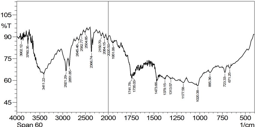
Development of amphotericin b based organogels against mucocutaneous fungal infections

plate method. The Petri plates were marked into four                       Experiments on Animals), skin irritation test was
equal parts. The first part was marked as S (standard),                    carried out for optimized formulations on Albino mice.
the second part was marked as T (test), the third part                     For the studies, eight mice of either sex were selected.
was marked as S6 (formulation), and the fourth part                        The selected mice were caged together and provided
was marked as P1 (formulation). The cup or holes                           with normal food. The mice were anaesthetized using
about 8mm in diameter were cut into the centre of                          anaesthetic ether. Hairs were depleted from the back of
each part of the medium with a sterile cork borer.                         mice by depilatories, and an area of 2 cm2 was marked.
The hole S (standard) was filled with the solution of                      After 24 h of depilation, the mice were subjected to
0.01mg amphotericin B in DMSO and T (test) was                             irritation studies. The animals were categorized into
filled with placebo gel. The optimized formulations                        four sets; each set contains two animals in which, and
S6 and P1 were weighed equivalent to 0.01mg of drug                        one was used as control, and the other one was used
and placed in the holes S1 and P1 respectively. The                        as a test. On the control, 100 mg of gel without drug
plates were transferred into biological oxygen demand                      was applied, and on the test, 100 mg of the optimized
incubator and maintained at 37±2 ºC for 24-48 h.                           formulation was applied. The skin surface was observed
After incubation, the Petri plates were observed for                       for any visible change such as erythema (redness) after
the zone of inhibition. The diameter of the zone of                        24, and 48 h of the formulation application. The mean
inhibition was reported in mm (EI Maghraby, Barry,                         erythemal scores were recorded depending on the
William,2008).                                                             degree of erythema: no erythema = 0, slight erythema
                                                                           (barely perceptible-light pink) = 0.5, moderate erythema
Ex-vivo permeation studies                                                 (dark pink) = 1 (Nava et al., 2011).

      The modified validated ex-vivo permeation                            Stability studies
apparatus was used for the ex-vivo drug release studies
through pig skin. The medium used in this study was                             The accelerated stability studies for the optimized
a phosphate buffer of pH 7.4. The pig ear skin was                         formulation were carried out as per the ICH guidelines
collected from the local slaughter house and cleaned                       (Nakhat et al., 2005). The optimized gels were filled in
it properly. The collected skin with suitable size was                     a collapsible tube and stored away from light at 25± two
stored at -20 ºC. Before the study, the pig ear skin was                   ºC and 40±2 ºC and 75% relative humidity (RH) for
taken out of the freezer and allowed to come into room                     three months. After storage, the samples were tested for
temperature. Weighed accurately 1g of the organogel                        their visual appearance, pH, % drug content and % drug
and placed in the centre of the pig ear skin and tied to                   release (Bhalodia, Shukla, 2011).
the specially designed glass cylinder, which was donor
compartment (Schwartz et al., 2014). The glass cylinder                    RESULTS AND DISCUSSION
was then attached to the metallic shaft and suspended
in 50 mL dissolution medium so that the membrane                           Compatibility studies
just touched to the receptor surface. The temperature of
the dissolution medium was maintained at 37+ 0.5 ºC                             FTIR spectrum of amphotericin B showed a
throughout the experiment, and the medium was stirred                      characteristic sharp peak at 3413.15 cm-1 due to the
at 50 rpm using a magnetic stirrer. In specified intervals,                amine group, peak at 2923.22 cm-1 representing hydroxyl
3 mL sample was taken from the receptor medium, and                        group and C-O stretching peak at 1181 cm-1. When
the same volume was replaced with phosphate buffer                         compared with the pure drug, no considerable change
of pH 7.4. The filtered samples were analyzed by UV                        in the IR peaks of the drug was observed in spectra of
spectrometer at 409 nm after suitable dilution (Tazrart                    optimized formulations; it indicates the absence of any
et al., 2017).                                                             incompatibility between drug and excipients.

Skin irritation test

    After the approval from the CPCSEA (Committee
for the Purpose of Control and Supervision of

Braz. J. Pharm. Sci. 2020;56: e17509                                                                                       Page 5/17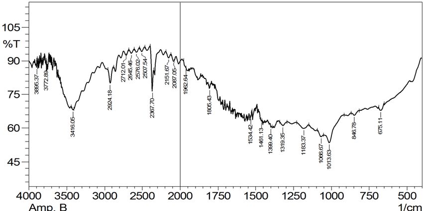
Kavya Gopalan, Jobin Jose

FIGURE 1 - FTIR spectrum of amphotericin B.

FIGURE 2 - FTIR spectrum of span 60.

Page 6/17                                     Braz. J. Pharm. Sci. 2020;56: e17509
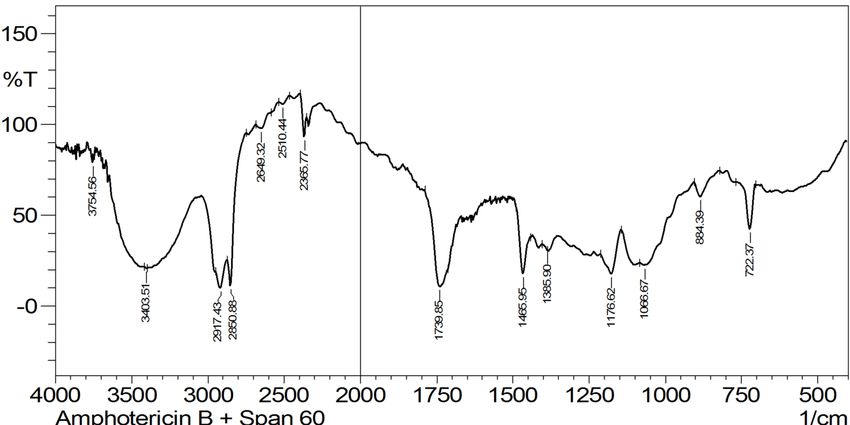
Development of amphotericin b based organogels against mucocutaneous fungal infections

FIGURE 3 - FTIR spectrum of optimized formulation S1.

FIGURE 4 - FTIR spectrum of pluronic F127.

Braz. J. Pharm. Sci. 2020;56: e17509                                                     Page 7/17
Kavya Gopalan, Jobin Jose

FIGURE 5 - FTIR spectrum of optimized formulation P1.

Physical appearance                                           from the formulation S1 to S6 due to the increased
                                                              concentration of span 60. In the case of pluronic
      All the developed organogel was creamy                  lecithin organogel, formulation P6 showed the highest
light yellowish, non-transparent and showed good              viscosity and P1 showed the least viscosity indicated
homogeneity. The span 60 based organogels was                 that the direct effect of polymer concentration on
appeared to be little more yellowish than the pluronic        viscosity. However, viscosities of all the pluronic
lecithin organogel. The results were depicted in Table III.   lecithin organogel were found to be more than that
      The pH of all the formulations was measured and         of the span 60 based organogel. The rheogram was
enlisted in Table III. The pH of all the formulations was     constructed for all the organogel formulations and it was
remained within the normal skin pH range, indicated           found that all the formulation exhibits shear thinning
that it would not cause any irritation upon administration    property (Essa, 2010). The values of the spreadability
(Essa, 2010).                                                 (Table IV) of the formulated organogel showed that
                                                              only a small amount of force is required to spread
Gel-sol transition temperature                                the gel easily. The entire span 60 based organogels
                                                              showed better spreadability when compared to
    The gel-sol transition temperature of all the             pluronic lecithin organogel (Gupta, Nappinnai, Gupta,
formulations was found to be in the range of 42 °C-           2010). Extrudability of all the formulations were
50 °C as enlisted in Table III. In the case, both types       determined and listed in Table IV. The results implied
of organogels the gel-sol transition directly depends         that more than 90% of the contents were extrudable
on the viscosity of the system. The gel-sol transition        indicating they have excellent extrudability. (>90%
temperature of all the pluronic lecithin organogels           extrudability: excellent, >80% extrudability: good,
was found to be more than that of the span 60 based           >70% extrudability: fair). The extrudability studies
organogel.                                                    indicated that suitable consistency of the gel was
                                                              required to extrude the gel from the tube uniformly.
Viscosity, Spreadability and Extrudability

     The viscosity of all the prepared formulation
was determined by Brookfield viscometer, and the
data were shown in Figure 6 and 7. The viscosity of
the span 60 based organogel was increased gradually

Page 8/17                                                                                  Braz. J. Pharm. Sci. 2020;56: e17509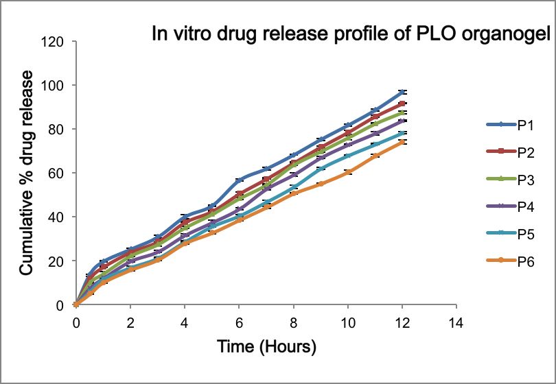
Development of amphotericin b based organogels against mucocutaneous fungal infections

TABLE III - Physical appearance, pH and gel-sol transition temperature

                                                                                                   Gel-sol transition
Formulation code                                 Appearance                              pH+ SD*
                                                                                                   temperature(°C)

S1                                                                                       6.9+0.1          42

S2                                                                                       6.4+0.3          43

S3                                                                                       6.3+0.1          44

S4                                                                                       6.7+0.2          45

S5                                                                                       6.1+0.1          46

S6                                                                                       6.5+0.2          47
                                       Non transparent, light yellow
P1                                                                                       7.1+0.1          45

P2                                                                                       6.8+0.2          46

P3                                                                                       7.4+0.2          47

P4                                                                                       7.3+0.1          48

P5                                                                                       7.0+0.3          49

P6                                                                                       7.1+0.2          50

(*SD=Standard Deviation)

FIGURE 6 - Rheogram of span 60 based organogel.

Braz. J. Pharm. Sci. 2020;56: e17509                                                                               Page 9/17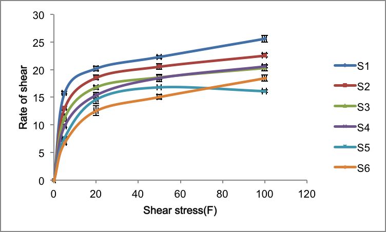
Kavya Gopalan, Jobin Jose

FIGURE 7 - Rheogram of pluronic lecithin organogel.

Drug content                                            no significant difference in the drug content among
                                                        the organogels, indicated the drug was uniformity
    The drug content of developed gels was found        distributed in the gel base.
to be in the range of 96-99% (Table IV). There was

TABLE IV - Spreadability, extrudability, drug content

Formulation code                  Spreadabilty (mm)        Extrudability            Drug content (%)

S1                                    25.12±0.01                                        99.46±0.002

S2                                     24.11±0.01                                       98.24±0.005

S3                                    23.02±0.05                                        98.55±0.011

S4                                    22.26±0.01                                        97.46±0.010

S5                                    20.10±0.20                                        97.10±0.003

S6                                    19.25±0.04                                        97.46±0.002
                                                             Excellent
P1                                    17.24±0.01                                        99.08±0.001

P2                                     16.56±010                                        96.26±0.010

P3                                    15.21±0.24                                        98.20±0.001

P4                                    13.56±0.03                                        97.72±0.004

P5                                    12.25±0.04                                        96.93±0.002

P6                                     9.10±0.02                                        97.45±0.001

Page 10/17                                                                        Braz. J. Pharm. Sci. 2020;56: e17509
Development of amphotericin b based organogels against mucocutaneous fungal infections
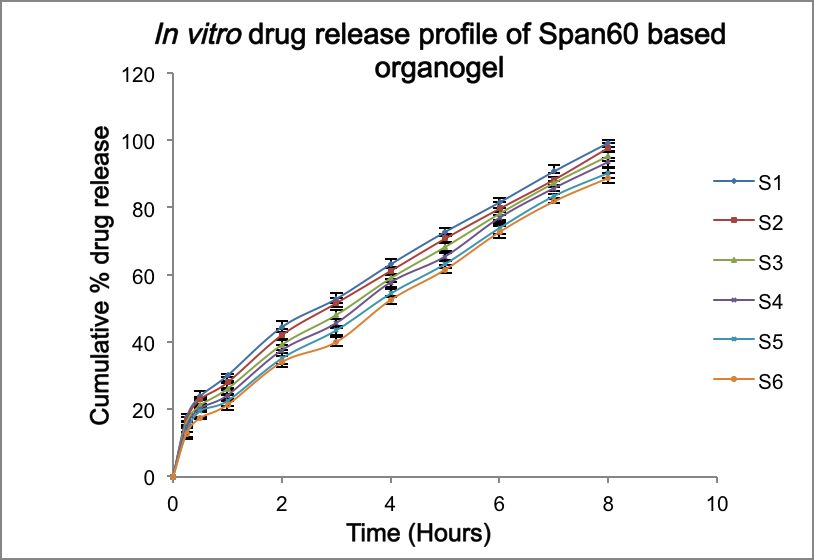
In vitro drug release studies due to its highest drug release within eight h (Tavano
et al., 2010).
In vitro drug release profile of all the formulated In vitro drug release profile of pluronic-lecithin
span 60 based organogel was given in Figure 8. The organogel was also carried, and results were shown
study was carried out for eight h using modified in vitro in Figure 9. The drug release from all the developed
permeation apparatus through the dialysis membrane. gel was extended to 12 h. The order of drug release
All the formulations showed extended drug release for from the formulation P1 to P6 varied as follows,
eight h. However, S1 showed the highest drug release P1>P2>P3>P4>P5>P6. The drug release values from
of 99.4% at the end of 8 h. The order of drug release P1 to P6 indicated that, as the polymer concentration
from the formulation S1 to S6 varied as follows, increased, the drug release was decreased, which
S1>S2>S3>S4>S5>S6. Among the formulations from might be due to extensive formation of network-like
S1 to S6, as the concentration of span 60 increased, structures with very high viscosity. The formulation
the drug release was decreased. The decrease in drug P1 was selected as the best due to its effective
release was due to an increase in alkyl chain length of prolonged release of the drug throughout 12 h (Srinivas
surfactant. The formulation S1 was selected as the best et al., 2010).
FIGURE 8 - Drug release profile of span 60 based organogel in phosphate buffer of pH 7.4 as a function of hours.
Braz. J. Pharm. Sci. 2020;56: e17509 Page 11/17Kavya Gopalan, Jobin Jose
FIGURE 9 - Drug release profile of pluronic lecithin organogel in phosphate buffer of pH 7.4 as a function of hours.
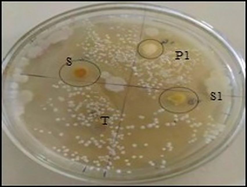
Kinetic studies S1 and P1 were compared with the zone of inhibition
produced by the standard pure drug. The formulation of
In the case of span 60 based organogel, the release S1 and P1 showed a zone of inhibition of 14.21±0.002 mm
kinetics best-fit model indicated that formulations S1 and 10.11±0.005 mm, respectively. So the formulation
to S4 followed the Higuchi model while S5 and S6 S1 had comparatively higher antifungal activity than
followed first order release. The optimized formulation formulations P1.
S1 followed the Higuchi model with the R2 value of
0.993 (as shown in Table V).
In the case of pluronic lecithin organogel, the release
kinetics best-fit model indicated that formulations P1 to
P4 followed Korsmeyer-Peppas model and P5 and P6
followed zero order release. The results were shown in
Table VI. The optimized formulation P1 best fit to the
Korsmeyer Peppas model with R2 value of 0.990. The
values of release exponent “n” obtained by applying the
Peppas equation was found to be less than 0.45, which
revealed that the drug release follows Fickian diffusion
(Ning et al., 2005).
In vitro antifungal activity against Candida albicans
The in the vitro antifungal study was carried out
by cup plate method to compare the effectiveness of FIGURE 10 - Comparison of antifungal activity of formulation
the optimized formulations to controls against Candida S1 and P1 against Candida albicans by cup plate method.
albicans and results were given in Figure 10. The zone
of inhibition produced by the optimized formulations
Page 12/17 Braz. J. Pharm. Sci. 2020;56: e17509Development of amphotericin b based organogels against mucocutaneous fungal infections
TABLE V - Kinetic models of formulations S1 to S6
Kinetic models
Formulation code Zero order First order Higuchi Korsmeyer-peppas
R2 k R2 k R2 k R2 k n
S1 0.960 -0.191 0.985 -0.0017 0.993 4.289 0.955 0.691 0.343
S2 0.961 -0.188 0.986 -0.0016 0.99 4.19 0.958 0.689 0.191
S3 0.971 -0186 0.985 -0.0015 0.986 4.13 0.963 0.687 0.174
S4 0.975 -0.184 0.986 -0.0014 0.982 4.06 0.968 0.686 0.157
S5 0.980 -0.178 0.983 -0.0013 0.978 3.94 0.973 0.684 0.142
S6 0.983 -0.177 0.980 -0.0013 0.972 3.88 0.977 0.683 0.125
TABLE VI - Kinetic models of formulations P1 to P6
Kinetic models
Formulation code Zero order First order Higuchi Korsmeyer-peppas
R2 K R2 k R2 k R2 k n
P1 0.978 -0.130 0.969 -0.0009 0.967 3.06 0.990 0.6615 0.051
P2 0.982 -0.122 0.984 -0.0008 0.969 2.88 0.995 0.6551 0.030
P3 0.989 -0.121 0.992 -0.0007 0.962 2.84 0.997 0.6576 -0.005
P4 0.993 -0.115 0.994 -0.0006 0.950 2.68 0.994 0.6544 -0.046
P5 0.993 -0.105 0.992 -0.0006 0.951 2.46 0.987 0.6453 -0.070
P6 0.996 -0.101 0.995 -0.0006 0.951 2.34 0.977 0.6448 -0.102
Ex vivo drug release studies 88.2%. The ex vivo drug release profile showed a better
release of formulation S1 when compared to P1. The
The ex vivo drug release studies of the optimized formulation S1 showed maximum drug release within ten
formulations (S1 and P1) were carried out using a modified h. The probable reason could be the stronger interaction
ex vivo permeation apparatus through the pig membrane, between S1 formulation and lipids in the pig membrane,
and the results were given in figure 11. The release of the causing their rearrangement and thus facilitated drug
drug from the formulation S1 through pig membrane was penetration. This result was in agreement with the previous
found to be 99.3%, but the formulation P1 showed only data reported by Staub (Staub, Schapoval, Bergold, 2005).
Braz. J. Pharm. Sci. 2020;56: e17509 Page 13/17Kavya Gopalan, Jobin Jose
FIGURE 11 - Ex vivo drug release profile of formulation S1 and P1 through pig membrane.
Skin irritation studies
The result obtained from the skin irritation study
was given in Table VII. The optimized formulations
were applied to the skin of mice as per the procedure
is given in methodology (Figure 12(a)) and observed
for sensitivity reaction up to 48 h. There was no sign
of development of erythema at the end of 24 h after
application of formulations (Figure 12(b)). From the set 2
and set 5 animals, the test a animal showed slight, patchy
erythema and graded as 0.5 at the end of 48 h (Figure FIGURE 12(b) - Mice skin for observation of erythema at 24 h.
12(c)). The results obtained were graded according to
the standard grading system, and the average primary
irritation was calculated. The primary irritation index
on each mouse was calculated. The average primary
irritation index was found to be 0.027. The results
indicated that optimized formulations S1 and P1 were
compatible with skin (Staub, Schapoval, Bergold, 2005).
FIGURE 12(c) - Mice skin for observation of erythema after
48 h.
Stability studies
Stability studies were conducted for the optimized
organogels for three months, and results were given
in the Table VIII. The optimized formulations were
FIGURE 12(a) - Mice skin for observation of erythema at 0 h.
evaluated for visual appearance, drug content, drug
Page 14/17 Braz. J. Pharm. Sci. 2020;56: e17509Development of amphotericin b based organogels against mucocutaneous fungal infections
TABLE VII - Results of skin irritation study
Parameter
Formulation code Study sample Type Erythema* Primary irritation index
0h 24 h 48 h
Control 0 0 0 P11=0/3=0
S1 Set 1
Test 0 0 0 P11=0/3=0
Control 0 0 0 P11=0/3=0
S1 Set 2
Test 0 0 0.5 P11=0.5/3=0.16
Control 0 0 0 P11=0/3=0
S1 Set 3
Test 0 0 0 P11=0/3=0
Control 0 0 0 P11=0/3=0
P1 Set 4
Test 0 0 0 P11=0/3=0
Control 0 0 0 P11=0/3=0
P1 Set 5
Test 0 0 0.5 PII=0.5/3=0.16
Control 0 0 0 P11=0/3=0
P1 Set 6
Test 0 0 0 P11=0/3=0
* Average primary irritation index = 0 +0 + 0 + 0,16 +0 + 0 + 0 + 0 + 0 + 0,16 + 0 +0,12 = 0.027
release and pH. There was no significant variation in were found to be physically and chemically stable
visual appearance, drug release, drug content and pH throughout the study period. This result was in
of both span 60 based and pluronic lecithin organogels. agreement with the previous data reported by Nesseem
The results indicated that the optimized formulations (Nesseem, 2001).
Braz. J. Pharm. Sci. 2020;56: e17509 Page 15/17Kavya Gopalan, Jobin Jose
TABLE VIII - Stability studies of optimized formulations S1 and P1
Storage temperatures
Formulation code Parameters evaluated
25+2 ºC±SD 40+2 ºC±SD
Visual appearance Yellowish creamy and no phase separation
Drug content* 99.46±0.002 99.98±0.012
S1
pH* 6.90.1±0.1 7.10±0.2
% Drug release 99.4±0.001 100.01±0.002
Visual appearance Yellowish creamy and no phase separation
Drug content 99.25±0.101 99.97±0.002
P1
pH 7.1±0.1 7.1±0.1
% Drug release 69.7±0.001 71.01±0.002
(*SD=Standard Deviation)
CONCLUSION REFERENCES
The development of safer and more effective drug Bhalodia NR, Shukla VJ. Antibacterial and antifungal activities
delivery systems of antifungal agents has radically from leaf extracts of Cassia fistula l.: An ethnomedicinal plant.
changed the treatment options of fungal infections. In J Adv Pharm Tech Res. 2011;2(2):104-9.
this study, different gel-based systems were developed Chandrasekar P. Management of invasive fungal infections: a
for amphotericin B and evaluated for its efficacy role for polyenes. J Antimicrob Chemother. 2011;66(3):457-465.
to prevent fungal infections. Based on the results,
organogels prepared by sorbitan monostearate showed Chen SC, Playford EG, Sorrell TC. Antifungal therapy
better antifungal activity, and also all the formulations in invasive fungal infections. Curr Opin Pharmacol.
2010;10(5):522-530.
were found to be stable and safe throughout the study
period. Certainly, these findings can be applied for the El Maghraby GM, Barry BW, Williams AC. Liposomes and
treatment of topical fungal infections. skin: from drug delivery to model membranes. Eur J Pharm
Sci. 2008;34(4-5):203-22.
ACKNOWLEDGEMENTS
Essa EA. Effect of formulation and processing variables on
the particle size of sorbitan monopalmitate niosome. Asian J
The authors acknowledge the invaluable support of
Pharm. 2010;4(4):227-33.
Nitte University, Mangalore for providing facilities to
carry out the work. Gendy AM, Jun HW, Kassem AA. In vitro release studies
of flurbiprofen from different topical formulations. Drug Dev
CONFLICT OF INTEREST Ind Pharm. 2002;28(7):823-31.
Gupta KS, Nappinnai M, Gupta VR. Formulation and
The authors disclose no conflict of interest.
evaluation of topical meloxicam niosomal gel. Int J Biopharm.
2010;1:7-13.
Page 16/17 Braz. J. Pharm. Sci. 2020;56: e17509This is an open-access article distributed under the terms of the Creative Commons Attribution License.
Development of amphotericin b based organogels against mucocutaneous fungal infections
Hammer KA, Carson CF, Riley TV. Antimicrobial activity Schwartz J, Moreno E, Fernández C, Navarro-Blasco I,
of essential oils and other plant extracts. J Appl Microbiol. Nguewa P, Palop J et al. Topical treatment of L. major infected
1999;86:985-990. BALB/c mice with a novel diselenide chitosan hydrogel
formulation. Eur J Pharm Sci. 2014;62:309-316.
Hosadurga RR, Rao SN, Jose J, Rompicharla NC Shakil
M, Shashidhara R. Evaluation of the efficacy of 2% Shaikh IM, Jadhav SL, Jadhav KR, Kadam VJ, Pisal SS.
curcumin gel in the treatment of experimental periodontitis. Aceclofenac Organogels: In vitro and in vivo Characterization.
Pharmacognosy Res.2014;6(4):326-333. Curr Drug Deli. 2009;6(1):1-7.
Jose J, Charyulu RN. Prolonged drug delivery system of Shivhar UD, Jain KB, Mathur VB. Formulation development
an antifungal drug by association with polyamidoamine and evaluation of diclofenac sodium gel using water soluble
dendrimers. Int J Pharm Investig. 2016;6(2):123-127. Poly acrylamide polymer. Digest J Nanomater Biostructures.
2009;4(2):285-90.
Khan AW, Kotta S, Ansari SH, Sharma RK, Kumar A, Ali
J. Formulation, development, optimization and evaluation Singh VK, Pramanik K, Pal K. Development and
of aloe vera gel for wound healing. Pharmacogn Mag. characterization of sorbitan monostearate and sesame oil
2013;9(1)6-10. based organogels for topical delivery of antimicrobials.
AAPS Pharm Sci Tech.2015; 16(2):293-305.
Mohamed MI. Optimization of chlorphenesin emulgel
formulation. AAPS J. 2004;6(3):81-87. Srinivas S, Anand Kumar Y, Hemanth A, Anitha M.
Preparation and evaluation of niosomes containing
Pandey M, Belgamwar V, Gattani S, Surana s, Tekade A. aceclofenac. Dig J Nanomater Bios. 2010;5(1):249-54.
Pluronic lecithin organogel as a topical drug delivery system.
Drug Deliv. 2010;17(1):38-47. Staub I, Schapoval EE, Bergold AM. Microbiological assay of
ketoconazole in shampoo. Int J Pharm. 2005;292(1-2):195-9.
Nava G, Pinon E, Mendosa L, Mendoza N Quintanar D,
Ganem A. Formulation and in vitro, ex vivo and in vivo Tavano L, Muzzalupo R, Cassano R, Trombino S, Ferrarelli
evaluation of elastic liposomes for transdermal delivery of T, Picci N. New sucrose cocoate based vesicles: Preparation,
ketorolac tromethamine. Pharmaceutics. 2011;3(4):954-970. characterization and skin permeation studies. Colloids Surf B
Biointerfaces. 2010;75(1):319-22.
Nesseem DI. Formulation and evaluation of itraconazole via
liquid crystal for topical delivery system. J Pharm Biomed Tazrart A, Bolzinger MA, Moureau A, Molina T, Coudert
Anal. 2001;26(3):387-99. S, Angulo JF, et al. Penetration and decontamination of
americium-241 ex vivo using fresh and frozen pig skin. Chem
Ning M, Guo Y, Pan H, Chen X, Gu Z. Preparation, in vitro Bio Inter. 2017;267:40-47.
and in vivo evaluation of liposomal/niosomal gel delivery
systems for clotrimazole. Drug Dev Ind Pharm. 2005;31(4- Voltan AR, Fusco-Almeida AM, Mendes–Giannini MJS.
5):375-83. Candiduria: epidemiology, resistance, classical and alternative
antifungals drugs. SOJ Microbiol Infect Dis. 2014;2(2):1-7.
Petrikkos G, Skiada A. Recent advances in antifungal
chemotherapy. Int J Antimicrob Agents. 2007;30(2):108-117. Xie JL, Polvi EJ, Shekhar-Guturja T, Cowen LE. Elucidating
drug resistance in human fungal pathogens. Future Microbiol.
Quindós G. Epidemiology of candidaemia and invasive 2014;9(4):523-542.
candidiasis. A changing face. Rev Iberoam Micol.
2014;31(1):42-8. Received for publication on 29th August 2017
Accepted for publication on 01st February 2019
Braz. J. Pharm. Sci. 2020;56: e17509 Page 17/17You can also read