Iridophoroma associated with the Lemon frost colour morph of the leopard gecko (Eublepharis macularius) - Nature
←
→
Page content transcription
If your browser does not render page correctly, please read the page content below
www.nature.com/scientificreports

            OPEN           Iridophoroma associated with
                           the Lemon Frost colour morph of
                           the leopard gecko (Eublepharis
                           macularius)
                           Paweł Szydłowski 1*, Jan Paweł Madej2, Magdalena Duda3, Janusz A. Madej4,
                           Agnieszka Sikorska-Kopyłowicz3, Anna Chełmońska-Soyta1, Lucyna Ilnicka5 &
                           Przemysław Duda6
                           The Lemon Frost is a new colour morph of the leopard gecko, which emerged in ca. 2015 as a result
                           of selective breeding and spontaneous mutation. According to multiple breeders observation of
                           Lemon Frost inbreeding with wild-type leopard geckos, Lemon Frost seems to be a codominant
                           trait. Additionally breeders observed another, presumably associated trait - tumour-like skin lesions.
                           Three private-owned Lemon Frost morph leopard geckos with tumour-like skin lesions were admitted
                           to our clinic for examination, which included histopathology, X-ray and ultrasonography. The
                           histopathological investigation of the biopsies indicated malignant iridophoroma; however, no changes
                           were observed in diagnostic imaging. This research is the first report of clinical and histopathological
                           findings of iridophoroma in leopard geckos.

                           The leopard gecko (Eublepharis macularius, Blyth 1845) is a nocturnal species naturally found in Afghanistan,
                           Pakistan, India, Iran and Nepal1,2. Additionally, the leopard gecko is one of the most popular breeding species and
                           has been kept by private owners for over thirty years. As the result of long-term breeding programmes, about one
                           hundred colour morphs have come into existence to date.
                               Reptile skin colouration depends on a distribution and presence of the chromatophores, which include the
                           melanophores, the xanthophores, the erythrophores and the iridophores3–5. These cells originate from a differen-
                           tiation of neural crest stem cells5. Melanophores are dark brown cells containing melanosomes filled with mela-
                           nin. Xanthophores and erythrophores contain vesicle structures with carotenoid or pteridine pigments ranging
                           from yellow to red4. Iridophores are light-reflecting cells containing light-reflecting platelets made up of crys-
                           talline guanine, adenine, hypoxanthine or uric acid inclusions6; the ultrastructure and arrangement results in
                           white, blue to red skin colouration7. The wild-type adult leopard gecko has a skin pigmentation pattern made up
                           of a yellow-and-black-spotted dorsal part, a greyish tail with white transversal stripes and black dots, a yellowish
                           head with black dots and a white/light cream ventral part of the body8. On account of different types of chromato-
                           phores and the morphology of leopard gecko colour morphs, a few categories exist to describe these morphs. The
                           basic colour morphs of leopard gecko can be found in Fig. 1. There are simple colour morphs as well as combina-
                           tions of them. Colour morphs describe the presence and distribution of melanophores and xathophores. In short:
                           hypo- and hyperxanthic morphs (respectively less or more intense yellow pigmentation, e.g. Tangerine, High
                           Yellow), axanthic (lack of yellow pigmentation, e.g. Mack Super Snow, Blizzard), hypomelanistic (less intense

                           1
                            Department of Immunology, Pathophysiology and Veterinary Preventive Medicine, Faculty of Veterinary Medicine,
                           Wroclaw University of Environmental and Life Sciences, Norwida 31, Wroclaw, 50-375, Poland. 2Department of
                           Histology and Embryology, Faculty of Veterinary Medicine, Wroclaw University of Environmental and Life Sciences,
                           Norwida 25, Wroclaw, 50-375, Poland. 3Department of Internal Diseases and Clinic of Diseases of Horses, Dogs
                           and Cats, Faculty of Veterinary Medicine, Wroclaw University of Environmental and Life Sciences, pl. Grunwaldzki
                           47, Wroclaw, 50-366, Poland. 4Department of Pathology, Faculty of Veterinary Medicine, Wroclaw University of
                           Environmental and Life Sciences, Norwida 31, Wroclaw, 50-375, Poland. 5Department of Epizootiology and Clinic
                           of Bird and Exotic Animals, Faculty of Veterinary Medicine, Wroclaw University of Environmental and Life Sciences,
                           pl. Grunwaldzki 45, Wroclaw, 50-366, Poland. 6Department of Molecular Physiology and Neurobiology, University of
                           Wroclaw, Sienkiewicza 21, Wroclaw, 50-335, Poland. *email: szydlowski.pawel@outlook.com

Scientific Reports |   (2020) 10:5734 | https://doi.org/10.1038/s41598-020-62828-9                                                           1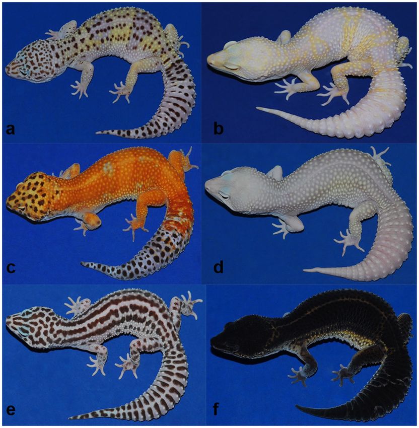
www.nature.com/scientificreports/ www.nature.com/scientificreports
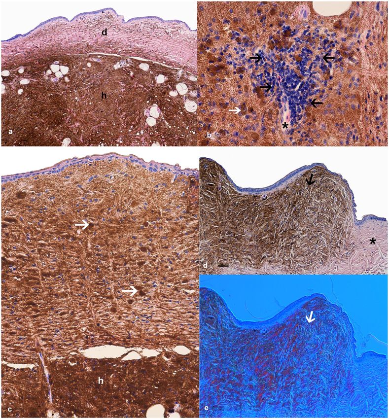
Figure 1. Examples of leopard gecko colour morphs. Wild-type (a) with a normal arrangement of dots
(melanophores), yellow colour (xathophores). The Tremper albino leopard gecko (b) with colourless dots and
stripes (lack of melanin in melanophores). Example of the hypomelanistic colour morph (c) – a lack of dots on
the dorsal part of body. Lack of any black coloured dots (amelanism) and the yellow pigmentation (axanthism)
in a non-albino colour morph called “Blizzard” (d). Axantic (lack of yellow pigmentation) colour morph (e).
Hipermelanistic colour morph (f). Photo by Steve Sykes - Geckos Etc.
black-brown pigmentation or less numerous dorsal dots and spots) and three different albino strains (Bell, Las
Vegas and Tremper albino). All names of colour morphs are derived from the breeder. Hence, there are no formal
and scientific rules to describe currently existing colour morphs of the leopard geckos. Selective breeding has led
to combinations of existing colour morphs and a development of new ones. One of them, the Lemon Frost, is
characterized by an increased white body colouration and a brightening of the yellow/orange areas of the body.
In ca. 2015 new colour morph called Lemon Frost, emerged as a result of selective breeding. Since that time the
breeders observed that large number of these geckos were affected by numerous tumour-like skin lesions.
The genetics of the leopard geckos are well developed. It is known that they have 38 chromosomes (2n), their
genome size is 4.91 picograms (2c) and it contains 43.66% of GC nucleotides9. Additionally, a genome with high
coverage sequenced what revealed that leopard geckos have 24,755 protein-coding genes10. Despite these facts
the genetic character of the colour morphs are still unclear and the only information is based on private breeders’
observations. In this case, according to breeders contest, the Lemon Frost phenotype seems to be a codominant
trait. Apart from their unique morphology, the Lemon Frost colour morph is associated with nodular skin lesions.
In veterinary practice, reptile neoplasms affecting any kind of tissue or organ are not frequently observed11, and
skin tumours seem to be particularly rare. The frequency of chromatophoromas in reptiles is estimated at 14.5%,
and melanophoromas (11.2%) are more often encountered than iridophoromas (3.3%)12. Melanophoromas have
been reported in several cases11–13. Iridophoromas have been found in a few cases, e.g in Pituophis melanoleucus
and Morelia viridis14. In lizards iridophoromas were reported and well-described in Pogona vitticeps12. There is
Scientific Reports | (2020) 10:5734 | https://doi.org/10.1038/s41598-020-62828-9 2
www.nature.com/scientificreports/                                                              www.nature.com/scientificreports

                           Figure 2. Example of the distribution of skin lesions. (a–c) Black arrows: from pale to white lesions. Black
                           asterisks: point-to-point range of skin lesions. Radiograms of leopard geckos with skin lesions which resemble
                           the typical soft tissue views (white arrows) (d,e).

                           much evidence that iridophoromas in reptiles may be either benign or malignant. Benign iridophoromas have
                           been reported in veiled chameleon (Chamaeleo calyptratus), a bearded dragons (Pogona vitticeps), and a savannah
                           monitor (Varanus exanthematicus)12. Cases of malignant iridophoromas were found in snakes14,15, in a dwarf
                           bearded dragon (Pogona henrylawsoni)16 and in green iguana (Iguana iguana)17.
                               Xanthophoromas in reptiles have been also observed15. The reports of melanophoromas in reptiles indicate
                           a visceral metastatic character of the tumour, whereas iridophoromas can occur with or without the visceral
                           metastases12 and affecting only the skin. The aim of this study is to describe iridophoromas in Eublepharis macu-
                           larius and to the best of the authors’ knowledge this is the first report of such kind of a tumour within Eublepharis
                           macularius.

                           Materials and Methods
                           Ethical note. This study did not perform any experiments on animals. All performed examinations and sam-
                           ples collection were done during routine veterinary practice and did not require local ethics committee approval.
                           All methods were performed in accordance with the relevant guidelines and regulations. Leopard geckos owner
                           gave a permission for treatment and the use of samples to subsequent diagnostics.

                           Materials.       In this report leopard geckos sourced from a private owner were examined in our clinic (Faculty
                           of Veterinary Medicine, Wroclaw University of Environmental and Life Sciences) in relation to the presence
                           of tumour-like skin lesions. Three geckos were selected for clinical examination: the male Snow Lemon Frost
                           (weight 86 g, age 10 months), the male Hypo Lemon Frost (weight 55 g, age 10 months) and the female Eclipse
                           (weight 57 g, age 3 years). All of the geckos were in good body condition, weight and size adequate to their age.
                           According to the patient history we found that the Snow Lemon Frost and the Hypo Lemon Frost were related
                           (via crossing different females with the same male), and that the Eclipse had a cross-fertilization with a Lemon
                           Frost in its lineage. The animals were kept in a rack-system type of enclosure under a temperature gradient (26–
                           30 °C), 12 hour day/night cycle and without UVB lightning. The animals were fed with mealworms supplemented
                           with vitamin powder. All of the geckos were affected by nodular lesions of the skin (Fig. 2a–c), which emerged
                           about two months earlier and affected 100% of leopard geckos of this colour morph from this owner. The lesions
                           were painless, immobile and located on different parts of the body: eyelids, neck and both ventral and dorsal
                           parts. The lesions had round or irregular shape varying from 0.5 to 2.0 cm in the longer diameter and ca 0.5 cm in
                           the shorter diameter. The animals showed no other clinical symptoms.

Scientific Reports |   (2020) 10:5734 | https://doi.org/10.1038/s41598-020-62828-9                                                            3
www.nature.com/scientificreports/ www.nature.com/scientificreports
Methods. The diagnostic procedure included X-ray imaging (GIERTH HF 200, Germany) and ultrasonogra-
® ™
phy (Supersonic Aixplorer MultiWave Ultrasound, France), which were performed to check their effectiveness
in the diagnosis of the lesions, the presence of putative metastases or any other concomitant internal pathologies.
Several biopsies were taken from the nodular-like oval-shaped lesions with healthy skin margins from the necks
or bodies of all three geckos. Not all of the skin lesions were qualified for an excision. Only the lesions located in a
body region where about 0.5 cm of a healthy skin margin could be taken were chosen for a procedure (e.g. lesions
located in a direct vicinity of an eyeball were excluded) (Fig. 2b). Sedation was applied using a standard protocol
with 5% isoflurane (Piramal Healthcare, UK). The biopsy wounds were stitched with absorbable sutures (4–0
polyglycolide monofilament). Analgesic meloxicam was administered at a dose of 0.1 mg/kg. The samples were
fixed in buffered 4% formaldehyde and routinely processed in paraffin. The 7 µm-thick cut sections were stained
in haematoxylin and eosin (H&E), then analysed histopathologically in the light microscopy (Eclipse 80i, Nikon,
Melville, NY, USA) and in the Nomarski contrast (Differential interference contrast, DIC) to provide evidence
of the iridophorous character of the cells. After the surgical treatment the leopard geckos were given back to the
owner.
Ethics approval and consent to participate. This study does not perform any experiments on animals.
All performed examinations and samples collection were done during routine veterinary practice and did not
require local ethics committee approval.
Results
X-ray analysis showed shading typical for soft tissues clearly visible as an enlarged contour of body and head soft
tissues. None of the three geckos presented any abnormalities suggestive of metastases (Fig. 2d,e).
Ultrasound was used to examine several organs: the gallbladder (which presented normal with correct wall
thickness, filled with clear bile), kidneys (correct shape and size), spleen (correct and homogeneous), liver paren-
chyma (homogeneous), liver vascular system (without abnormalities), stomach and intestines (normal, with food
content). The ultrasound findings listed above apply to all the specimens.
Histopathological investigation revealed tumorous changes that consisted of iridophores, localised in the der-
mis and hypodermis of all the three geckos (Fig. 3). The cells contained considerable amounts of anisotropic
crystalline material that caused characteristic polarization in the Nomarski contrast (DIC), (Fig. 3e). In all three
individuals iridophores almost exclusively occupied the hypodermis, while in the dermis number of these cells
varied between samples and individuals, that they cover from part (Fig. 3a) to almost whole field of view (Fig. 3c).
Iridophores were generally chaotically distributed, locally in a vortex arrangement (Fig. 3a–c). The cells were
spindle-shaped, with marked pigmentation and mostly without mitotic figures in a high power field (HPF). The
cell nuclei were oval in shape, with mean diameters of 8 × 4 μm, but some of them revealed hyperpigmentation
and irregular shape of the nucleus (heteronucleosis). Some iridophores presented atypical morphology: larger
diameter, oval shape, eccentrically located nucleus and brown cytoplasm. In the dermis the cells were surrounded
by connective tissue with a typical structure and arrangement of collagen fibres. The processes of iridophore-like
cells spread among collagen fibres. However, in the hypodermis, connective tissues were scant and iridophore
processes interwoven. This histopathological view indicated the malignant iridophoroma. These changes were
accompanied by scant lymphohistiocytic cell infiltrates. The margins of the healthy skin did not reveal any pres-
ence of iridophores.
Although there are no widely accepted standards for a convalescence time after a skin surgery in geckos, it is
reasonable to proceed veterinary check-ups twice: after a wound healing time (about a week), and after the very
next moulting. Unfortunately, the owner of the affected leopard geckos did not agree to clinical control exami-
nations. However, after 6 months he assured, that surgically removed skin lesions did not reveal recurrence, but
new ones appeared in other body localizations. Furthermore, he did not observe any other abnormal symptoms
like weight or appetite loss.
Discussion
Leopard geckos are a very popular species that are kept in captivity. To date there is no information in a literature
about iridophore-derived type of tumour in a wild-type or other leopard gecko colour morphs.
Histopathological analyses of the presented cases indicated that the tumorous-like lesions contained
iridophore-like cells, which could not be found in the healthy skin margins. Light microscopy and DIC exami-
nation confirmed that the observed chromatophores were iridophores because of their light-reflecting character
and the presence of anisotropic crystalline material in the cytoplasm what is characteristic for this cell type14,18,19.
The morphology of the iridophores and infiltrative nature of the tumour indicates the malignant character of
these changes. Surprisingly, our previous studies indicated that there is lack of iridophores in the skin of healthy
animals of this species8. This phenomenon was also confirmed the present study by observation of healthy skin
around the tumorous changes. The malignancy of the tumour-forming cells was determined pathomorpholog-
ically by the cells morphology, the infiltrative character of the changes that invade dermis and hypodermis12.
Observed lymphohistiocytic cell infiltrations were small and scant and clinical observations revealed no signs
such as inflammation, pain or any other discomfort in the patients. What more, the geckos arriving at our clinic
in satisfying body condition displayed normal behaviour. The findings and the observations were not an indica-
tion for a euthanasia. Additionally, X-ray and ultrasonography revealed no signs of visceral metastases presence.
According to our observations, surgical procedure is strongly recommended for operable lesions. The proce-
dure is an effective method for a complete removal of the lesions, and no relapses have been observed so far.
Nonetheless, it does not prevent the appearing of new ones in other body parts.
The presented cases are interesting for a few reasons. First, Heckers et al. (2012) suggested that chromato-
phoromas occur significantly more frequently in day-active reptiles like bearded dragons (Pogona sp.)12 than in
Scientific Reports | (2020) 10:5734 | https://doi.org/10.1038/s41598-020-62828-9 4www.nature.com/scientificreports/ www.nature.com/scientificreports
Figure 3. Histopathology of the leopard gecko skin iridophoroma. (a–d) H&E. (e) Nomarski contrast (DIC).
(a) Large mass of iridophores in hypodermis (h) and scattered clusters of iridophores in dermis (d), regular
collagen fibre arrangements were noted. (b) Infiltration of lymphohistiocytic cells (black arrows) around a
skin capillary (black asterisk), atypical oval shaped iridophore with eccentrically located nucleus and brown
cytoplasm (white arrow). (c) Tumour cells present both in the epidermis, dermis and hypodermis (h), round
and spindle shaped cells filled with guanine crystals (white arrows), regular collagen fibre arrangement. (d)
Overview of the affected skin with a tumour lesion (black arrow) and healthy margin without iridophores and
other chromatophores (black asterisk). (e) Nomarski contrast used to confirm the presence of iridophores in the
skin sections; note the characteristic change in colour of light-reflecting cells (white arrow).
nocturnal, cryptic species like leopard geckos. To the best of our knowledge this is the first study describing the
iridophoroma in leopard geckos, a popular species bred in captivity. Second, our findings indicate that a selective
inbreeding influences the disease incidence. Additionally, it cannot be excluded that the cross-fertilization of
morphs with the Lemon Frost may be associated with an increased incidence of iridophoroma in next generations
(e.g. the Eclipse patient). Our findings are the first observation of iridophoromas in leopard geckos and further
research should consider statistical analysis of different colour morphs crossing with Lemon Frost to fully estab-
lish which crosses yield offspring with iridophoromas.
In view of a putative connection between iridophoromas and the genome, further breeding of the Lemon
Frost leopard gecko line is not recommended until the pathogenesis of the lesions will be fully recognised and
described.
Data availability
All data generated or analysed during this study are included in this published article.
Scientific Reports | (2020) 10:5734 | https://doi.org/10.1038/s41598-020-62828-9 5www.nature.com/scientificreports/ www.nature.com/scientificreports
Received: 10 June 2019; Accepted: 20 March 2020;
Published: xx xx xxxx
References
1. Bonke, E., Böhme, W., Opiela, K. & Rödder, D. A remarkable case of cannibalism in juvenile Leopard Geckos, Eublepharis
macularius (Blyth, 1854) (Squamata: Eublepharidae). Herpetology Notes. 4, 211–212 (2011).
2. Rawat, Y. B., Thapa, K. B., Bhattarai, S. & Shah, K. B. First Records of the Common Leopard Gecko, Eublepharis macularius (Blyth
1854) (Eublepharidae), in Nepal. IRCF Reptiles and Amphibians. 26(1), 58–61 (2019).
3. Alibardi, L. Ultrastructural features of skin pigmentation in the lizard Heloderma suspectum with emphasis on xanthomelanophores.
Acta Zool. 96, 154–159 (2013).
4. Bagnara, J. T. Cytology and cytophysiology of non-melanophore pigment cells. Int. Rev. Cytol. 20, 173–205 (1966).
5. Bagnara, J. T. et al. Common origin of pigment cells. Science. 203, 410–415 (1979).
6. Saenko, S. V., Teyssier, J., van der Marel, D. & Milinkovitch, M. C. Precise colocalization of interacting structural and pigmentary
elements generates extensive colour pattern variation in Phelsuma lizards. BMC Biol. 11, 105 (2013).
7. Teyssier, J., Saenko, S. V., van der Marel, D. & Milinkovitch, M. C. Photonic crystals cause active colour change in chameleons. Nat.
Commun. 6, 6368 (2015).
8. Szydłowski, P., Madej, J. P. & Mazurkiewicz-Kania, M. Ultrastructure and distribution of chromatophores in the skin of the leopard
gecko (Eublepharis macularius). Acta Zool. 97, 370–375 (2016).
9. Vinogradov, A. E. Genome size and GC-percent in vertebrates as determined by flow cytometry: the triangular relationship.
Cytometry. 31, 100–109 (1998).
10. Xiong, Z. et al. Draft genome of the leopard gecko, Eublepharis macularius. GigaSience. 5, 47 (2016).
11. Hernandez-Divers, S. M. & Garner, M. M. Neoplasia of reptiles with an emphasis on lizards. Vet. Clin. North. Am. Exot. Anim. Pract.
6, 251–73 (2003).
12. Heckers, K. O., Aupperle, H., Schmidt, V. & Pees, M. Melanophoromas and Iridophoromas in Reptiles. J. Comp. Pathol. 146, 258–268
(2012).
13. Elkan, E. Malignant melanoma in a Snake. J. Comp. Pathol. 84, 51–57 (1974).
14. Jacobson, E. R., Ferris, W., Bagnara, J. T. & Iverson, W. O. Chromatophoromas in pine snake. Pigm Cell Res. 2, 225–239 (1989).
15. Muñoz-Gutiérrez, J. F., Garner, M. M. & Kiupel, M. Cutaneous Chromatophoromas in Captive Snakes. Vet. Pathol. 53, 1213–1219
(2016).
16. de Brot, S., Sydler, T., Nufer, L. & Ruetten, M. Histologic, immunohistochemical, and electron microscopic characterization of a
malignant iridophoroma in a Dwarf Bearded dragon (Pogona henrylawsoni). J. Zoo. Wildl. Med. 46, 583–587 (2015).
17. Rousselet, E. et al. Cutaneous iridophoroma in a Green iguana (Iguana iguana). Vet. Clin. Pathol. 46, 625–628 (2017).
18. Irizarry-Rovira, A. R., Wolf, A. & Ramos-Vara, J. A. Cutaneous melanophoroma in a green iguana (Iguana iguana). Veterinary
Clinical Pathology. 35, 101–105 (2006).
19. Szydłowski, P., Madej, J. P. & Mazurkiewicz-Kania, M. Histology and ultrastructure of the integumental chromatophores in tokay
gecko (Gekko gecko) (Linnaeus, 1758) skin. Zoomorphology. 136, 233–240 (2017).
Acknowledgements
We would like to express our gratitude to Steve Sykes - Geckos Etc. (http://www.geckosetc.com) for permission
to use photographs of leopard geckos from his collection. Publication language correction supported by Wroclaw
Center of Biotechnology, programme the Leading National Research Center (KNOW) for the years 2014–2018.
Author contributions
P.S. Principal investigator, study conception, samples collection, data acquisition, analysis and interpretation,
major manuscript draft, histological methodology, figures preparation. J.P.M. Secondary investigator, data
acquisition, manuscript draft, histological methodology, critical revision. M.D., J.A.M., A.S.K., A.C.S., L.I., P.D.
samples collection, minor manuscript corrections, critical revision, participation in a manuscript preparation.
J.A.M. histopathology images interpretation. All authors read and approved the final manuscript.
Competing interests
The authors declare no competing interests.
Additional information
Correspondence and requests for materials should be addressed to P.S.
Reprints and permissions information is available at www.nature.com/reprints.
Publisher’s note Springer Nature remains neutral with regard to jurisdictional claims in published maps and
institutional affiliations.
Open Access This article is licensed under a Creative Commons Attribution 4.0 International
License, which permits use, sharing, adaptation, distribution and reproduction in any medium or
format, as long as you give appropriate credit to the original author(s) and the source, provide a link to the Cre-
ative Commons license, and indicate if changes were made. The images or other third party material in this
article are included in the article’s Creative Commons license, unless indicated otherwise in a credit line to the
material. If material is not included in the article’s Creative Commons license and your intended use is not per-
mitted by statutory regulation or exceeds the permitted use, you will need to obtain permission directly from the
copyright holder. To view a copy of this license, visit http://creativecommons.org/licenses/by/4.0/.
© The Author(s) 2020
Scientific Reports | (2020) 10:5734 | https://doi.org/10.1038/s41598-020-62828-9 6You can also read