3D Cell Culture 101: An Introduction to 3D Cell Culture Tools and Techniques
←
→
Page content transcription
If your browser does not render page correctly, please read the page content below
3D Cell Culture 101:
                                        An Introduction to
                                      3D Cell Culture Tools
                                          and Techniques
                                                                                              White Paper

                                   If you are new to 3D, what do you
                                   choose?

In this white paper                Introduction

                                   For decades, three-dimensional (3D) cell culture has been employed by tissue
     Scaffold-free platforms for
 3                                 engineers, stem cell scientists, cancer researchers and cell biologists, largely in
     spheroid growth
                                   university settings. The development of new materials or methods has been
                                   driven by the desire of these scientists to incorporate experimental systems that
 5   Scaffolds
                                   better represent the in vivo environment into their research. Early adopters of
                                   3D cell culture technology have reaped the benefits of better data with

 7   Gels                          groundbreaking knowledge of tissue and cancer behavior.

                                   3D cell culture methods were once expensive, messy, laborious, and difficult to

     Bioreactors                   adapt to existing procedures. Today, researchers can pick among an array of
 8
                                   3D cell culture tools, ranging from simple to complex, to fit their specific needs.
                                   Segmentation of the 3D cell culture choices into discrete categories
 9   Microchips                    demonstrates a new maturation in the market. As the number of 3D cell culture
                                   options has grown, publications proving their importance have exploded in
                                   numbers (Figure 1).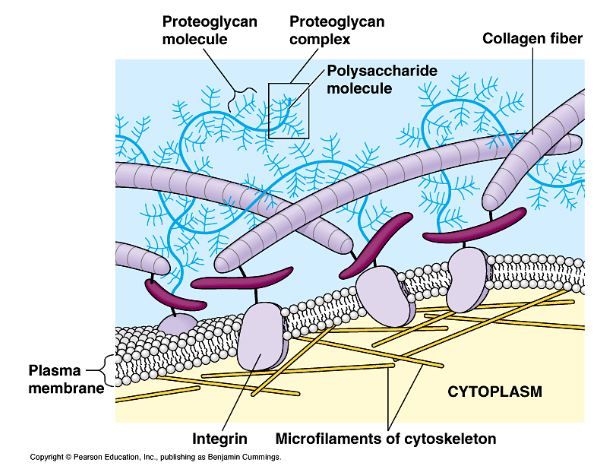
3D Cell Culture 101
Figure 1. Publications referencing 3D cell culture have grown dramatically in the
last several years. Each line on the graph represents a separate search for three-
dimensional cell culture plus the additional listed search term. A small degree of
overlap is possible.
If you are new to 3D, what do you choose?
An important consideration in 3D cell culture is replicating or mimicking the
extracellular matrix (ECM) (Figure 2). The ECM provides physical structure,
sequesters and secretes growth factors, and facilitates cellular
communication. Consideration of the ECM composition, structure, and
density in your target tissue, or whether or not you would like to provide ECM
at all, may dictate the 3D cell culture format choice.
In this tutorial, we discuss 3D cell culture methods that rely on cells to secrete
their own ECM and others that utilize natural or artificial materials to mimic
the ECM until cells create their own. Five types of 3D cell culture options are
reviewed: scaffold-free platforms for spheroid growth, scaffolds, gels,
bioreactors, and microchips. For your convenience, select review articles
are included at the end of each section.
Figure 2. Schematic of various components of the ECM.
2 3D Biomatrix White Paper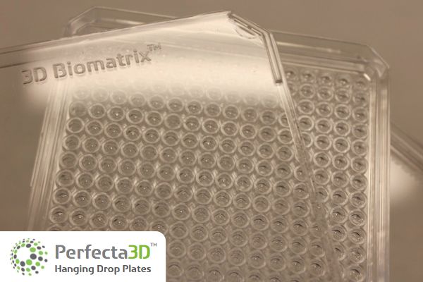
Scaffold-Free Platforms
for Spheroid Growth
Scaffold-free platforms for spheroid growth
Spheroids are self-assembled spherical clusters of cell colonies. They were
first documented in 1944 by Johannes Holtfreter who worked with spherical
aggregates of embryonic cells. Spheroids naturally mimic solid tissues,
avascular tumors, and embryoid bodies, and have found application
among researchers in cancer and stem cell research. With inherent
metabolic (oxygen, carbon dioxide, nutrients, wastes) and proliferative
gradients, spheroids serve as excellent physiologic models.
Scaffold-free platforms for spheroid growth do not contain added
biomaterials or ECM, and cells grown in them generate and organize their
"After struggling to generate own 3D ECM, so spheroids closely resemble in vivo tissues. Co-cultures with
reproducible spheroids in other cell types (i.e., endothelial, stromal, epithelial cells) extend the
spinner cultures and agarose- predictive cytotoxicity capabilities of this 3D cell culture system.
coated plates, the Scaffold-free platforms have no support structure or porosity. The overall
Perfecta3D® Hanging Drop spheroid size is limited beyond a critical size of 500 – 600 µm in diameter,
Plates have finally given me after which central secondary necrosis develops in most, but not all,
consistent cell growth and spheroids grown from permanently-transformed cell lines.
morphology, and the Standardized mass production of 3D spheroids makes them applicable for
spheroids size and shape are both basic laboratory research and high-throughput screening (HTS)
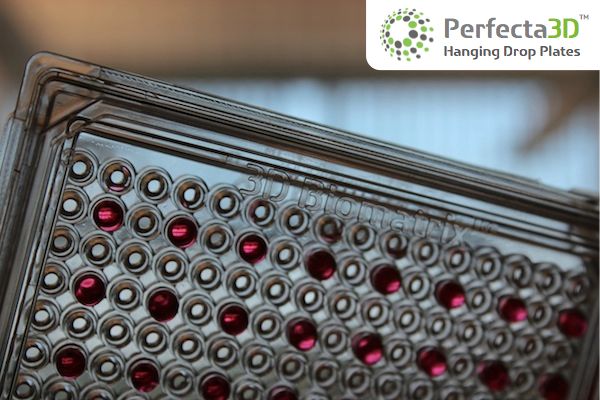
remarkably reproducible." applications. The Perfecta3D® Hanging Drop Plate from 3D Biomatrix™ is
designed to enable consistent formation of spheroids using conventional
Research scientist at a major Canadian liquid handling tools. This scaffold-free platform is simple to use and
university generates spheroids with consistent sizes and shapes so that testing is
controllable and reliable. Adjusting the seeding density, from as few as 50
cells to as many as 15,000 cells, allows production of varying spheroid sizes.
3 3D Biomatrix White Paper
The plate consists of the main hanging drop culture plate and a
complementary lid and tray, which serve to maintain sterility and reduce
evaporation. Access holes in the culture plate allow manipulation of fluids
and spheroids from the topside. A water reservoir constructed around the
periphery of the culture plate also helps to alleviate evaporation (Figure 3).
Figure 3. A schematic of the Perfecta3D Hanging Drop Plate.
Hanging drops are created by dispensing small volumes of cell
suspensions, using standard pipette tips, into the access holes on the top
of the plate, just like pipetting into conventional multi-well plates. In a
similar fashion, reagents and drugs can be added to or removed from
each hanging drop.
A plateau structure on the bottom of the plate stabilizes the hanging
drops (Figure 4). The Perfecta3D Hanging Drop Plates do not have a
bottom substrate for cells to eventually attach to, therefore cells in
suspension aggregate into a spheroid.
Figure 4. Spheroids are created by dispensing cell suspensions into the access
holes of the Perfecta3D Hanging Drop Plate, just like pipetting into
conventional multi-well plates.
Spheroids can be harvested and analyzed using colorimetric, fluorescence,
and luminescence assays measured with a plate reader. Microscopic
imaging of spheroids can be performed directly with the transparent plate,
lid and tray assembled. The platform also offers simplified liquid handling
procedures and compatibility with HTS instruments, such as liquid handling
robots like the Biomek® FX and epMotion automated pipetting systems.
4 3D Biomatrix White Paper
Articles:
The inventing University of Michigan research team discusses the Perfecta3D
Hanging Drop Plate.
• Tung, YC, Hsiao, AY, Allen, SG, Torisawa, Y, Ho, M, Takayama, S, High
throughput 3D spheroid culture and drug testing using a 384 hanging
drop array, Analyst, 136 (2011), 473-478.
Demonstration of Z-factors and co-cultures with the Perfecta3D Hanging
Drop Plate.
• Hsiao, AY, Tung, YC, Qu, X, Patel, LR, Pienta, KJ and Takayama, S, 384
hanging drop arrays give excellent Z-factors and allow versatile
formation of co-culture spheroids, Biotechnology and Bioengineering,
109 (2012), 1293-1304.
Scaffolds
An overview of 3D in vitro cancer models as they pertain to drug discovery,
with a specific focus on those that have been developed from a tissue
engineering perspective.
• Burdett E, Kasper FK, Mikos, AG and Ludwig, JA, Engineering Tumors: A
Tissue Engineering Perspective in Cancer Biology, Tissue Engineering:
Part B Vol 16, No. 3 (2010), 351-9.
Scaffolds
Scaffolds, also commonly called 3D matrices, are available in a large
variety of materials with different porosities, permeabilities and mechanical
characteristics designed to reflect the in vivo ECM of the specific tissues
being modeled. Scaffolds are manufactured using a variety of techniques,
such as 3D printing, particulate leaching, or electrospinning, each of which
introduces different porosities, pore sizes, scaffold materials and features.
Scaffolds are typically divided into two main application categories:
functional implants for clinical and regenerative medicine applications and
in vitro 3D scaffolds for laboratory applications.
Though the characteristics of implantable scaffolds may vary greatly
depending on the tissue being mimicked, the ultimate goal for many
implantable scaffolds is to provide support to a wound site and aid eventual
replacement of the scaffold by natural tissue. As such, the requirements for
functional implant scaffolds differ from those for in vitro 3D laboratory
applications. Functional implants must match the defect site, support and
promote desired cell growth, and biodegrade without harmful effects.
When scaffolds are used for in vitro laboratory applications, geometric
match and biodegradability are less necessary. In fact, degradability may
introduce an undesirable variable into experiments as byproducts may
change the chemistry and pH of the culture system. Furthermore, as the
scaffold degrades and cells re-organize the matrix, the cells may not retain
their 3D configuration. In vitro scaffolds should represent a more stable
structure and function similar to the natural in vivo environment.
5 3D Biomatrix White Paper
3D scaffolds for in vitro laboratory applications are available in a variety of
materials: metals, ceramics, polymers - natural and synthetic, and
composites. Properties to consider include biocompatibility, wettability,
mechanical properties, and surface chemistry. The method of fabrication
must also be considered as it may introduce a random or ordered structure
to the scaffold (Figure 5). When utilizing a scaffold with a random structure,
such as those that result from particulate leaching methods, scaffold-to-
scaffold variability, as well as isolation of areas within the scaffold, may be a
problem.
Due to the variety of material and structural choices for scaffolds, they are
widely used in many applications. Furthermore, as they provide a surface on
which cells can grow, they can easily impart 3D growth with little alteration
to cell culture procedures. The porosity of scaffolds aids mass transport of
nutrients, oxygen, and wastes, allowing for larger culture growth than the
scaffold-free platform discussed earlier; it can be difficult to extract all cells
for analysis with increased scaffold size and tortuosity. Imaging may also
become difficult depending on the scaffold size, transparency of material,
and depth of the microscope.
In a 2008 review article, Lee et al. discuss the macro-, micro-and nano-scale
elements of 3D scaffolds.
• Macro-scale: Overall size and shape, dependent on application.
• Micro-scale: Porosity, pore interconnectivity, pore geometry, pore size
distribution and elements of surface topography. Micro-scale elements
may be customized for different tissue types. Micro-scale elements
facilitate mass transport, diffusion of nutrients, metabolic wastes and
soluble molecules and can activate certain genes and modulate Figure 5. Examples of random (left) and
cellular behavior in differentiation and proliferation. Micro-scale features ordered (right) scaffold structures.
Image adapted from Lee et al.
also affect the overall robustness of the scaffolds and hence the desired
application, such as use in a bioreactor, multiwell plate or human body.
• Nano-scale: Nutrient supply and functional effects due to the size of
many cell-signaling molecules.
Articles:
A review of 3D cell-growth techniques and scaffolds analyzed from the
perspective of materials properties, manufacturing and functionality.
• Lee J, Cuddihy M and Kotov NA, Three-Dimensional Cell Culture
Matrices: State of the Art, Tissue Engineering: Part B, Vol 14, Number 1
(2008), 61-86.
General review of 3D cell culture approaches and techniques.
• Haycock JW (ed.), 3D Cell Culture: Methods and Protocols, Methods in
Molecular Biology, Springer Science+Business Media, LLC , Vol 695
(2011), DOI 10.1007/978-1-60761-948-0_1.
6 3D Biomatrix White Paper
Gels
In general, gels have a soft tissue-like stiffness and aim to mimic the ECM.
Gels made from ECM mixtures of natural origin, such as collagen, and
alginate, have been used for decades as substrates for 3D cell culture. The
most well-known gel is Matrigel™, first documented in 1972 by Hynda
Kleinman. Matrigel is a reconstituted basement membrane preparation
extracted from the Engelbreth-Holm-Swarm mouse sarcoma, a tumor rich in
ECM proteins, such as laminin and collagen, plus growth factors and
enzymes.
Gels
Gels made from animal-sourced natural ECM extracts may contain residual
growth factors, undefined constituents, such as animal viruses, or non-
quantified substances. Batch-to-batch variations make it difficult to
compare and correlate work from different scientific groups. Gels may also
change in structure over time as they are organized by cells in culture.
The desire to impart controllability in gels drove the development of
synthetic gels, such as the poly-ethylene glycol (PEG)-based hydrogels from
QGel™, which are modified to obtain desired characteristics. Modification
of the bulk material may be performed through hybridization of natural and
synthetic materials, incorporation of different types of proteins or molecules
within the matrix, and hybridization of biomaterials with functional nano-
materials.
A drawback to many 3D gels is the difficulty of use, which often stems from
the gelling mechanism. In the case of Matrigel, the gel must be kept on ice
to keep its viscosity low enough for manipulating and mixing with cells. pH-
based gelling mechanisms are also common, which can expose sensitive
cells to adverse conditions.
The ability to choose from a variety of natural and synthetic materials, the
soft tissue-like material properties, and the ease of converting cultures to 3D
have made gels widely popular. As well, 3D cell cultures in gels have led to
important findings, specifically in cancer research.
Lastly, gels can be combined with other methods, such as spheroid cultures,
scaffolds, and microchips.
Article:
Three current technologies are presented in this review: membranes,
sponges/gels and microcarriers.
• Justice, BA, Badr, NA and Felder RA, 3D cell culture opens new
dimensions in cell-based assays, Drug Discovery Today, Volume 14,
Numbers 1/2 (2009), 102-107.
7 3D Biomatrix White Paper
Bioreactors
Bioreactors have been adapted for 3D cell culture by the addition of
scaffolds and are most ideal for high volume cell production and ex vivo
tissue engineering applications (Figure 6). Many bioreactors include media
flow, allowing for circulation of nutrients, removal of wastes, and
homogeneity of the environment within the reactor. As such, they are well-
suited for cell expansion applications or scaled production of cellular
products, such as antibodies.
Bioreactors for 3D cell culture fall into 4 main categories:
Bioreactors
• The spinner flask is typically composed of a glass media reservoir with
side arms that can be opened and may have porous covers for gas
exchange. Scaffolds are suspended on a structure or placed within the
media, which is stirred by a stir bar.
• Low-shear-stress rotating wall vessels, originally designed by the National
Aeronautics and Space Administration to simulate microgravity, are
composed of two concentric cylinders, an inner stationary cylinder that
provides for gas exchange and an outer rotating cylinder. Free-moving
scaffolds and media are placed in the space between the two
cylinders.
• Perfusion bioreactors use a pump system to perfuse media, directly or
indirectly, through a scaffold. The basic design consists of a media
reservoir, a pump, a tubing circuit, and a perfusion cartridge, which
houses the scaffold. Hollow-fiber systems, a sub-group, are composed of
small tube-like filters, approximately 200 µm in diameter with molecular
weight cut-offs, sealed into a cartridge shell.
• When combined with scaffolds, bioreactors can be used for specialty
purposes that utilize mechanical load or electrical or pulsed electrical
fields.
A large variety of scaffolds or microcarriers, which can be matched to
experimental and cellular needs, are available for use in bioreactors. Cells
and/or supernatants must be harvested for analysis. The major restrictions for
Figure 6. Examples of bioreactor systems: the use of bioreactors may be the cost and throughput. Although the simple
A. Spinner flask, B. Rotating-wall vessel and
spinner flask is an inexpensive option, many bioreactors require specialized
C. Perfusion system. Images adapted from
Yeatts et al. equipment. Bioreactors may not be the ideal choice for HTS unless a custom
automated system is designed.
8 3D Biomatrix White Paper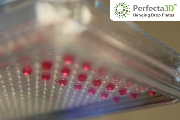
Articles:
An overview of the concepts, advantages, challenges, and potential future
applications associated with current bioreactor systems for bone tissue
engineering.
• Rauh J, Milan F, Gunther KP, and Stiehler, M, Bioreactor Systems for Bone
Tissue Engineering, Tissue Engineering: Part B, Volume 17, Number 4
(2011), 263-280.
A summary of in vitro bioreactors for bone tissue engineering as well as
commercial bioreactor systems.
• Yeatts AB, Fisher JP, Bone tissue engineering bioreactors: Dynamic
culture and the influence of shear stress, Bone 48 (2011), 171–181. Microchips
Microchips
Microchips, frequently called ‘organ on a chip’ or microsystems, are the next
wave of 3D cell culture models. ‘Organ on a chip’ integrate microfluidics
technologies with cells cultured within microfabricated 3D devices, using
techniques from the microchip industry.
Although not expected to be widely commercially available for some time,
advances in microfabrication and microfluidic techniques open the door to
this option. Recent advances include development of integrated ‘organ on
chip’ microsystems that reproduce key architectural, functional,
biochemical and mechanical features such as mechanical strain and shear
forces, of living organs, including lung, liver (Figure 7), kidney, gut, bone,
breast, brain and eye. The eventual goal is to link the individual organ
microsystems together to develop a human-on-a-chip.
Scale-up and manufacturing of microchips is a crucial milestone, which
needs to be overcome to make the technology an affordable,
commercially-viable option. Today’s microchips use photo or soft
lithography and replica molding techniques along with silicone rubber,
poly(dimethylsiloxane) (PDMS), microfluidics systems. These systems are less Figure 7. A micro-engineered liver-on-a-
expensive, easier to fabricate, have high gas permeability and are optically chip reconstitutes hepatic
microarchitecture. The microsystem
transparent. Although PDMS has desirable properties it has poor chemical consists of a central liver-cell culture
resistance to some solvents and can absorb small hydrophobic molecules chamber and a surrounding nutrient flow
channel separated by microfabricated
limiting its effectiveness in commercial uses. Other challenges involve poorly-
barrier structures that mimic the highly
understood effects of polymers and microfluidics on cellular behavior. HTS permeable endothelial barrier between
hepatocytes and the liver sinusoid. Image
options are currently limited; the minute sample volumes make collection
adapted from Huh et al.
and analysis difficult.
Although microchips may be the next wave of 3D tools, many of the current
versions require considerable expertise to operate and troubleshoot, limiting
widespread adoption.
9 3D Biomatrix White Paper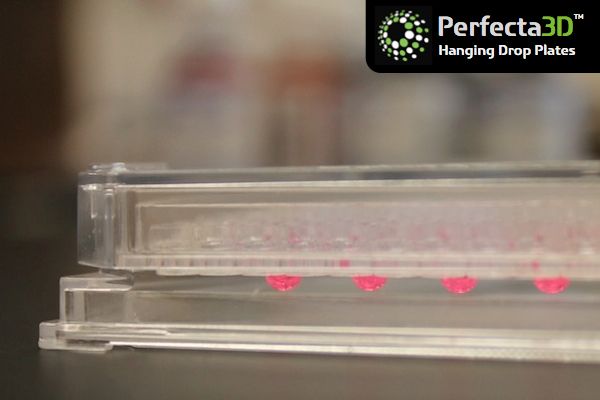
Articles:
                                           A review of new advances in 3D culture, ‘organs-on-chips’.
                                           •      Huh D, Hamilton GA and Ingber DE, From 3D cell culture to organs-on-
                                                  chips, Trends in Cell Biology, Vol. 21, No. 12 (2011), 745-754
                                           For years, scientists have struggled to reconstruct tissues and organs by
                                           combining cells and nanotechnology. These devices are now edging from
                                           cool concept to practical application.
                                           •      Baker M, A Living System on a Chip, Nature, Vol 471 (2011), 661-665

3D Cell Culture Options                    How to decide what fits your lab

                                           If you are just starting out, deciding on the best option for your specific
                                           application can be frustrating. Each available option has pros and cons
                                           (Table 1), as well as best-suited applications.

 Option          Pros                                  Cons                                         Research Stage

                 • No added materials
                 • Consistent spheroid
                   formation; control over size                                                     • Basic research
                                                       • No support or porosity
 Scaffold-free   • Co-cultures possible                                                             • Drug discovery
                                                       • Limited flexibility
 systems         • Transparent                                                                      • Personalized
                                                       • Size of spheroid limiting
                 • HTS capable; compatible                                                            medicine
                   with liquid handling tools
                 • Inexpensive

                 • Large variety of materials
                                                       • Possible scaffold-to-scaffold variation
 In vitro 3D       possible for desired properties                                                  • Basic research
 scaffolds for                                         • May not be transparent
                 • Customizable                                                                     • Drug discovery
 laboratory                                            • Cell removal may be difficult
                 • Co-cultures possible                                                             • Cell expansion
 applications                                          • HTS options limited
                 • Medium cost

                                                       • Gelling mechanism
                 • Large variety of natural or
                   synthetic materials                 • Gel-to-gel variation and structural
                                                         changes over time                          • Basic research
 Gels            • Customizable
                                                       • Undefined constituents in natural gels     • Drug discovery
                 • Co-cultures possible
                                                       • May not be transparent
                 • Inexpensive
                                                       • HTS options limited

                 • Several options available                                                        • Basic research
                                                       • Cost
 Bioreactors     • High volume cell production                                                      • Tissue engineering
                                                       • HTS options limited
                 • Customizable                                                                     • Cell expansion

                                                       • Commercial availability
                 • In vitro organ specific systems
                                                       • Required expertise                         • Basic research
 Microchips      • High gas permeability
                                                       • Cost                                       • Drug discovery
                 • Transparent
                                                       • HTS options limited

                                    Table 1. Summary of 3D cell culture options.
A perusal of the review articles listed throughout this paper is a good start.
Initially a comparison of one or more options may be required. The best-fit
3D cell culture option will depend on many factors (Figure 8).
3D Biomatrix
1600 Huron Parkway
Building 520, 2nd Floor
Ann Arbor, MI 48109-2590
USA
www.3DBiomatrix.com
service@3DBiomatrix.com
Phone: 734.272.4688
Fax: 734.818.1999
©2012 3D Biomatrix, Inc.
3D Biomatrix is a trademark of 3D Biomatrix, Inc.
Perfecta3D is a registered trademark of 3D Biomatrix, Inc.
Figure 8. Factors to consider when choosing 3D cell culture options.
What are you waiting for? Take the leap to 3D cell culture today.
As we have demonstrated in this short tutorial there are many 3D cell culture
options available today. One, or more, options may be right for your
application.
3D cell culture options:
• Scaffold-free platforms for spheroid growth
• Scaffolds
• Gels
• Bioreactors
• Microchips
The sooner the median level of 3D cell culture adoption rises, the sooner
companies will introduce new products, and the faster the technique will
progress, especially in analytic techniques, such as imaging, assay
validation, correlation to historical 2D culture results, and automation. 3D cell
culture may have taken decades to reach its current level of growing
acceptance, but there is no mistaking this: the technique is here to stay.
11 3D Biomatrix White PaperYou can also read