Calumenin contributes to epithelial-mesenchymal transition and predicts poor survival in glioma
←
→
Page content transcription
If your browser does not render page correctly, please read the page content below
Translational Neuroscience 2021; 12: 67–75
Research Article
Ying Yang, Jin Wang*, Shihai Xu*, Fei Shi, Aijun Shan
Calumenin contributes to epithelial-
mesenchymal transition and predicts poor
survival in glioma
https://doi.org/10.1515/tnsci-2021-0004 key markers, including N-cadherin, vimentin, snail,
received July 20, 2020; accepted December 14, 2020 slug, and TWIST1. Survival and Cox regression analysis
Abstract showed that higher CALU predicted worse survival, and
Background ‒ Calumenin (CALU) has been reported to the prognostic value was independent of WHO grade
be associated with invasiveness and metastasis in some and age.
malignancies. However, in glioma, the role of CALU Conclusions ‒ CALU was correlated with more malig-
remains unclear. nant phenotypes in glioma. Moreover, CALU seemed to
Methods ‒ Clinical and transcriptome data of 998 glioma serve as a pro-EMT molecular target and could contribute
patients, including 301 from CGGA and 697 from TCGA to predict prognosis independently in glioma.
dataset, were included. R language was used to perform Keywords: calumenin, glioma, epithelial-mesenchymal
statistical analyses. transition
Results ‒ CALU expression was significantly upregu-
lated in more malignant gliomas, including higher
grade, IDH wildtype, mesenchymal, and classical subtype.
Gene Ontology analysis revealed that CALU-correlated 1 Introduction
genes were mainly enriched in cell/biological adhesion,
response to wounding, and extracellular matrix/structure In central nervous system, glioma is the most prevalent
organization, all of which were strongly correlated with and fatal primary cancer in adults [1]. Despite a substan-
the epithelial-mesenchymal transition (EMT) phenotype. tial body of improvements in therapy, the prognosis
GSEA further validated the profound involvement of for most glioma patients is still dismal. Particularly for
CALU in EMT. Subsequent GSVA suggested that CALU patients who suffered from higher grade glioma (WHO
was particularly correlated with three EMT signaling grade IV, glioblastoma, GBM), which is the most malig-
pathways, including TGFβ, PI3K/AKT, and hypoxia path- nant and lethal type, the median survival remains less
way. Furthermore, CALU played synergistically with EMT than 15 months [2,3]. There is a growing recognition that
epithelial-mesenchymal transition (EMT) plays a key
role in mediating tumorigenesis, stemness, invasiveness,
* Corresponding author: Jin Wang, Department of Emergency,
resistance to radiochemotherapy, and early recurrence in
Shenzhen People’s Hospital (The Second Clinical Medical College, glioma [4–7]. It is therefore imperative to identify novel
Jinan University; The First Affiliated Hospital, Southern University of EMT-related molecules for potential glioma diagnosis
Science and Technology), Shenzhen 518020, China, and intervention.
e-mail: szph3022@szhospital.com, tel: +86-159-1414-1979 Calumenin (CALU) has been widely reported in a
* Corresponding author: Shihai Xu, Department of Emergency,
range of malignancies including head and neck cancer
Shenzhen People’s Hospital (The Second Clinical Medical College,
Jinan University; The First Affiliated Hospital, Southern University of [8], endometrial cancer [9], colon [10] and colorectal
Science and Technology), Shenzhen 518020, China, cancer [11], lung cancer [10,12], melanoma [13], hepato-
e-mail: heykojnu@163.com cellular and pancreatic carcinoma [14], and breast cancer
Ying Yang: Department of Pediatrics, Futian Women and Children [15]. CALU, a calcium-binding protein localized in the
Health Institute, Shenzhen 518045, China
endoplasmic reticulum (ER), is mainly involved in such
Fei Shi, Aijun Shan: Department of Emergency, Shenzhen People’s
Hospital (The Second Clinical Medical College, Jinan University; The
ER functions as protein folding and sorting. Besides,
First Affiliated Hospital, Southern University of Science and CALU has recently been shown to influence cell mobility,
Technology), Shenzhen 518020, China migration, invasion, and metastasis during particular
Open Access. © 2021 Ying Yang et al., published by De Gruyter. This work is licensed under the Creative Commons Attribution 4.0
International License.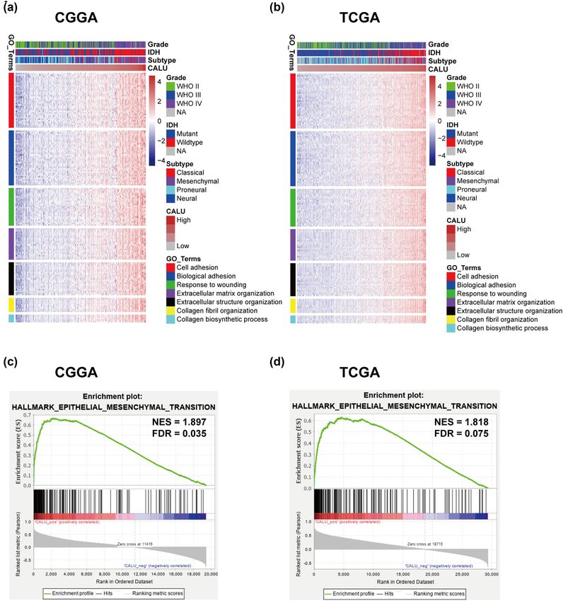
68 Ying Yang et al.
events, such as tumorigenesis, wound healing, immune of survival package. Gaussian test was performed before
response, and coagulation [16–21]. Several studies have data analysis that required Gaussian distribution. We
explored the relationship between CALU expression and performed Pearson correlation to calculate the correla-
survival and yielded relatively consistent results. In most tion coefficient between CALU and every gene. Genes
types of cancer, a higher level of CALU in lesions indi- that strongly correlated with CALU were screened out
cated a more malignant phenotype and a shorter survival with Pearson |r| > 0.6 in each dataset. Gene Ontology
for patients. analysis (GO) of CALU-correlated genes was implemented
However, the expression patterns and biological func- based on DAVID [25] website (version 6.8, https://david-
tions of CALU in gliomas have rarely been described. Only d.ncifcrf.gov/). For Gene Set Enrichment Analysis (GSEA)
one study presented by Sreekanthreddy et al. [22] investi- [26] and Gene Set Variation Analysis (GSVA) [27], a series
gated the prognostic potential of serum CALU in GBM, of gene sets were obtained from the GSEA network (http://
which accounted for about 40% of pan-glioma. Here, we software.broadinstitute.org/). A p-value less than 0.05 was
analyzed clinical and transcriptome data of 998 patients, considered to be statistically significant. Two-sided signif-
aiming at exploring the role of CALU in gliomas. icance tests were adopted throughout.
2 Materials and methods 3 Results
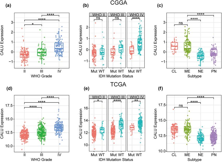
2.1 Sample and data collection 3.1 CALU was significantly upregulated in
GBM, IDH wildtype, mesenchymal, and
From Chinese Glioma Genome Atlas website (CGGA, http:// classical subtype
www.cgga.org.cn/), we selected 301 glioma samples with
mRNA microarray data. From The Cancer Genome Atlas
website (TCGA, http://cancergenome.nih.gov/), we obtained According to the WHO grade system, CALU expression
697 glioma patients with RNA-sequencing data. Clinical was analyzed in both CGGA and TCGA datasets, and
data, including WHO grade, IDH mutation status, molecular the results congruently showed a significantly positive
subtype, and prognosis, were also available. Thus, a total of correlation between WHO grade and CALU expression
998 samples were included in the present study. Baseline (Figure 1a and d). Moreover, when IDH mutation status
characteristics of glioma samples in both datasets were sum- was defined as a subclassifier, we observed that IDH wild-
marized in Table S1. In CGGA_301 dataset, microarray data, type GBM exhibited the highest expression pattern of
which had already been normalized and centered (using CALU in both CGGA and TCGA datasets. Besides, CALU
GeneSpring GX 11.0 platform) by data provider, were directly expression in IDH mutant glioma seemed to be univer-
utilized. However, in TCGA_697 dataset, RNAseq data (RSEM sally lower than that in IDH wildtype, across different
normalized, level 3) were log2 transformed before data ana- WHO grade, except for lower grade glioma (LGG) in
lysis. Because this study used online databases, it did not CGGA, which exhibited apparent trends although not
require approval of the Ethics Committee. significant (Figure 1b and e). Subsequently, the distri-
bution of CALU expression among different molecular
Ethical approval: The conducted research is not related to subtypes (defined by TCGA network) was investigated.
either human or animals use. As shown in Figure 1c and f, CALU was significantly upre-
gulated in classical and mesenchymal subtype compared
to neural and proneural subtype. These findings indi-
cated that higher CALU expression was usually accom-
2.2 Statistical analysis panied by higher malignancy potential of glioma.
Statistical analyses were primarily performed with R lan-
guage (version 3.6.2). A set of R packages, such as
ggplot2, pROC [23], pheatmap, corrgram, circlize [24], 3.2 CALU-related biological process
and gsva, were used to handle corresponding calcula-
tions and to produce figures. Cox proportional hazard In total, 621 genes in CGGA chort and 965 in TCGA cohort
regression analyses were performed with coxph function were identified as CALU-related genes. To ensure the
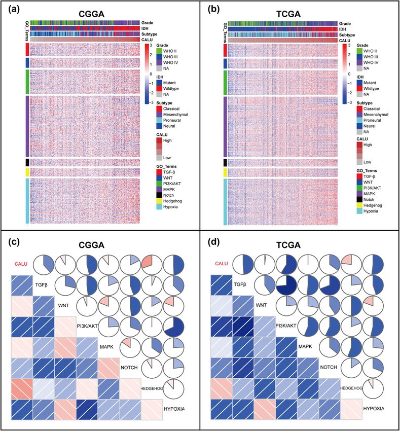
Role of CALU in glioma 69 Figure 1: CALU expression in CGGA and TCGA dataset according to WHO grade (a and d), IDH mutation status (b and e), TCGA molecular subtype (c and f). * indicates p value < 0.05, **indicates p value < 0.01, *** indicates p value < 0.001, **** indicates p value < 0.0001. accuracy of the analysis, we subsequently identified 3.3 CALU-related EMT signaling pathways 203 genes that overlapped between two independent cohorts, all of which were positively correlated with To get further understanding of the association between CALU (Table S2). Based on these genes, GO analysis CALU and EMT, seven gene sets, representing distinct EMT revealed that genes that significantly correlated with signaling pathways [28], were obtained from GSEA network CALU were highly enriched in a set of biological pro- (Table S3). Through cluster analyses, we identified 3 EMT cesses that correlated with EMT, including cell/biological signaling pathways (TGF-β, PI3K/AKT, and hypoxia), adhesion, response to wounding, extracellular matrix/ which might be strongly correlated with CALU (Figure 3a structure organization, collagen fibril organization, and and b). Moreover, seven gene sets were transformed into collagen biosynthetic process (Figure 2a and b). More- seven metagenes with GSVA analysis, which were subse- over, the association between CALU expression and quently put into Pearson correlation together with CALU. EMT was revealed by GSEA analysis. CALU expression According to Pearson r among seven metagenes and was found to be positively associated with the gene set CALU, Corrgrams were plotted to assess their interrela- of HALLMARK_EPITHELIAL_MESENCHYMAL_TRANSI- tionships. CALU was found to be positively correlated TION in both CGGA dataset (NES = 1.897, FDR = 0.035) with TGF-β, PI3K/AKT, and hypoxia, in line with what and TCGA dataset (NES = 1.818, FDR = 0.075) (Figure 2c we observed in clusters. However, only a very weak cor- and d). These findings suggested that CALU might be relation was revealed between CALU expression and four particularly involved in EMT process during glioma other pathways (WNT, MAPK, NOTCH, and HEDGEHOG), progression. which might be ascribed to signal noise (Figure 3c and d).
70 Ying Yang et al.
Figure 2: Functional enrichment of CALU in glioma. Gene Ontology analysis (a and b) and Gene set enrichment analysis (c and d).
3.4 CALU was synergistic with EMT key As shown in Figure 4a and b, CALU expression showed
markers high agreement with N-cadherin, snail, slug, and vimentin.
In contrast, a weak relationship between CALU and E-
Assuming that CALU played a vital role in regulating cadherin was found in Circos plots, which could be defined
glioma EMT, we investigated the association between as a noise. Heretofore, some other members, including
CALU and EMT markers, including N-cadherin, E-cad- TWIST1/2, β-catenin, and ZEB1/2, have been reported as
herin, snail, slug, and vimentin. Pearson correlation tests key markers in EMT [29]. Thus, we additionally put them
were performed with CALU and the above five EMT mar- into analysis together with CALU. CALU expression was
kers in both CGGA and TCGA. Circos plots were derived tightly associated with TWIST1 in both CGGA and TCGA
from Pearson r-values between CALU and five markers. datasets (Figure 4c and d).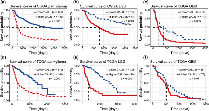
Role of CALU in glioma 71
Figure 3: Cluster (a and b) and GSVA (c and d) of CALU-related EMT signaling pathways in glioma.
3.5 Higher CALU was related to a worse as shown in Figure 5a and d. Moreover, glioma patients
prognosis were further divided into LGG and GBM subgroup. In both
subgroups, patients with higher CALU exhibited univer-
To evaluate the prognostic value of CALU in glioma, sally worse survival than those with relatively lower
Kaplan-Meier (KM) survival curves were plotted. In pan- CALU (Figure 5b, c, e and f), except for TCGA GBM, which
glioma samples, when comparing the two groups defined also showed an apparent trend. To identify the indepen-
by median CALU expression, we observed that higher dent effect of CALU on glioma prognosis, Cox regression
CALU expression predicted a significantly shorter survival, analyses were performed with covariates including CALU72 Ying Yang et al.
Figure 4: Correlation of CALU and EMT biomarkers in glioma (a and b, key biomarkers; c and d, other biomarkers).
expression, age, and WHO grade. Multivariate analyses subtype. Survival analyses revealed that higher CALU
revealed that CALU expression was a significant prognos- was related to a worse prognosis, independent of age
ticator independent of age and WHO grade in both CGGA and WHO grade. These results concordantly indicated
and TCGA (Table 1). that CALU might contribute to malignant progression of
glioma, which were in line with the results from a previous
GBM study [22]. Thus, unveiling the regulative mechanism
of CALU may facilitate to develop a novel gene for poten-
4 Discussion tial glioma diagnosis and treatment.
CALU is one of the members of CREC protein family.
We explored CALU expression at transcriptional level via This molecule family mainly consists of Cab45, Reticulocalbin
a cohort of 998 glioma samples and demonstrated that 1, ERC-55, and CALU and is characterized by multiple EF-
CALU expression was positively correlated with WHO hand motifs with low affinity of Ca2+-binding [30]. Under
grade. In addition, upregulation of CALU was usually par- normal physiological conditions, CALU primarily partici-
alleled with a more malignant and aggressive phenotype, pates in regulating Ca2+-dependent protein folding, sorting
such as IDH wildtype, classical subtype, and mesenchymal and maturation in the ER [31], Ca2+ homeostasis [32,33],Role of CALU in glioma 73
Figure 5: Survival analysis for CALU in pan-glioma (a and d), LGG (b and e), and GBM (c and f).
and muscle contraction/relaxation [34]. However, in tumor recurrence and resistance to therapy in glioma [5,6,36].
microenvironment, CALU was reported to play a critical role These findings suggested that CALU might facilitate the
in promoting a series of malignant phenotypes including malignant progression of glioma primarily via modulating
cancer cell survival [21], filopodia formation and cell migra- EMT process, which has not yet been reported previously.
tion [20], invasiveness [12], metastasis [15,35], cancer devel- Despite no report with regard to the pro-EMT effect of CALU,
opment [10], and resistance to chemotherapy [13]. So far, the other two members (Cab45 [37] and EFHD2 [38]) from
very little is known about the biological function of CALU in the same protein family have been described in EMT regu-
glioma. In the current study, GO analysis was performed to lation, which indirectly supported the potential role of
elucidate the biological function of CALU in glioma and it CALU in glioma EMT.
revealed that CALU showed high association with multiple We then chose a panel of EMT pathways and markers
EMT-related biological processes, including cell adhesion, and examined their interrelationships with CALU. CALU
biological adhesion, extracellular matrix/structure organi- was revealed to be highly associated with TGFβ, PI3K/
zation, collagen fibril organization, and collagen biosyn- AKT, as well as hypoxia pathway, indicating that CALU
thetic process. GSEA in both CGGA and TCGA further exhib- might regulate glioma EMT through these signaling path-
ited a remarkable relationship between CALU and EMT. ways. Furthermore, most of the EMT biomarkers showed
EMT has been extensively reported to act as a critical robust correlation with CALU, suggesting a synergistic
mechanism not only in invasiveness, but also in early effect among CALU and these members during EMT
Table 1: Cox regression analysis of overall survival in glioma
Covariates CGGA_301 TCGA
Univariate Multivariate Univariate Multivariate
HR (95% CI) P HR (95% CI) P HR (95% CI) P HR (95% CI) P
Age 1.041 (1.027–1.055) 0.000 1.017 (1.004–1.031) 0.010 1.075 (1.062–1.087) 0.000 1.046 (1.032–1.060) 0.000
Grade 2.670 (2.221–3.210) 0.000 2.292 (1.867–2.814) 0.000 5.057 (3.915–6.532) 0.000 3.033 (2.273–4.047) 0.000
CALU 2.123 (1.746–2.581) 0.000 1.300 (1.033–1.636) 0.025 2.159 (1.892–2.463) 0.000 1.295 (1.094–1.534) 0.00374 Ying Yang et al.
process. These findings further validated the potential [2] Yang W, Wu PF, Ma JX, Liao MJ, Wang XH, Xu LS, et al. Sortilin
role of CALU during EMT process in glioma. promotes glioblastoma invasion and mesenchymal transition
In conclusion, CALU was upregulated in more malig- through GSK-3β/β-catenin/twist pathway. Cell Death Dis.
2019;10(3):208.
nant gliomas and predicted much worse prognosis.
[3] Wei J, Ouyang X, Tang Y, Li H, Wang B, Ye Y, et al. ER-stressed
Furthermore, CALU seemed to be mainly involved in MSC displayed more effective immunomodulation in RA
EMT process of glioma, potentially through modulating CD4(+)CXCR5(+)ICOS(+) follicular helper-like T cells through
TGFβ, PI3K/AKT, and hypoxia pathway. However, limita- higher PGE2 binding with EP2/EP4. Mod Rheumatol.
tions still exist in this study. First, no biological validation 2019;30(3):509–16.
[4] Ma YS, Wu ZJ, Bai RZ, Dong H, Xie BX, Wu XH, et al. DRR1
was performed, which might compromise the robustness
promotes glioblastoma cell invasion and epithelial-mesench-
of results. Further researches focusing on in vivo/in vitro ymal transition via regulating AKT act ivation. Cancer Lett.
experimental studies are warranted. Second, despite the 2018;423:86–94.
large sample in our study, data from TCGA and CGGA [5] Li C, Zheng H, Hou W, Bao H, Xiong J, Che W, et al. Long non-
were mainly retrospectively collected, the control of coding RNA linc00645 promotes TGF-β-induced epithelial-
mesenchymal transition by regulating miR-205-3p-ZEB1 axis in
data quality was heterogeneous, and some data were
glioma. Cell Death Dis. 2019;10(10):717.
unavailable, which might lead to potential bias.
[6] Li H, Li J, Zhang G, Da Q, Chen L, Yu S, et al. HMGB1-induced
p62 overexpression promotes snail-mediated epithelial-
Acknowledgments: We appreciate the generosity of CGGA mesenchymal transition in gliobla stoma cells via the degra-
project and TCGA project for sharing data. dation of GSK-3β. Theranostics. 2019;9(7):1909–22.
[7] Sun H, Long S, Wu B, Liu J, Li G. NKCC1 involvement in the
epithelial-to-mesenchymal transition is a prognostic bio-
Funding: This work was supported by Medical Scientific
marker in gliomas. Peer J. 2020;8:e8787.
Research Foundation of Shenzhen Health Commission [8] Wu W, Tang X, Hu W, Lotan R, Hong WK, Mao L. Identification
(szfz2018022), Shenzhen Science and Technology Innova- and validation of metastasis-associated proteins in head and
tion Foundation (JCYJ20190806150005453), and Futian neck cancer cell lines by two-dimensional electrophoresis and
Public Welfare Scientific Research Project (FTWS2020099). mass spectrometry. Clin Exp Metastasis. 2002;19(4):319–26.
[9] Voisin SN, Krakovska O, Matta A, DeSouza LV, Romaschin AD,
Colgan TJ, et al. Identification of novel molecular targets for
Author contributions: Ying Yang performed the analysis
endometrial cancer using a drill-down LC-MS/MS approach
and wrote the manuscript. Shihai Xu, Fei Shi, and Aijun with iTRAQ. PLoS One. 2011;6(1):e16352.
Shan provided technical support and analyzed the data. [10] Nasri Nasrabadi P, Nayeri Z, Gharib E, Salmanipour R,
Jin Wang designed the study and reviewed the manu- Masoomi F, Mahjoubi F, et al. Establishment of a CALU,
script. Ying Yang and Shihai Xu provided financial sup- AURKA, and MCM2 gene panel for discrimination of metastasis
from primary colon and lung cancers. PLoS One.
port. All of the authors read and approved the final
2020;15(5):e0233717.
manuscript. [11] Torres S, Bartolome RA, Mendes M, Barderas R, Fernandez-
Acenero MJ, Pelaez-Garcia A, et al. Proteome profiling of
Conflict of interest: Authors state no conflict of interest. cancer-associated fibroblasts identifies novel proinflamma-
tory signatures and prognostic markers for colorectal cancer.
Data Availability Statement: The datasets analyzed Clin Cancer Res. 2013;19(21):6006–19.
[12] Nagano K, Imai S, Zhao X, Yamashita T, Yoshioka Y, Abe Y,
during the current study are available in the Chinese
et al. Identification and evaluation of metastasis-related pro-
Glioma Genome Atlas repository, http://www.cgga.org. teins, oxysterol binding protein-like 5 and calumenin, in lung
cn/, and in the The Cancer Genome Atlas repository, tumors. Int J Oncol. 2015;47(1):195–203.
http://cancergenome.nih.gov/. All data generated during [13] Tang H, Ma M, Dai J, Cui C, Si L, Sheng X, et al. miR-let-7b and
this study are included in this published article and its miR-let-7c suppress tumourigenesis of human mucosal mela-
noma and enhance the sensitivity to chemotherapy. J Exp Clin
supplementary information files.
Cancer Res. 2019;38(1):212.
[14] Wang Q, Shen B, Chen L, Zheng P, Feng H, Hao Q, et al.
Extracellular calumenin suppresses ERK1/2 signaling and cell
migration by protecting fibulin-1 from MMP-13-mediated pro-
teolysis. Oncogene. 2015;34(8):1006–18.
References [15] Kurpinska A, Suraj J, Bonar E, Zakrzewska A, Stojak M,
Sternak M, et al. Proteomic characterization of early lung
[1] Meng X, Zhao Y, Han B, Zha C, Zhang Y, Li Z, et al. Dual response to breast cancer metastasis in mice. Exp Mol Pathol.
functionalized brain-targeting nanoinhibitors restrain temo- 2019;107:129–40.
zolomide-resistant glioma via attenuating EGFR and MET sig- [16] Voora D, Koboldt DC, King CR, Lenzini PA, Eby CS, Porche-
naling pathways. Nat Commun. 2020;11(1):594. Sorbet R, et al. A polymorphism in the VKORC1 regulatorRole of CALU in glioma 75
calumenin predicts higher warfarin dose requirements in expression profiles. Proc Natl Acad Sci USA.
African Americans. Clin Pharmacol Ther. 2010;87(4):445–51. 2005;102(43):15545–50.
[17] Wang Y, Cui X, Wang Y, Fu Y, Guo X, Long J, et al. Protective [27] Hanzelmann S, Castelo R, Guinney J. GSVA: Gene set variation
effect of miR378* on doxorubicin-induced cardiomyocyte analysis for microarray and RNA-seq data. BMC Bioinforma.
injury via calumenin. J Cell Physiol. 2018;233(10):6344–51. 2013;14:7.
[18] Wang Y, Sun Y, Fu Y, Guo X, Long J, Xuan LY, et al. Calumenin [28] Gonzalez DM, Medici D. Signaling mechanisms of the epithe-
relieves cardiac injury by inhibiting ERS-initiated apoptosis lial-mesenchymal transition. Sci Signal. 2014;7(344):re8.
during viral myocarditis. Int J Clin Exp Pathol. [29] Xu J, Zhang Z, Qian M, Wang S, Qiu W, Chen Z, et al. Cullin-7
2017;10(7):7277–84. (CUL7) is overexpressed in glioma cells and promotes tumor-
[19] Zhao L, Wang Y, Shao L, Gu J, Long J, Zhao M, Calumenin DNA. igenesis via NF-κB activation. J Exp Clin Cancer Res.
Methylation and gene expression in viral myocarditis. Int J Clin 2020;39(1):59.
Exp Pathol. 2018;11(2):808–12. [30] Mazzorana M, Hussain R, Sorensen T. Ca-dependent folding of
[20] Feng H, Chen L, Wang Q, Shen B, Liu L, Zheng P, et al. human calumenin. PLoS One. 2016;11(3):e0151547.
Calumenin-15 facilitates filopodia formation by promoting TGF- [31] Suzuki N, Ban S, Itoh E, Chen S, Imai FL, Sawano Y, et al.
beta superfamily cytokine GDF-15 transcription. Cell Death Dis. Calcium-dependent structural changes in human reticulo-
2013;4:e870. calbin-1. J Biochem. 2014;155(5):281–93.
[21] Ren Y, Yeoh KW, Hao P, Kon OL, Sze SK. Irradiation of epithelial [32] Sahoo SK, Kim DH. Calumenin interacts with SERCA2 in rat
carcinoma cells upregulates calcium-binding proteins that cardiac sarcoplasmic reticulum. Mol Cell. 2008;26(3):265–9.
promote survival under hypoxic conditions. J Proteome Res. [33] Jung DH, Mo SH, Kim DH. Calumenin, a multiple EF-hands Ca2+-
2016;15(12):4258–64. binding protein, interacts with ryanodine receptor-1 in rabbit
[22] Sreekanthreddy P, Srinivasan H, Kumar DM, Nijaguna MB, skeletal sarcoplasmic reticulum. Biochem Biophys Res
Sridevi S, Vrinda M, et al. Identification of potential serum Commun. 2006;343(1):34–42.
biomarkers of glioblastoma: Serum osteopontin levels corre- [34] Sahoo SK, Kim DH. Characterization of calumenin in mouse
late with poor prognosis. Cancer Epidemiol Biomarkers Prev. heart. BMB Rep. 2010;43(3):158–63.
2010;19(6):1409–22. [35] Zheng P, Wang Q, Teng J, Chen J. Calumenin and fibulin-1 on
[23] Robin X, Turck N, Hainard A, Tiberti N, Lisacek F, Sanchez JC, tumor metastasis: Implications for pharmacology. Pharmacol
et al. pROC: An open-source package for R and S+ to Res. 2015;99:11–5.
analyze and compare ROC curves. BMC Bioinforma. [36] Li H, Li J, Chen L, Qi S, Yu S, Weng Z, et al. HERC3-mediated
2011;12:77. SMAD7 ubiquitination degradation promotes autophagy-
[24] Gu Z, Gu L, Eils R, Schlesner M, Brors B. Circlize Implements induced EMT and chemoresistance in glioblastoma. Clin
and enhances circular visualization in R. Bioinformatics. Cancer Res. 2019;25(12):3602–16.
2014;30(19):2811–2. [37] Luo J, Li Z, Zhu H, Wang C, Zheng W, He Y, et al. A novel role of
[25] Huang da W, Sherman BT, Lempicki RA. Systematic and inte- Cab45-G in mediating cell migration in cancer cells. Int J Biol
grative analysis of large gene lists using DAVID bioinformatics Sci. 2016;12(6):677–87.
resources. Nat Protoc. 2009;4(1):44–57. [38] Fan CC, Cheng WC, Huang YC, Sher YP, Liou NJ, Chien YC, et al.
[26] Subramanian A, Tamayo P, Mootha VK, Mukherjee S, Ebert BL, EFHD2 promotes epithelial-to-mesenchymal transition and
Gillette MA, et al. Gene set enrichment analysis: A correlates with postsurgical recurrence of stage I lung ade-
knowledge-based approach for interpreting genome-wide nocarcinoma. Sci Rep. 2017;7(1):14617.You can also read