Transgenic expression of epidermal growth factor and keratinocyte growth factor in -cells results in substantial morphological changes
←
→
Page content transcription
If your browser does not render page correctly, please read the page content below
167

Transgenic expression of epidermal growth factor and keratinocyte
growth factor in â-cells results in substantial morphological changes
M L Krakowski, M R Kritzik, E M Jones, T Krahl, J Lee,
M Arnush, D Gu, B Mroczkowski and N Sarvetnick
Department of Immunology, The Scripps Research Institute, 10550 North Torrey Pines Road, La Jolla, California 92037, USA
(Requests for offprints should be addressed to N Sarvetnick)

Abstract
The upregulation of a limited number of growth factors in                      profound changes in pancreatic morphology including
our interferon-ã transgenic model for regeneration within                      proliferation of pancreatic cells and extensive intra-islet
the pancreas lead us to propose that these factors are                         fibrosis. Insulin-producing â-cells were found in some of
important during pancreatic regeneration. In this study,                       the ducts of older Ins-EGF and Ins-EGFKGF trans-
we have assessed the influence of two growth factors                           genic mice, and amylase-producing cells were observed
within the pancreas, epidermal growth factor (EGF) and                         within the islet structures of the double transgenic mice.
keratinocyte growth factor (KGF), by ectopically express-                      These data suggest that both EGF and KGF are capable of
ing these proteins under the control of the human insulin                      affecting pancreatic differentiation and growth, and that
promoter in transgenic mice. This â-cell-targeted expres-                      co-expression of these molecules in islets has a more
sion of either EGF or KGF resulted in significant                              substantial impact on the pancreas than does expression of
morphological changes, including cellular proliferation and                    either growth factor alone.
disorganized islet growth. Intercrossing the individual                        Journal of Endocrinology (1999) 162, 167–175
Ins-EGF and Ins-KGF transgenic mice resulted in more

Introduction                                                                      The growth factors required for pancreatic differentia-
                                                                               tion are not yet fully defined. A survey of potentially
The healthy adult pancreas is a developmentally stable                         critical growth factors, including fibroblast growth
organ with limited mitotic activity. Models allowing the                       factor-2, recombinant human hepatocyte growth factor,
study of pancreatic growth and differentiation are of                          insulin-like growth factor-II, platelet-derived growth fac-
particular interest because the elimination or dysfunction                     tor, and nerve growth factor did not identify any factors
of pancreatic cells is associated with a number of disease                     that could influence the growth of pancreatic bud cultures
states. In the human autoimmune disease insulin-                               in vitro. However, EGF and transforming growth factor
dependent diabetes mellitus, there is selective and                            â-1 (TGFâ-1) were able to induce ductal and endocrine
permanent destruction of the insulin-producing â-cells in                      cell development respectively (Sanvito et al. 1994). While
the islets of Langerhans. Pancreatic disorders also affect the                 the ability of EGF to stimulate epithelial cell and fibroblast
duct system, as is observed in chronic pancreatitis and                        proliferation is well documented, it has also demonstrated
cystic fibrosis (Lack 1989, Sessa et al. 1990, Hootman & de                    mitogenic properties for pancreatic growth (Dembinski
Ondarza 1993). It appears from these examples of disease                       et al. 1982, Logsdon 1986, 1987, Marti et al. 1989, Verme
that the lost cells are not restored in vivo. Nevertheless, the                & Hootman 1990, Mangino et al. 1992). In addition,
potential for such cells to regenerate has been shown                          evidence exists linking the overexpression of EGF and its
experimentally (Sarvetnick et al. 1988, Brockenbrough                          receptor to both chronic pancreatitis and malignant pan-
et al. 1988, Rosenberg & Vinik 1989, Bonner-Weir et al.                        creatic growth (Barton et al. 1991, Korc et al. 1992, 1994,
1993, Wang et al. 1993, Wang & Bouwens 1995). We                               Yamanaka et al. 1993, Friess et al. 1996). Indeed, over-
have shown that the ectopic expression of interferon-ã                         expression of this growth factor could potentially confer a
(IFN-ã) in the mouse results in regeneration within the                        direct growth advantage to pancreatic cancer cells (Korc
pancreas (Gu & Sarvetnick 1993). Further investigation                         1998).
revealed that the expression of the growth factor epidermal                       The highly related member of the fibroblast growth
growth factor (EGF) was upregulated in the islets of these                     factor family, keratinocyte growth factor (KGF), has also
transgenic mice (Arnush et al. 1996).                                          been shown to stimulate the proliferation of pancreatic

Journal of Endocrinology (1999) 162, 167–175                                                              Online version via http://www.endocrinology.org
0022–0795/99/0162–167  1999 Society for Endocrinology Printed in Great Britain

                                                                                                           Downloaded from Bioscientifica.com at 03/14/2020 11:40:05AM
                                                                                                                                                         via free access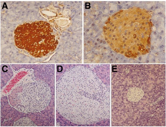
168 M L KRAKOWSKI and others · Expression of EGF and KGF in the pancreas
ductal epithelial cells in rats after daily systemic injection Use Committee. Mice were housed under a controlled
for 1–2 weeks (Yi et al. 1994). KGF is also involved in 12 h light: 12 h darkness cycle and provided with food and
wound healing (Werner et al. 1992) and in the differen- water ad libitum.
tiation of many epithelial tissues (Aaronson et al. 1990,
Alarid et al. 1994). Additionally, KGF is known to
profoundly upregulate epidermal cell proliferation
Histological analysis and immunocytochemistry
(Aaronson et al. 1991). More specifically, we have pre-
viously observed that ectopic expression of KGF under the Pancreata were fixed overnight in 10% neutral buffered
control of the insulin promoter initiated significant intra- formalin (3·6% formaldehyde) and embedded in paraffin.
islet proliferation of ductal cells in the pancreata of Paraffin sections (5 µm) were either conventionally stained
transgenic mice (Krakowski et al. 1999). with hematoxylin and eosin (H&E) for histological evalu-
For our IFN-ã transgenic model, a screen for factors ation or stained for the presence of insulin, glucagon,
potentially involved in the regenerating phenotype yielded somatostatin, amylase, EGF, EGF receptor or bromo-
both EGF and KGF. Therefore, we chose to study how deoxyuridine (BrdU) using immunocytochemical tech-
these two factors might be involved by generating trans- niques (see Gu & Sarvetnick 1993, Arnush et al. 1996).
genic mice. In this study, we have investigated the Sections were deparaffinized and blocked with 10% nor-
influence of these growth factors on pancreatic differen- mal goat serum before applying the primary antibodies for
tiation by generating transgenic mice which overexpress insulin, glucagon, amylase or somatostatin (all from
both EGF and KGF in the â-cells of the islets of DAKO, Carpentaria, CA, USA), or BrdU (Accurate/
Langerhans. Here we report that co-expression of these Sera-Lab, Westbury, NY, USA). Binding of the primary
growth factors acted in concert to produce striking mor- antibody was detected using the appropriate secondary
phological changes in the pancreas, without causing any antibody (Vector Laboratories, Burlingame, CA, USA or
apparent abnormalities in pancreatic function. Boehringer-Mannheim, Indianapolis, IN, USA), and the
horseradish peroxidase (HRP)-labeled avidin-biotin
complex (ABC kit; Vector Laboratories). HRP was
Materials and Methods visualized using 3,3 -diaminobenzidine as a substrate.
Gill’s hematoxylin was used as a counterstain. Masson’s
Transgenic mouse generation trichrome staining was completed at the Scripps Research
Institute Department of Histology. Briefly, paraffin-
The 280 bp EGF cDNA was used to generate the embedded sections were fixed in Bouin’s fixative, stained
Ins-EGF transgenic mouse. The cDNA was cloned into a successively with Weigert’s iron hematoxylin and with
vector containing the human insulin promoter and the Biebrich scarlet-acid fuchsin acid then counterstained with
hepatitis B 3 -untranslated sequence. The Ins-EGF frag- aniline blue. This results in collagen and mucin staining
ment was isolated by low melt agarose, purified using blue indicating the presence of fibrosis while cytoplasm
Geneclean (BIO101 Inc., La Jolla, CA, USA) and NACS stains red. Islet size was measured as in Krakowski et al.
Prepac DNA purification columns (BRL, Gaithersburg, (1999). Briefly, anti-insulin-stained sections from mice
MD, USA), and microinjected into fertilized zygotes from of various ages were examined at 10power (Zeiss
(BALB/cC57BL/6) F2 mice. Progeny were screened Axioscope), using the 100 µm size of the crosshairs
for the presence of the transgene by PCR typing of tail for comparison. Islets were scored as small (400 µm). Sixteen
overnight. PCR was performed using two 24-mer primers Ins-KGF, sixteen Ins-EGF, eleven Ins-EGFKGF and
specific for the human insulin promoter and EGF se- twelve non-transgenic mice, including all ages, were
quence. Transgene-positive mice were further bred with examined. Statistical analyses of both young (3 months) mice for mean islet size (...)
analogous and has been described elsewhere (Krakowski were compared using the program Statview as described
et al. 1999). Once both lines had been backcrossed to below.
BALB/c mice for four generations, individual homozygote
Ins-EGF and Ins-KGF mice were mated and progeny
interbred to create homozygous, double transgenic
(Ins-EGFIns-KGF) mice. Blood glucose measurement
At regular intervals (approximately every 2 weeks), blood
was obtained from the tail and blood glucose levels were
Animal husbandry
determined using Glucofilm blood glucose test strips
All mice were maintained in a specific pathogen-free (Miles Diagnostic, Elkhart, IN, USA). Non-fasting blood
facility at the Scripps Research Institute according to the glucose values of non-transgenic BALB/c mice in our
rules and regulations of the Institutional Animal Care and colony ranged from 80 to 160 mg/dl.
Journal of Endocrinology (1999) 162, 167–175
Downloaded from Bioscientifica.com at 03/14/2020 11:40:05AM
via free access
Expression of EGF and KGF in the pancreas · M L KRAKOWSKI and others 169
Glucose tolerance testing islets (Fig. 1A). As expected, EGF was expressed at low
levels in the islets of non-transgenic littermate controls
To determine glucose tolerance over time, transgenic and
(Fig. 1B). Furthermore, while the level of the EGF
control littermate mice were fasted overnight and an initial
receptor (EGF-R) appeared comparable to the low con-
blood glucose reading taken (time zero) as described
stitutive level of expression previously observed for this
above. Mice were then injected intraperitoneally with
receptor (Damjanov et al. 1986, Chabot et al. 1987), we
0·2 g -glucose/100 g body weight (0·1 g/ml in sterile
did detect a modest upregulation of the EGF-R in the
PBS), and blood glucose measured after 10 min and again
ductal epithelia of the transgenic pancreas (data not
after 60 min.
shown).
BrdU labeling
Morphological changes of Ins-EGF mice
Cellular proliferation was quantified by injecting 100 µg/g
Ins-EGF transgenic mice had dramatic morphological
body weight BrdU (Serva, Heidelberg, Germany) intra-
changes within their pancreata. While the pancreatic ducts
peritoneally into mice 15 h prior to their being killed.
appeared normal in the transgenic mice, the islets were
Paraffin-embedded pancreata were sectioned and stained
found to increase in size as the mice aged. After 3 months
with an anti-BrdU antibody (Accurate Chemical,
of age, these mice had significantly more islets of larger size
Westbury, NY, USA) as described above after treatment
(>400 µm in diameter) than did their non-transgenic
with 2·8 M HCl for 15 min. The extent of proliferation
littermates, in which most islets were170 M L KRAKOWSKI and others · Expression of EGF and KGF in the pancreas
Figure 1 Overexpression of EGF within islets of transgenic Ins-EGF mice leads to disorganization of islet architecture and increases with
age. Three-month-old Ins-EGF (A) and age- and sex-matched non-transgenic control littermate (B) both stained with anti-EGF antibody as
shown in brown chromagen staining against blue hematoxylin counterstain. Three-month-old (C) and 17-month-old (D) Ins-EGF mice
stained with H&E, revealing rare and sparse mononuclear cell infiltration to the irregularly shaped islets that increased in size and
complexity with age. Age- and sex-matched non-transgenic control littermate stained with H&E (E), demonstrating typical islet morphology
and no immune infiltration. Original magnification of A: 20 and B: 32, C–E: 20.
resulted in enlarged islets, with substantial proliferation of differentiation and function, Ins-EGF mice were crossed
duct cells within the islet mass. In addition, we observed to Ins-KGF mice, and Ins-EGFKGF progeny were
the presence of hepatocyte-like (albumin and á- interbred to homozygosity for both transgenes. Charac-
fetoprotein-producing) cells in the islets of the KGF- terization of these double transgenic mice revealed unusual
expressing transgenic mice (Krakowski et al. 1999). These changes in pancreatic morphology which did not simply
pancreatic heptatocytes are easily recognizable as the reflect the effects of each individual transgene.
non-insulin-producing cells that are extremely large in size Ins-EGFKGF mice developed enlarged, distended,
compared with the other islet cells. Additionally, they are and non-confluent islets at earlier ages than was seen for
found on the periphery of the islet and are often binucleate either the single Ins-EGF or Ins-KGF transgenic mice.
(Fig. 2F, see arrowheads). Despite the morphological Additionally, all of the transgenic mice, aged 3 months or
differences in the pancreata of KGF transgenic mice, no greater, had significantly more islets of larger size
pathology, hyperglycemia, or hypoglycemia were found to (>400 µm diameter) (Fig. 2A–F) than did non-transgenic
be associated with KGF expression in the islets of trans- mice (Fig. 2G and H), in which most islets wereExpression of EGF and KGF in the pancreas · M L KRAKOWSKI and others 171
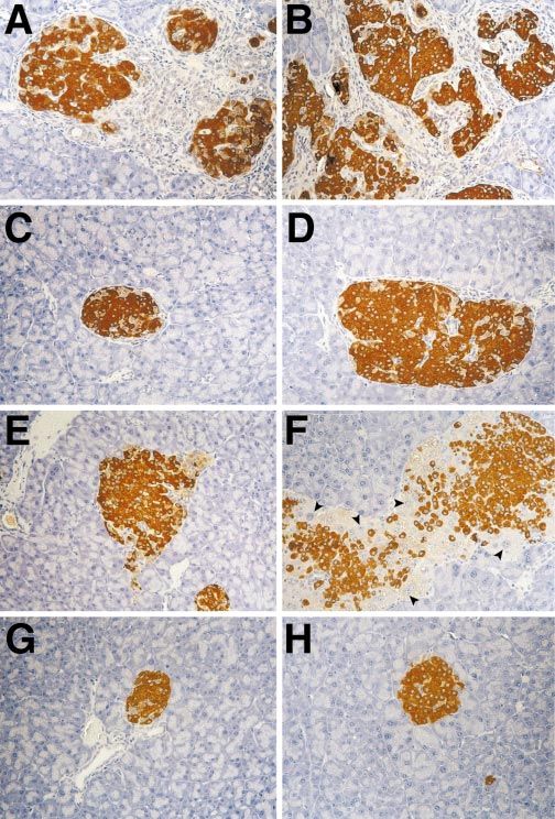
Figure 2 Morphological changes occur at an earlier age in Ins-EGFKGF mice, and are more severe than
in single transgenic mice. Three-month-old (A) and 6-month-old (B) Ins-EGFKGF mice stained with
anti-insulin antibody (shown in brown) and counterstained with blue hematoxylin showing increasingly
larger, and more disorganized networks of islets. Compare the effects of increasing age from 3 (left panel)
to 6 months (right panel) on islet of Langerhans morphology in C, D: Ins-EGF, E, F: Ins-KGF and G, H:
non-transgenic (all age-matched) demonstrating delayed and lesser phenotypic changes in single transgenic
mice and normal morphology in non-transgenic controls. Arrowheads in panel F indicate pancreatic
hepatocytes within lns-KGF mouse. Original magnification for all photos was 20.
Journal of Endocrinology (1999) 162, 167–175
Downloaded from Bioscientifica.com at 03/14/2020 11:40:05AM
via free access172 M L KRAKOWSKI and others · Expression of EGF and KGF in the pancreas
Figure 3 Ins-EGFKGF mice have intra-islet ducts, ductal insulin-positive cells, fibrosis and amylase-positive cells within islets of
Langerhans. Seventeen-month-old female Ins-EGF mouse stained as in Fig. 2 shows insulin-producing cells within the duct wall (A).
Intra-islet duct (designated D’) identified by CAII staining in a 5·5-month-old male mouse (B). Trichrome staining reveals a severe degree
of fibrosis within the Ins-EGFKGF transgenic mice (4-month-old male) (D) in contrast to the non-transgenic age- and sex-matched
littermate (C). Fibrosis within the islets appears as bright blue spindle shaped cells with elongated nuclei, and increases with age (data not
shown). Anti-amylase antibody stained with brown chromogen against a blue hematoxylin counterstain reveals atypical endocrine as well
as the expected exocrine staining within the pancreata of Ins-EGFKGF mice. (E) Early stages of engulfment of acinar tissue by ongoing
fibrotic reaction occurring around the islet. (F) Acinar cells staining positive for amylase surrounded by islet tissue and fibrotic cells. (G)
Amylase-positive cells without distinct acinar cell morphology within islet tissue. Original magnification of A: 40, B: 20, C and D:
40, E: 20 and F and G: 40.
Ins-KGF 2·70·6 (n=8), Ins-EGF 3·30·6 (n=13), ducts of the Ins-EGFKGF transgenic mice at a later age
Ins-EGFKGF 5·50·7 (n=4) and non-transgenic (6 months) (data not shown).
0·90·4 (n=8). In this regard, the double transgenic mice Normal, non-fasting blood glucose levels were
(Fig. 2A and B) had more islets of larger size than did the measured over the animal’s lifespan (90–150 mg/dl). Yet
Ins-KGF transgenic mice (PExpression of EGF and KGF in the pancreas · M L KRAKOWSKI and others 173
Table 1 Typical pattern of fluctuations in blood glucose. Values are double transgenic mice; such positive staining was not
means S.E.M. observed within the islets of either single transgenic mouse
Blood glucose (mg/dl)
or non-transgenic control. Figure 3E–G illustrates the
morphology of amylase-positive islet sections, in what we
Transgene 0 min 10 min 60 min believe to represent progressive stages in the evolution of
Ins-KGF (n=3) 673 29694 11629 this phenomenon. That is, it appears that amylase-positive
Ins-EGFKGF (n=4) 11913 348105 12216 cells are engulfed by the ongoing fibrotic reaction occur-
Ins-EGF (n=3) 1087 46632 141109 ring adjacent to islets (Fig. 3E-G), perhaps due to dis-
BALB/c (n=4) 10913 20526 12812 rupted matrix connections or altered adhesion between
cells, after which the membranes of the engulfed cells
Fasting blood glucose concentration (mg/dl) in all three transgenic and break down to leave a residual fragment of exocrine tissue,
control mice during an intraperitoneal glucose tolerance test of male
mice of various ages (range between 3 and 14·5 months) Data are given which stains positive for amylase (Fig. 3G). The presence
as the mean and standard error for each type of transgenic. Blood for of these exocrine cells within the endocrine (islet) tissue is
determination of the blood glucose was taken from the retro-orbital venous
plexus during anaesthesia.
at least partially responsible for the non-confluent insulin
staining we observed in the islets of double transgenic
mice, as the cells which produce amylase likely displace
per section at 20magnification. This measurement was the insulin-producing cells.
representative of the number of BrdU-positive cells per
pancreatic slice. On average, we found that Ins-EGF and
Ins-KGF mice had 6·82·0 (n=5 mice) and 11·03·8 Discussion
(n=6 mice) BrdU-positive cells respectively. In contrast,
the double transgenic mice had 15·33·1 (n=8 mice) and We have generated a model system in which the expres-
the non-transgenic littermates had 0·70·3 (n=6 mice) sion of KGF and EGF has been targeted to the â-cells in
positive cells. Thus all three lines of transgenic mice had the islets of Langerhans. KGF and EGF are members of a
significantly more pancreatic cell proliferation than did the large family of very similar growth factors. Members of this
control mice (P174 M L KRAKOWSKI and others · Expression of EGF and KGF in the pancreas
â-cells generates an accelerated and extensive series of several uncharacteristic cell types exist in the transgenic
changes to both endocrine and exocrine tissues. For mice overexpressing EGF and KGF in islets also suggests a
example, we have observed significant intra-islet duct cell possible role for these growth factors in pancreatic
proliferation in the pancreata of Ins-KGF mice. In differentiation. We have detected the presence of
addition, we have found that hepatocyte cells exist within insulin-producing cells in the ducts of the Ins-EGF and
the islets of these transgenic mice. Pancreatic cell prolifer- Ins-EGFKGF transgenic mice and of hepatocyte-like
ation was also observed in the Ins-EGF and Ins-EGF cells in the islets of KGF transgenic mice. Thus, EGF and
KGF mice. The Ins-EGF and Ins-EGFKGF transgenic KGF might contribute to pancreatic differentiation by
mice also exhibited disorganized islets and intra-islet promoting cellular lineage commitment along specific
fibrosis as well, both of which were more extensive in the pathways. However, as in any model system, it is possible
double transgenic mouse. We also found that many that the phenotypes exhibited by our transgenic mouse
features shared by both single transgenic mice, such as models are due to targeted overexpression in islets and are
pancreatic cell proliferation, increased size and disorgan- not reflective of innate physiological influences. Future
ization of islets, and fibrosis, were seen to a greater extent studies designed to assess the influence of these growth
in the double transgenic mice. Interestingly, the amylase- factors on pancreatic growth and differentiation will
positive cells seen in the double transgenic mice are not therefore be important in addressing these critical issues.
found in either of the single transgenic mice, indicating In summary, we have developed a transgenic mouse
that localized overexpression of both EGF and KGF in system which will enable us to study the effects of EGF
â-cells is required to produce this unique phenotype. and KGF on pancreatic growth, differentiation, and on the
Despite the extensive morphological changes that are pathologies associated with aberrant overexpression of
observed in the pancreata of growth factor transgenic these growth factors. In addition, the transgenic mice we
mice, only very minor deficiencies in pancreatic function have produced will enable us to study the generation
were detected. All of the endocrine hormones and exo- of the distinct cell types described here, such as the
crine enzymes were present in these transgenic mice, and hepatocyte-like cells in the Ins-KGF mice and the ductal-
blood glucose levels remained normal throughout the lives endocrine cells in the Ins-EGF and Ins-EGFKGF mice.
of the animals. Thus the physical changes apparent in these As such, these studies will enhance our understanding of
mice did not interfere with normal pancreatic function. how these critical growth modulators contribute to the
Interestingly, some of the pathologies observed also char- growth and differentiation of the pancreas.
acterize several pancreatic diseases. For example, the GK
rat model of non-insulin-dependent diabetes is character- Acknowledgements
ized by disorganized islets, significant fibrosis, and clusters
of â-cells separated by strands of connective tissue The authors would like to heartily thank Gail Patson and
(Movassat et al. 1995). In addition, chronic pancreatitis is Augusta Good who maintained and screened the mouse
characterized by inflammation and fibrosis (Steer 1989). colony. We would also like to thank Margaret A Chadwell
Interestingly, in chronic pancreatitis and in some human for completing the Trichome stains. The administrative
pancreatic cancers, EGF, EGF-R, and KGF are often assistance of Joanne Dodge and Jackie Soto is always
overexpressed (Barton et al. 1991, Korc et al. 1992, 1994, gratefully appreciated. Drs Marc Horwitz and Malin
Yamanaka et al. 1993, Siddiqi et al. 1995, Friess et al. Flodstrom are thanked for providing provocative scientific
1996). These observations suggest that these factors might discussion.
play a role in disease progression and pathology. For Grant numbers and sources of support: M L K is
instance, it is possible that altered expression of these supported by an NMSS postdoctoral fellowship, E M J was
critical growth modulators could confer a significant supported by NIH postdoctoral fellowship DK09355–01,
growth advantage to pancreatic cancer cells. D G was supported by a postdoctoral fellowship from the
The physiological roles of KGF and EGF in pancreatic Juvenile Diabetes Foundation, and N S is supported by a
development are not clear. Indeed, KGF knockout mice Diabetes Interdisciplinary Research Center from the
do not appear to have any abnormalities in pancreatic Juvenile Diabetes Foundation and by NIH grant
development or function (Guo et al. 1996). However, HD-29764 and JDFI 995010. This is publication
there is likely to be redundancy within the large family of 11669-IMM from the Department of Immunology, the
highly related proteins to which these proteins belong, Scripps Research Institute.
perhaps masking the contribution of individual factors
during development. Such influences would be expected
to become more apparent when overexpression of these References
proteins is localized, perhaps manifest in the phenotypes
Aaronson SA, Rubin JS, Finch PW, Wong J, Marchese C, Falco J,
we observe. Indeed, as discussed earlier, these growth Taylor WG & Kraus MH 1990 Growth factor-regulated pathways
factors do appear to influence pancreatic growth and in epithelial cell proliferation. American Review of Respiratory Diseases
differentiation, in vivo and in vitro. The observation that 142 S7–S10.
Journal of Endocrinology (1999) 162, 167–175
Downloaded from Bioscientifica.com at 03/14/2020 11:40:05AM
via free accessExpression of EGF and KGF in the pancreas · M L KRAKOWSKI and others 175
Aaronson S, Bottaro D, Miki T, Ron D, Finch P, Fleming T, Ahn J, Mangino M, Hubchak S & Scarpelli D 1992 Stimulation of DNA
Taylor W & Rubin J 1991 Keratinocyte growth factor. A fibroblast synthesis in pancreatic duct cells by gastrointestinal hormones:
growth factor family member with unusual target cell specificity. interaction with other growth factors. Pancreas 7 271–279.
Annals of the New York Academy of Sciences 638 62–77. Marti U, Burwen S & Jones A 1989 Biological effects of epidermal
Alarid ET, Rubin JS, Young P, Chedid M, Ron D, Aaronson SA & growth factor, with emphasis on the gastrointestinal tract and liver:
Cunha GR 1994 Keratinocyte growth factor functions in epithelial an update. Hepatology 9 126–138.
induction during seminal vesicle development. Proceedings of the Movassat J, Saulnier C & Portha B 1995 Beta-cell mass depletion
National Academy of Sciences of the USA 91 1074–1078. precedes the onset of hyperglycaemia in the GK rat, a genetic
Arnush M, Gu D, Baugh C, Sawyer SP, Davis D, Mroczkowski B, model of non-insulin diabetes mellitus. Diabetes/Metabolism Reviews
Krahl T & Sarvetnick N 1996 Growth factors in the regenerating 21 365–370.
pancreas of gamma-interferon transgenic mice. Laboratory Rosenberg L & Vinik A 1989 Induction of endocrine cell
Investigation 74 985–990. differentiation: a new approach to management of diabetes. Journal
Barton C, Hall P, Hughes C, Gullick W & Lemoine N 1991 of Laboratory and Clinical Medicine 114 75–83.
Transforming growth factor alpha and epidermal growth factor in Sanvito F, Herrera P-L, Huarte J, Nichols A, Montesano R, Orci L &
human pancreatic cancer. Journal of Pathology 163 111–116. Vassalli J-D 1994 TGF-b1 influences the relative development of
Bonner-Weir S, Baxter L, Schuppin G & Smith F 1993 A second the exocrine and endocrine pancreas in vitro. Development 120
pathway for regeneration of adult exocrine and endocrine pancreas: 3451–3462.
a possible recapitulation of embryonic development. Diabetes 42
1715–1720. Sarvetnick N, Liggitt D, Pitts SL, Hansen SE & Stewart TA 1988
Brockenbrough J, Weir G & Bonner-Weir S 1988 Discordance of Insulin-dependent diabetes mellitus induced in transgenic mice by
exocrine and endocrine growth after 90% pancreatectomy in rats. ectopic expression of class II MHC and interferon-gamma. Cell 52
Diabetes 37 232–236. 773–782.
Brown KD 1995 The epidermal growth factor/transforming growth Sessa F, Bonato M, Frigerio B, Capella C, Solcia E, Prat M, Bara J &
factor-alpha family and their receptors. European Journal of Samloff I 1990 Ductal cancers of the pancreas frequently express
Gastroenterology and Hepatology 7 914–922. markers of gastrointestinal epithelial cells. Gastroenterology 98
Chabot J, Walker P & Pelletier G 1987 Demonstration of epidermal 1655–1665.
growth factor binding sites in the adult rat pancreas by light Siddiqi I, Funatomi H, Kobrin MS, Friess H, Buchler MW & Korc M
microscopic autoradiography. Pancreas 2 653–657. 1995 Increased expression of keratinocyte growth factor in human
Damjanov I, Mildner B & Knowles B 1986 Immunohistochemical pancreatic cancer. Biochemical and Biophysical Research Communications
localisation of the epidermal growth factor receptor in normal 215 309–315.
human tissues. Laboratory Investigation 55 588–592. Steer ML 1989 Classification and pathogenesis of pancreatitis. Surgical
Dembinski A, Gregory H, Konturek S & Polanski M 1982 Trophic Clinics of North America 69 467–480.
action of epidermal growth factor on the pancreas and Verme TB & Hootman SR 1990 Regulation of pancreatic duct
gastroduodenal mucosa in rats. Journal of Physiology 325 35–42. epithelial growth in vitro. American Journal of Physiology 258
Friess H, Berberat P, Schilling M, Kunz J, Korc M & Buchler M G833–G840.
1996 Pancreatic cancer: the potential clinical relevance of alterations Vinter-Jensen L, Juhl CO, Teglbjaerg PS, Poulsen SS, Dajani EZ &
in growth factors and their receptors. Journal of Molecular Medicine 74 Nexo E 1997 Systemic treatment with epidermal growth factor in
35–42. pigs induces ductal proliferations in the pancreas. Gastroenterology
Gu D & Sarvetnick N 1993 Epithelial cell proliferation and islet 113 1367–1374.
neogenesis in IFN-ã transgenic mice. Development 118 33–46.
Guo L, Degenstein L & Fuchs E 1996 Keratinocyte growth factor is Wang R & Bouwens L 1995 Duct- to islet-cell differentiation and
required for hair development but not for wound healing. Genes islet growth in the pancreas of duct-ligated adult rats. Diabetologia
and Development 10 165–175. 38 1405–1411.
Hootman SR & de Ondarza J 1993 Overview of pancreatic duct Wang T, Bonner-Weir S, Oates P, Chulak M & Simon B 1993
physiology and pathophysiology. Digestion 54 323–330. Pancreatic gastrin stimulates islet differentiation of transforming
Korc M 1998 Role of growth factors in pancreatic cancer. Surgical growth factor a-induced ductular precursor cells. Journal of Clinical
Oncology Clinics of North America 7 25–41. Investigation 92 1349–1356.
Korc M, Chandrasekar B, Yamanaka Y, Friess H, Buchier M & Werner S, Peters K, Longaker M, Fuller-Pace F, Banda M &
Berger H 1992 Overexpression of the epidermal growth factor Williams L 1992 Large induction of keratinocyte growth factor
receptor in human pancreatic cancer is associated with concomitant expression in the dermis during wound healing. Proceedings of the
increases in the levels of epidermal growth factor and transforming National Academy of Sciences of the USA 89 6896–6900.
growth factor alpha. Journal of Clinical Investigation 90 1352–1360. Wogensen L, Ma Y-H, Grodsky G, Robertson R, Burton F, Sutcliffe
Korc M, Friess H, Yamanaka Y, Kobrin M, Buchler M & Beger H J & Sarvetnick N 1993 Functional effects of transgenic expression of
1994 Chronic pancreatitis is associated with increased concen- cholera toxin in pancreatic beta-cells. Molecular and Cellular
trations of epidermal growth factor receptor, transforming growth Endocrinology 98 33–42.
factor alpha, and phospholipase C gamma. Gut 35 1468–1473. Yamanaka Y, Friess H, Kobrin M, Buchler M, Beger H & Korc M
Krakowski ML, Kritzik MK, Jones EM, Krahl T, Rubin JS, Gu D & 1993 Coexpression of epidermal growth factor receptor and ligands
Sarvetnick N 1999 Pancreatic expression of KGF leads to in human pancreatic cancer is associated with enhanced tumor
differentiation of islet hepatocytes and proliferation of duct cells. aggressiveness. Anticancer Research 13 565–569.
American Journal of Pathology 154 683–692. Yi ES, Yin S, Harclerode DL, Bedoya AA, Bikhazi NB, Housley
Lack E 1989 Primary tumors of the exocrine pancreas. American Journal RM, Aukerman SL, Morris CF, Pierce GF & Ulich TR 1994
of Surgery and Pathology 13 66–88. Keratinocyte growth factor induces pancreatic ductal epithelial
Logsdon C 1986 Stimulation of pancreatic acinar cell growth by CCK, proliferation. American Journal of Pathology 145 80–85.
epidermal growth factor, and insulin in vitro. American Journal of
Physiology 251 G487–G494.
Logsdon C 1987 Effects of calcium mediated secretagogues on the Received 12 January 1999
growth of pancreatic acinar cells in vitro. Gut 28 (Suppl 1) 117–120. Accepted 23 March 1999
Journal of Endocrinology (1999) 162, 167–175
Downloaded from Bioscientifica.com at 03/14/2020 11:40:05AM
via free accessYou can also read