Original Article Characterization of Tibetan Mastiff pancreatic progenitor cells and differentiation into insulin-secreting cells
←
→
Page content transcription
If your browser does not render page correctly, please read the page content below
Int J Clin Exp Med 2018;11(3):1632-1643
www.ijcem.com /ISSN:1940-5901/IJCEM0061374

Original Article
Characterization of Tibetan Mastiff pancreatic
progenitor cells and differentiation into
insulin-secreting cells
Xiaohong He1*, Shuang Zhang2*, Tengfei Lu1, Wenhua Pei1, Yanjie Zheng1, Xiao Han1, Weijun Guan1
1
  Institute of Animal Science, Chinese Academy of Agricultural Sciences, Beijing 100193, PR China; 2Scientific
Exploreation Research Center, Harbin Sport University, Nangang District, Harbin, Heilongjiang Province, PR China.
*
  Equal contributors.
Received July 12, 2017; Accepted December 30, 2017; Epub March 15, 2018; Published March 30, 2018

Abstract: It is the first time to explore the biological characterization and differentiation of Tibetan Mastiff derived
pancreatic progenitor cells. In this investigation, we isolated, purified, and cultured the pancreatic progenitor cells
(PPCs) from islet tissue. PPCs could be cultured until passage 13 with a typical spindle shape and they were posi-
tive to the mesenchymal stem cells surface markers, while negative to the epithelial cells specific marker CK19.
Immunofluorescence staining analysis was conducted and the results demonstrated that PPCs expressed NESTIN,
NKX6.1, PDX1 and VIMENTIN. Results of karyotype indicates a stable property for PPCs. Differentiated insulin-
secreting cells were detected through morphology observation, RT-PCR, immunofluorescence staining and dithizone
staining. All results revealed the differentiation was successful and efficient.

Keywords: Tibetan mastiff, islet cells, insulin-secreting cells, pancreas tissue

Introduction                                                    cause of the long course of disease, the high
                                                                cost, the high mortality rate and the disability
Tibetan Mastiff (TM) is kind of a dog, which                    of current treatment methods, it is urgent to
mainly lived in the Qing-Tibet Plateau. The TM                  find a new treatment direction for diabetes.
is considered to be the oldest and most fero-                   Studies presented that transplantation of pan-
cious and nowdays has developed as pet [1].                     creas and expansion of functional beta cells
Due to the hard living conditions, settled in                   can both regulate glucose homeostasis in vi-
the Qing-Tibet Plateau for animals and hum-                     vo [12]. Organ transplantation is limited owing
ans are all challenges [2]. Therefore, the TM                   to the lack of donor organs and immune reje-
has a vital significance for the people living                  ction, resulting in the expansion of functional
there. The TM can adapt well to high altitude                   beta cells in vivo as the best choice for the
and have outstanding strength and endurance                     treatment of diabetes and other related diseas-
[3]. The TM also has a powerful metabolic abi-                  es [6]. Beta cells are the only effective factors
lity to endure the plateau hypoxia response [4].                to regulate glucose homeostasis. They can al-
                                                                so effectively repair the disorder of insulin pr-
Diabetes mellitus (DM) is a high blood sugar                    oduction in diabetes mellitus and provide a
disorder, which is caused by the failure insulin                new direction for the treatment [13].
supply of beta cells [5]. It can be divided into
two types: type 1 diabetes mellitus (T1DM) and                  Pancreas is consisted of endocrine region and
type 2 diabetes mellitus (T2DM) [6]. T1DM is                    exocrine region. There are many hormones
an autoimmune disease characterized by en-                      such as insulin, glucose, somatostatin and pa-
docrine beta cells, whereas T2DM is an insu-                    ncreatic polypeptide secreted by pancreas [6].
lin resistant disease [7-10]. The deficiency of                 The physiological and pathological changes
functional beta cells is the major cause of                     of pancreatic tissue are closely related to life.
hyperglycemia and related diseases [11]. Be-                    Undifferentiated cells in pancreas are with self-
Induction of PPCs into insulin-secreting cells

Table 1. Primer Sequences Used In RT-Polymerase Chain                   Animal Care Committee at Chine-
Reaction                                                                se Academy of Agricultural Scien-
                                                     Produce Tm         ces. All experimental protocols
Gene      Sequence (5’ to 3’)                                           were approved by the Experimen-
                                                       size  (°C)
C-myc    Forward: GCGACTCGGAGGAAGAACAA                 333    60        tation Committee of the Chinese
                                                                        Academy of Agricultural Sciences.
         Reverse: CGTTGTGTGTTCGCCTCTTG
CD44     Forward: CCCCATTACCAAAGACCACGA               408      60        To separate the islets from the pa-
         Reverse: TTCTGCAGGTTCCGTGTCTC                                   ncreas, we isolated the islets us-
Vimentin Forward: TTGGCACGTCTTGACCTTGA                431      59        ing enzymatic treatment and me-
         Reverse: TAGTTAGCAGCTTCGACGGC                                   chanic operation under sterile con-
Nestin   Forward: TCCGGGAAGGAGTCTGTAGG                267      60        dition. The islets were washed with
         Reverse: ACCCTCTGGGGACTCATCTC                                   phosphate-buffered saline (PBS)
Insulin  Forward: CGGCTTCTTCTACACGCCTA                202      59
                                                                         for more than 6 times. The islets
                                                                         were cut into small pieces after
         Reverse: GCGCCCCTAGTTGCAGTAAT
                                                                         removing excess membranes and
CK19     Forward: GGGCGATGTGCGAGCTGATAGTGA            189      58        blood vessels. Then they were di-
         Reverse: AAAGGACAGCAGAAGCCCCAGAGC                               gested at 37°C for 15~20 min by
KRT7     Forward: GCAGGCTGAGATCGACAACA                296      60        2.5 g/l trypsin (Amresco, USA). Du-
         Reverse: TCACCACAGAGATGTTCACGG                                  ring digestion, the tissues were
GAPDH Forward: TCCATCTTCCAGGAGCGAGA                   578      60        gently shaken every 5 minutes to
         Reverse: TCCGATGCCTGCTTCACTAC                                   make sure that the digestion was
                                                                         complete. We neutralized the dig-
                                                                         estion with DF12 (Gibco, USA) con-
renewal ability and multipotency [14]. Current              tained with 10% (v/v) fetal bovine serum (FBS;
research indicated that pancreatic stem cells               Gibco). After centrifuging, the pellets were ob-
were mainly originated from ducal tissue, islets            tained. We re-suspended them with a comp-
and acinus. Cells derived from ducal tissue                 lete medium which was composed of 90%
appeared as cobblestone, while the cells whi-               DF12, 10% FBS, 10 ng/ml leukaemia inhibitory
ch were isolated from islets were spindle [15].             factor (LIF, PeproTech, USA) and 1% penicillin/
Cells derived from pancreas have large advan-               streptomycin. The medium was refreshed every
tages on diabetes treatment compared with                   2 days.
other tissue cells. Firstly,the cells derived from
pancreas could be differentiated into functi-               RT-PCR analysis
onal insulin-secreting cells more easily than
other tissue cells. Secondly, the cells derived             Cellular mRNAs at three different passages
from pancreas could be with lower possibility               were obtained by Trizol reagent (Invitrogen).
of organ rejection. Thirdly, pancreas tissue has            Then the mRNAs were reversed as cDNA. The
no ethical issues [6].                                      products employed as templates were ampli-
                                                            fied by polymerase chain reaction (PCR). Pri-
Cells studied in this research were isolated                mers were presented in Table 1. The results
from pancreatic islets and expressed Nestin.                were visualised through 2.5% agarose gel el-
Those cells named as pancreatic progenitor
                                                            ectrophoresis.
cells (PPCs) have a great significance in the
development of islets [14, 16-18]. In this stu-             Immunofluorescence detection
dy, we characterized the PPCs of TM and eva-
luated the efficiency of insulin-secreting func-            Immumofluorescence (IF) staining was condu-
tional differentiation. We also supplied a new              cted on passage 3 cells. Cells were fixed with
cell resource for the treatment of diabetes.                4% paraformaldehyde at ambient temperature
                                                            for 20 min. Then the cells were permeabilized
Materials and methods
                                                            for 10 min at room temperature with 0.1%
Isolation and characterization of PPCs from                 Triton X-100 (Sigma) and blocked with 10%
Tibetan Mastiff pancreas                                    goat serum for 60 mim. The cells were stain-
                                                            ed with the following antibodies: NESTIN (1:
Tibetan Mastiff (TM) was supplied by Beijing                200, Bioss), NKX6.1 (1:200, Abcam, USA), duo-
breeding center according to the provisions of              denal homeobox 1 (PDX1; 1:200, Abcam, USA)

1633                                                             Int J Clin Exp Med 2018;11(3):1632-1643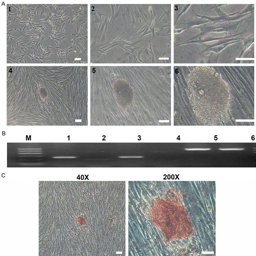
Induction of PPCs into insulin-secreting cells

                  Figure 1. Morphology of PPCs at different passages. Photomicrographs
                  presented that PPCs at passage 0 to passage 13 were all with a typical
                  fibroblast-like shape. While, PPCs at passage 10 appeared senescent.
                  Scale bars represent 50 μm.

1634                                       Int J Clin Exp Med 2018;11(3):1632-1643
Induction of PPCs into insulin-secreting cells

                                                                               plated at 50% confluence. The
                                                                               method for induction was al-
                                                                               ong with previous research
                                                                               (6.87 diabetes). Firstly, the ce-
                                                                               lls were exposed to high glu-
                                                                               cose Dulbecco’s modified Ea-
                                                                               gle medium (H-DMEM) 25
                                                                               mmol/l, 10% FBS and 10-6
                                                                               mol/l retinoic acid (Sigma Al-
                                                                               drich) for 24 h and followed
                                                                               by 2 day treatment with H-
Figure 2. Specific surface markers of PPCs were detected by RT-PCR. Re-        DMEM and 10% FBS. Secon-
sults showed the PPCs at passage 1 expressed C-myc, CD44, Vimentin,            dly, the cells were changed in-
Nestin, Insulin, while did not express CK19. PPCs at passage 6 and 10 only     to the medium containing low
expressed C-myc, CD44, Vimentin, Nestin.
                                                                               glucose DMEM 5.56 mmol/l,
                                                                               10% FBS, 10 mmol/l nicotin-
and VIMENTIN (1:200, Abcam, USA) at 4°C                       amide (Sigma-Aldrich), 20 ng/ml epidermal
overnight. FITC-conjugated secondary antibod-                 growth factor (PeproTech, Rocky Hill, NJ), 50
ies (1:200) were used at room temperature                     ng/ml of FGF-10 (R&D, Minneapolis, MN),
for 60 min. DAPI was used to counter stain                    and 300 nmol/l of (-)-indolactam V (LC labo-
nuclei under black condition. Results were pre-               ratories, Woburn, MA) for 9 days. Thirdly, the
sented by a con-focal optical system (Nikon,                  cells were cultured with the medium contain-
TE2000).                                                      ing 90% L-DMEM, 10% FBS, 10 nmol/l ex-
                                                              endin-4 (Sigma-Aldrich), and 50 ng/ml Activin
Karyotype analysis                                            A (PeproTech) for 7 days. For the control gr-
                                                              oup, the cells were cultured with 90% DM-
PPCs were evaluated at 30% confluence. Firs-                  EM/F12, 1% penicillin/streptomycin, 1% amph-
tly, cells were treated with hypotonic medium                 otericin B, and 10% FBS.
(0.075 mol/l KCl, 0.017 mol/l Na-citrate). Th-
en the cells were fixed with ethanol: acetic ac-              RT-PCR analysis and dithizione staining
id (1:2:1) and stained with Giemsa (1-s2.0).
Finally, counting the numbers of the chromo-                  Cellular mRNAs for the induced cells and the
some was performed with Cytovision Applied                    control group were obtained by Trizol reagent
Imaging.                                                      (Invitrogen). Then the mRNAs were reversed
                                                              as cDNA. The products employed as templa-
Flow cytometry                                                tes were amplified by polymerase chain reac-
                                                              tion (PCR). Primers were presented in Table.
Surface antigen expression of PPCs and dif-                   The results were visualized through 2.5% aga-
ferentiation were analyzed using flow cytome-                 rose gel electrophoresis.
try. Cells were digested with 0.25% trypsin-
EDTA and centrifugated. Then, the cells were                  Dithizone (DTZ, Sigma) were used to assess
fixed, washed and permeabilized. Cells were                   the differentiation. The induced cells were fix-
stained with the following antibodies: at 4°C                 ed with 4% paraformaldehyde at room tem-
over night. After washing with PBS, the cells                 perature for 30 min, then they were treated
were incubated with FITC-conjugated second-                   with DTZ solution for 1 h at 37°C. DTZ solution
ary antibody at 4°C for 1 h. For the control                  were consisted of 50 mg DTZ and 5 ml dime-
group, cells were incubated with PBS. Finally,                thyl sulfoxide (DMSO, Sigma). Results were ob-
the cells were washed and stained with DAPI                   served by microscope.
at room temperature under black condition
for 20 min. Results were evaluated by FACS                    Immunofluorescence detection
using CellQuest Research Software.
                                                              Immunofluorescence (IF) staining was perfor-
Differentiation into insulin-secreting cells                  med for induced cells and the control cells.
                                                              Both cells were fixed with 4% paraformalde-
To be induced into functional insulin-secreting               hyde at room temperature for 20 min. Then the
cells, PPCs at passage 6 were digested and re-                cells were permeabilized for 10 min at room

1635                                                              Int J Clin Exp Med 2018;11(3):1632-1643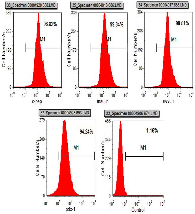
Induction of PPCs into insulin-secreting cells

1636                                  Int J Clin Exp Med 2018;11(3):1632-1643
Induction of PPCs into insulin-secreting cells
Figure 3. Immunofluorescence staining for PPCs antigens. DAPI was used to stain the nuclei. Results presented that
PPCs were positive to the antigens of NKX6.1, NESTIN, PDX1 and VIMENTIN. Scale bars represent 200 um.
Statistical analysis
Statistical analyses of the data were perform-
ed with a one-way ANOVA followed by the Tuk-
ey-Kramer honestly significant difference test
for the three sets of results. A P-value of less
than 0.05 was considered significant. Statisti-
cal analyses were conducted with a JMP Sta-
tistical Discovery Software (SAS Institute, Cary,
NC).
Results
Isolation and purification of PPCs
Cells isolated from islets formed a cell layer
and presented an obvious fibroblast-like mor-
phology (Figure 1). Cells first plated on the pl-
astic after 16 h inoculating. In the beginning,
PPCs at passage 0 took 3 days to proliferate
to passage 1. Along with the time, PPCs grew
rapidly and could be passaged every 1 or 2
days. Until PPCs proliferated to passage 10,
they appeared as blebbing and karyopyknosis
and had a lower proliferate ability. PPCs at
passage 11 took 4 day to be proliferated to
passage 12.
Reverse transcription-polymerase chain reac-
tion of PPCs
Results were presented by agarose gel. We
detected PPCs at passage 0, passage 6 and
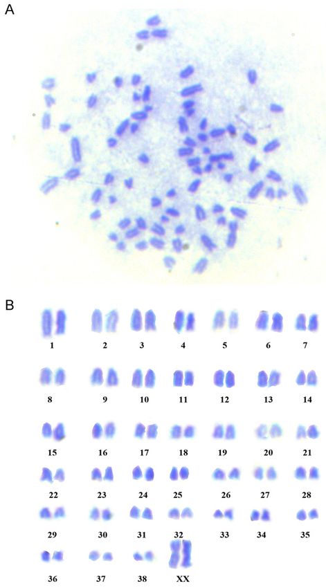
Figure 4. Karyotype analysis of Tibetan Mastiff de- passage 10. The cells at passage 0 expressed
rived PPCs. Tibetan Mastiff derived PPCs had 2n=78
diploid chromosome numbers. They were consisted
C-MYC, CD44, VIMENTIN, NESTIN, INSULIN, but
of 38 pair of chromosomes and 1 pair of sex chro- did not express epithelial marker CK19. The
mosomes. A XX type spread in metaphase was pre- cells at passage 6 and passage 10 expressed
sented (n=100). C-MYC, CD44, VIMENTIN, NESTIN, but did not
express INSULIN and epithelial marker CK19
(Figure 2).
temperature with 0.1% Triton X-100 (Sigma)
and blocked with 10% goat serum for 60 Immunofluorescent detection of cell surface
mim. For the induced cells, they were stain- markers
ed with the following antibodies: C-CEPTIDE
(1:200, Abcam, USA) and GLUCOSE (1:200, Surface marker antigens for PPCs were ana-
Abcam, USA) at 4°C overnight. For the con- lyzed by immunofluorescence staining. Results
trol group, the cells were treated with PBS at presented that PPCs were positive for NESTIN,
4°C overnight. FITC-conjugated secondary an- NKX6.1, duodenal homeobox 1 and VIMENTIN
(Figure 3).
tibodies (1:200) were used at room tempera-
ture for 60 min. DAPI was used to counter sta- Karyotype analysis
in nuclei under black condition. Results were
presented by a con-focal optical system (Nikon, Tibetan Mastiff derived PPCs were diploid (2n
TE2000). =78), consisting of 38 pair of euchromosomes
1637 Int J Clin Exp Med 2018;11(3):1632-1643Induction of PPCs into insulin-secreting cells Figure 5. PPCs were stained with surface specific markers as C-Pep, insulin, nestin and pdx1 and analyzed by flow cytometry. The positive population was enclosed by the line. and 1 pair of sex chromosomes. Results indi- PPCs to insulin was 99.84%, the positive rate cated that the hereditary property of the cells of PPCs to nestin was 98.51%, a positive rate cultured in vitro was stable (Figure 4). of PPCs to pdx1 was 94.24%. Positive rate detetction of PPCs Differentiation of insulin-secreting cells Surface specific antigens of PPCs were as- The differentiation of PPCs into insulin-secret- sessed by flow cytometry (Figure 5). Results ing cells was evaluated through morphology presented that the positive rate of PPCs to observation, RT-PCR analysis and immunofl- C-Peptide was 98.82%, the positive rate of uorescence staining (Figures 6, 7). There was 1638 Int J Clin Exp Med 2018;11(3):1632-1643
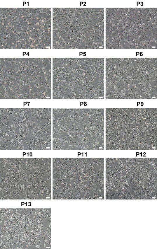
Induction of PPCs into insulin-secreting cells

Figure 6. Differentiation of PPCs into insulin-secreting cells. A. Morphology detection. The induced cells formed an
islet-like cluster at 7 day after induction. 1, Magnification, ×40; 2, Magnification, ×100; 3, Magnification, ×200. B.
RT-PCR analysis. Electrophoretogram revealed that the induced cell both expressed KRT7 and Insulin genes, while
the control group cells only expressed KRT7 gene. 1, 2 were the induced group; 3, 4 were the control group; 5, 6
were GAPDH (an internal control). C. Dithizone staining of induced cells. Islet-like cluster was stained with dithizone
and dyed scarlet. Pictures were magnificated to 40× and 200×.

no obvious change observed for the first we-                    to dithizone and dyed scarlet. C-PEPTIDE and
ek. Until the 10th day, there was little islet-like             INSULIN were specific antigens of insulin-se-
clusters appeared. Along with the differentia-                  creting cells and they were detected by im-
tion, more and larger islet-like clusters were                  munofluorescence staining (Figure 7). Results
observed, which was typical islet morphology                    indicated that the induced cells were both
(Figure 5A). We also detected the differentia-                  positive to C-PEPTIDE and INSULIN.
tion through RT-PCR and dithizone staining
(Figure 6B, 6C). Results presented that the                     Discussion
induced group cells both expressed KRT7 and
INSULIN, while the control group cells only                     Stem cells should fulfill the following two stan-
expressed KRT7. Dithizone staining results de-                  dards: (1) Cells could proliferate themselves
monstrated that the induced cells were positive                 without induction for a long time; (2) They could

1639                                                                  Int J Clin Exp Med 2018;11(3):1632-1643
Induction of PPCs into insulin-secreting cells

1640                                  Int J Clin Exp Med 2018;11(3):1632-1643
Induction of PPCs into insulin-secreting cells

Figure 7. Detection of differentiation by immunofluorescence staining. C-PEPTIDE and GLUCOSE staining were both
located in the cytoplasm. DAPI was used to stain the nuclei (blue), and C-PEPTIDE and GLUCOSE were used to stain
the cytoplasm (red). They were both magnificated to 100× and 200×.

be differentiated into multiple cell types [19].            analog and it could induce PPCs into insu-
In this study, we isolated PPCs from islet tis-             lin-secreting cells and inhibit insulin-secreting
sue with trypsin and inoculated them in the                 cells apoptosis [25, 26]. Active A diverted the
plate with a aleukaemia inhibitory factor (LIF).            induction into a PAX-4 expressing pancreatic
LIF could inhibit cell differentiation and pro-             lineage and not a glucagon producing cells
mote self-renewal ability [20]. LIF was usually             (αcells) [27]. The successful induction of PPCs
used in stem cells medium to restrain spon-                 into insulin-secreting cells was assessed by
taneous differentiation. Because pancreas tis-              RT-PCR detection, DTZ staining and immuno-
sue contained many different cells, it was im-              fluorescence staining.
portant for us to isolate islet tissue. PPCs co-
uld renew themselves from passage 0 to pas-                 The first pancreas transplantation was con-
sage 13 with a stable spindle-shape. Results                ducted in 1966. At present, the pancreas tran-
of RT-PCR presented that PPCs expressed                     splantation was performed with a kidney tran-
the mesenchymal stem cells surface markers,                 splantation. Though the transplantation has a
while did not express the epithelioid cells spe-            77% survival rate for the 1st year, it was still
cific marker. PPCs derived from Tibetan Mastiff             accompanied by immunosuppressive and or-
also had a normal karyotype as well as an abi-              gan injection. Islet transplantation provided a
lity to differentiate into insulin-secreting cells.         safe method. However, it was still restricted by
All results revealed that PPCs we isolated we-              the shortage of donor organs and unable to
re highly homogenous and multipotent.                       proliferate in vitro. Diabetes could be cured
                                                            through stem cell treatment. Therefore, PPCs
PPCs could also be differentiated into insulin-
                                                            were considered as the most perfect stem
secreting cells by a three-step method in this
study. Owing to many different protocols for                cell resource for the treatment of diabetes and
insulin-secreting cells differentiation, this thr-          degenerative diseases. Functional differentia-
ee-step method was high efficient and able to               tion into insulin-secreting cells of PPCs has
prevent transfection. The reprogramming pro-                been considered as a remarkable breakth-
tocols were immediate and effective, but due                rough. Through stem cell therapy was a hot
to the unsafe exogenous genes, they were                    spot in the current research, there were still
unsafe. In this three-step method, H-DMEM                   lots of challenges need to overcome. How to
and many other factors were used. High glu-                 obtain mature insulin-secreting cells efficient-
cose was an inducer in islet induction, and                 ly? How to obtain high inductivity to prevent
H-DMEM was a medium consisted of DMEM                       teratoma formation? How to figure out the
and 23.3 mmol/l glucose [21]. H-DMEM was                    molecular mechanisms under the differentia-
added at the first step to begin induction. At              tion? Hence, we should focus on the mecha-
the second step, the medium was added by                    nisms of differentiation and figure out each
L-DMEM, nicotinamide, epidermal growth fac-                 stage of differentiation both in morphology and
tor (EGF), FGF-10 and indolactam V (LV). Islet              molecular level. It was of great significance
clusters were appeared and aggregated main-                 for us to explore the differentiation mechani-
ly at this stage. LC could maintain the differ-             sm and signial pathways contained in it.
entiation more efficient [8]. Nicotinamide and
lower glucose could avoid the cells dying or                Conclusion
differentiating [11]. Nicotinamide could prom-
ote the production of insulin-secreting cells               In this article, we isolated, purified and char-
and protect cells from high concentration glu-              acterized PPCs from Tibetan Mastiff derived
cose [24]. EGF was a factor which was fit for               pancreatic islet tissue and differentiated them
PPCs proliferation, maturation and PDX-1 po-                into insulin-secreting cells using a three-step
sitive cells growth [25]. At the third stage, ex-           method. This was the first time to explore Ti-
endin-4 and Activin A were employed as indu-                betan Mastiff derived PPCs. It had great signifi-
cers. Exendin-4 was a glucagon-like peptide-1               cance to the diabetes patients.

1641                                                              Int J Clin Exp Med 2018;11(3):1632-1643
Induction of PPCs into insulin-secreting cells
Acknowledgements [11] Nyenwe EA, Jerkins TW, Umpierrez GE and
Kitabchi AE. Management of type 2 diabetes:
The Agricultural Science and Technology Inno- Evolving strategies for the treatment of pa-
vation Program (ASTIP (cxgc-ias-01). Project tients with type 2 diabetes. Metabolism 2011;
supported by the National Natural Science 60: 1-23.
[12] Couri CE and Voltarelli JC. Stem cell-based
Foundation of China (Grant No: 31472099;
therapies and immunomodulatory approaches
31672404). in newly diagnosed type 1 diabetes. Curr Stem
Cell Res Ther 2011; 6: 10-15.
Disclosure of conflict of interest [13] Mitutsova V, Yeo WWY, Davaze R, Franckhaus-
er C, Hani EH, Abdullah S, Mollard P, Schaeffer
None. M, Fernandez A, Lamb NJC. Adult muscle-de-
rived stem cells engraft and differentiate into
Address correspondence to: Weijun Guan, Institute insulin-expressing cells in pancreatic islets of
of Animal Science, Chinese Academy of Agricultural diabetic mice. Stem Cell Res Ther 2017; 8: 86.
Sciences, No.2 Yuanmingyuan West Road, Haidian [14] Selander L and Edlund H. Nestin is expressed
District, Beijing 100193, PR China. Tel: +86-10- in mesenchymal and not epithelial cells of the
62815992; Fax: +86-10-62815884; E-mail: wjguan- developing mouse pancreas. Mech Dev 2002;
86@163.com 113: 189-192.
[15] Suzuki A, Nakuchi H and Taniguchi H. Prospec-
References tive isolationof multipotent pancreatic progeni-
tors using flow-cytometric cell sor-ting. Diabe-
[1] Wang M, Wang YH, Ye Q, Meng P, Yin H and tes 2004; 8: 2143-2152.
Zhang DL. Serological survey of toxoplasma [16] Bai CY, Li XC, Gao YH, Wang KF, Fan YN, Sh-
gondii in tibetan mastiffs (canis lupus familia- uang Z, Ma YH and Wei JG. Role of microR-
ris) and yaks (bos grunniens) in Qinghai, China. NA-21 in the formation of insulin-producing
Parasit Vectors 2012; 5: 35. cells from pancreatic progenitor cells. Biochim
[2] Li Y, Wu DD, Boyko AR, Wang GD, Wu SF, Irwin Biophys Acta 2016; 2: 280-293.
DM and Zhang YP. Population variation re- [17] Lardon J, Rooman I and Bouwens L. Nestin ex-
vealed high-altitude adaptation of Tibetan pression in pancreatic stellate cells and angio-
mastiffs. Mol Biol Evol 2014; 31: 1200-1205. genic endothelial cells. Histochem Cell Biol
[3] Caspermeyer J. A tale of dog and man: tibetan 2002; 117: 535-540.
mastiff found to gain high-altitude. Adaptation [18] Treutelaar MK, Skidmore JM, Dias-Leme CL,
Hara M, Zhang L, Simeone D, Martin DM and
after domestication by interbreeding with the
Burant CF. Nestin-lineage cells contribute to
tibet gray wolf. Mol Biol Evol 2017; 34: 775-
the microvasculature but not endocrine cells
776
of the islet. Diabetes 2003; 52: 2503-2512.
[4] Li Y and Zhang YP. High genetic diversity of Ti-
[19] Okere B, Lucaccioni L, Domonici M and Lughet-
betan Mastiffs revealed by mtDNA sequences.
ti L. Cell therapies for pancreatic beta-cell re-
Chin Sci Bull 2012; 13: 1483-1487.
plenishment. Ital J Pediatr 2016; 42: 62.
[5] Calafiore R and Basta G. Stem cells for the cell
[20] Kawahara Y, Manabe T, Matsumoto M, Kaji-
and molecular therapy of type 1 diabetes mel-
ume T, Matsumoto M and Yuge L. LIF-free em-
litus (T1D): the gap between dream and reality. bryonic stem cell culture in simulated micro-
Am J Stem Cells 2015; 4: 22-31. gravity. PLoS One 2009; 7: e6343.
[6] Zhang S, Bai CY, Ma YH, Li XC, Gao YH, Fan YN, [21] Bonner-Weir S, Deery D, Leahy JL and Weir GC.
Guan WJ And Zheng D. The characterisation Compensatory growth of pancreatic beta-cells
and functional beta-cell differentiation of duck in adult rat safter short-term glucose infusion.
pancreas-derived mesenchymal cells. Br Poult Diabetes 1989; 1: 49-53.
Sci 2016; 2: 201-210. [22] Chen S, Borowiak M, Fox JL, Maehr R, Osafune
[7] Acharjee S, Ghosh B, Al-Dhubiab BE and Nair K, Davidow L, Lam K, Peng LF, Schreiber LF,
AB. Understanding type 1 diabetes: etiology Rubin LL and Melton D. A small molecule that
and models. Can J Diabetes 2013; 37: 269-76. directs differentiation of human ESCs into the
[8] Wallberg M and Cooke A. Immune mecha- pancreatic lineage. Nat Chem Biol 2009; 5:
nisms in type 1 diabetes. Trends Immunol 258-265.
2013; 34: 583-91. [23] Kolb H and Burkart V. Nicotinamide in type 1
[9] Wenzlau JM and Hutton JC. Novel diabetes au- diabetes. Mechanism of action revisited. Dia-
toantibodies and prediction of type 1 diabetes. betes care 1999; 22 Suppl 2: B16-20.
Curr Diab Rep 2013; 13: 608-15. [24] Otonkoski T, Beattie GM, Mally MI, Ricordi C
[10] Butler AE, Janson J, Bonner-Weir S, Ritzel R, and Hayek A. Nicotinamide is a potent inducer
Rizza RA and Butler PC. Beta-cell deficit and of endocrine differentiation in cultured human
increased beta-cell apoptosis in humans with fetal pancreatic cells. J Clin Investig 1993; 92:
type 2 diabetes. Diabetes 2003; 52: 102-10. 1459-1466.
1642 Int J Clin Exp Med 2018;11(3):1632-1643Induction of PPCs into insulin-secreting cells
[25] Zhang D, Jiang W, Liu M, Sui X, Yin X, Chen S, [27] Santamaria X, Massasa EE, Feng Y, Wolff E
Shi Y and Deng H. Highly efficient differentia- and Taylor HS. Derivation of insulin producing
tion of human ES cells and iPS cells into ma- cells from human endometrial stromal stem
ture pancreatic insulin-producing cells. Cell cells and use in the treatment of murine diabe-
Res 2009; 19: 429-438. tes. Molecular Therapy 2011; 11: 2065-2071.
[26] Capela A and Temple S. LeX is expressed by
principle progenitor cells in the embryonic ner-
vous system, is secreted into their environ-
ment and binds Wnt-1. Dev Biol 2006; 291:
300-313.
1643 Int J Clin Exp Med 2018;11(3):1632-1643You can also read