Increased Internal Porosity and Surface Area of Hydroxyapatite Accelerates Healing and Compensates for Low Bone Marrow Mesenchymal Stem Cell ...
←
→
Page content transcription
If your browser does not render page correctly, please read the page content below

applied
           sciences
Article
Increased Internal Porosity and Surface Area of
Hydroxyapatite Accelerates Healing and
Compensates for Low Bone Marrow Mesenchymal
Stem Cell Concentrations in Critically-Sized
Bone Defects
Eileen R. Dawson 1,2 , Richard K. Suzuki 2 , Melissa A. Samano 1,2 and Matthew B. Murphy 1,2,3, *
 1    Department of Biomedical Engineering, The University of Texas at Austin, Austin, TX 78705, USA;
      eileen.dawson@gmail.com (E.R.D.); melissa.samano@utexas.edu (M.A.S.)
 2    Celling Biosciences, Austin, TX 78701, USA; RichardKSuzuki@gmail.com
 3    Murphy Lifescience Consulting, Austin, TX 78702, USA
 *    Correspondence: mbmurphy@utexas.edu

 Received: 6 July 2018; Accepted: 6 August 2018; Published: 14 August 2018

 Featured Application: Volume and rate of tissue regeneration may be enhanced with increased
 scaffold porosity as well as increased numbers of non-cultured bone marrow cells.

 Abstract: For clinical treatment of skeletal defects, osteoinductive scaffolds must have the ability to
 conform to the unique geometry of the injury site without sacrificing biologically favorable properties,
 including porosity. This investigation seeks to combine the osteoinductive properties of porous
 hydroxyapatite (HA) scaffolds with the beneficial handling characteristics of granules or putties,
 while evaluating the effects of mesenchymal stem cell (MSC) concentration on the composite grafts’
 ability to regenerate bone in vivo. The results demonstrate that porous HA granules regenerate
 significantly larger volumes of bone compared to non-porous HA. Increased MSC concentrations in
 autologous bone marrow aspirate (BMA) contributed to greater bone regeneration. This effect was
 most predominant with non-porous HA. While the extent of bone regeneration using non-porous HA
 was strongly correlated with MSC concentration of the marrow, porous HA microparticles combined
 with autologous BMA were successful in faster treatment of critically-sized bone defects and with
 less dependence on the MSC concentration than non-porous HA.

 Keywords: hydroxyapatite; biomaterial porosity; mesenchymal stem cell; bone marrow aspirate;
 bone regeneration

1. Introduction
     Critically-sized bone defects arise from traumatic injury, tumor resection, autologous bone graft
harvesting, and surgical procedures, including spinal fusion. Autografts, considered the gold standard
in bone substitutes, are impractical for use in larger defects, as graft size would be limited to patient
sample availability. Additionally, pain and local donor site morbidity are commonly reported at the site
of graft harvesting [1]. Nonetheless, autografts are successful in defect treatment because they combine
a number of key components necessary for tissue growth. Bone regeneration requires contributions
from all aspects of the “tissue engineering paradigm”, including progenitor cells, which are a scaffold
to guide tissue formation and remodeling, and biochemical/biomechanical stimuli.
     Significant research has investigated various biomaterials and scaffolding techniques with
osteoconductive or osteoinductive properties. Among these biomaterials is hydroxyapatite (HA),

Appl. Sci. 2018, 8, 1366; doi:10.3390/app8081366                                www.mdpi.com/journal/applsci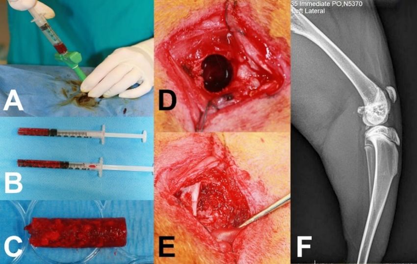
Appl. Sci. 2018, 8, 1366                                                                              2 of 11

the primary crystalized form of calcium phosphate comprising the inorganic portion of bones,
which has consistently demonstrated the ability to promote bone growth in vivo [2,3]. Monolithic
scaffolds are commonly utilized, usually conforming to the pre-determined geometry of controlled
defects created in animal models [4,5]. In clinical application, this type of scaffold would be impractical.
The implanted scaffold must span the full volume of the defect—thus, having a pre-sized scaffold
would limit its application to defects of specific sizes, or require the physician to modify the graft or the
graft site. Graft modification requires costly operating-room time and introduces the risk of damaging
the biomaterial. Graft site modification could have deleterious effects to the patient, in that use of
a reamer or burr causes heat generation and local tissue necrosis. Ultimately, a moldable formulation
may reduce time in surgery and avoid additional bone loss or trauma to the surrounding tissue area [6].
It would therefore be advantageous for the scaffolding material to be moldable to irregular geometries
presented in most clinical cases to completely fill the defect and bridge the native bone. There are,
however, a number of physical characteristics of bone grafts that need to be retained. The inclusion of
porosity in the HA scaffolds cannot be understated, as there is typically a correlation between the extent
and interconnectivity of pores and the scaffold’s ability to regenerate bone [7–10]. Interconnected
porosity encourages the in vivo infiltration of autologous cells and vasculature. High porosity may be
achieved with ceramics by employing a granular or microparticle preparation. This approach has been
validated in limited studies without specific tailoring of granule porosity or surface area, nor thorough
characterization of the cellular component of the graft [11–13].
      The contribution of cells is often overlooked in biomaterials-based approaches for orthopedic
healing. However, it is ultimately the cells, whether they be transplanted with the graft or recruited
endogenously, that are responsible for new tissue generation and remodeling [14,15]. We recently
reported that many Food and Drug Administration (FDA)-cleared or approved biomaterials for bone
healing are not efficient at retaining bone marrow-derived cells and, in many instances, were cytotoxic,
with pH values less than 7 or greater than 10 when reconstituted in plasma or saline [16]. Materials
that were not easily soluble (allograft bone and calcium phosphates) were most successful at retaining
bone marrow mesenchymal stem or stromal cells (MSCs) and inducing osteogenic gene expression
in an in vitro simulation of surgical graft preparation. In addition to the effects of materials on cells,
the source and number of cells must be considered. Many in vivo studies combine biomaterials with
culture-expanded autologous or allogeneic cells as an implantable graft. Although this is convenient
to standardize “doses” of therapeutic agents and seemingly control one variable of the regenerative
paradigm, researchers often fail to recognize the inherent variability of cell potency from donor to
donor, the effects of culture conditions/passage number, or the value of a heterogenous population,
including multiple progenitor phenotypes (mesenchymal, hematopoietic, endothelial, etc.). The clinical
translation of an in vitro expanded cell approach is further in question due to regulatory restrictions
of the United States FDA and other agencies. The usage of autologous cells at the point-of-care is
an appealing alternative, with less regulatory requirements and a decreased risk of cell contamination
or rejection. A growing amount of data has suggested strong correlations in clinical outcomes
with non-union fracture, degenerative disc disease, rotator cuff tear, avascular necrosis, and other
orthopedic injuries, based on the concentration of MSCs present in bone marrow at physiological
levels, or concentrated up to 10× [17–19]. The effects of non-cultured MSC concentration on bone
formation when combined with HA granular particles are presently unreported in the literature.
      In the present study, we investigated the combination of autologous bone marrow aspirate with
porous or non-porous HA granules. Both of these materials were prepared with a collagen-derived
carrier to quickly absorb the bone marrow and form a moldable putty, to pack into bilateral,
critically-sized femoral chondyle defects in adult rabbits. An aliquot of each animal’s bone marrow
was evaluated for nucleated cell concentration and MSC content to retrospectively examine the effects
of MSC concentration in fresh marrow, as well as the effects of granular porosity with identical cell
populations in the same animal.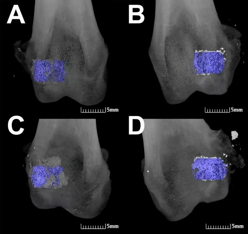
Appl. Sci. 2018, 8, x                                                                                         3 of 11

was evaluated for nucleated cell concentration and MSC content to retrospectively examine the
effects of MSC concentration in fresh marrow, as well as the effects of granular porosity with
identical
Appl.        cell8,populations
      Sci. 2018,    1366       in the same animal.                                       3 of 11

2. Materials and Methods
2. Materials and Methods
2.1. Implant Materials
2.1. Implant Materials
      Non-porous (Nanoss® Bioactive, Pioneer Surgical, Marquette, MI, USA) and porous (Solum IV,
      Non-porous    (Nanoss   ®
Celling Biosciences,    Austin,Bioactive,
                                 TX, USA)Pioneer      Surgical,
                                             HA particles     areMarquette,  MI, USA)
                                                                  a combination        and porous
                                                                                  of sintered        (Solum IV,
                                                                                               nanocrystalline
Celling   Biosciences,  Austin,  TX,   USA)  HA   particles   are a combination   of sintered
(
Appl. Sci. 2018, 8, x 4 of 11
non-porous
Appl. Sci. 2018, 8,HA
1366granules into a critical size defect created on the femoral condyle. First, 4bone of 11
marrow aspirate was harvested from the iliac crests of each animal (2 cc per rabbit). The porous and
non-porous HA grafts were separately mixed with the autologous bone marrow aspirate (BMA) of
prior to implantation
each animal prior to by combining 0.5
implantation by cc BMA with0.5
combining 0.5ccccBMA
porous or non-porous
with 0.5 cc porous HAorgranules (1:1 ratio
non-porous HA
by
granules (1:1 ratio by volume), respectively. Each biomaterial contained a gelatin carrier with
volume), respectively. Each biomaterial contained a gelatin carrier that dissolved when wetted that
BMA and when
dissolved formed a viscous
wetted withgelBMA around each HA
and formed particle.gel
a viscous The resulting
around eachputty was loaded
HA particle. Theinto 1 mL
resulting
syringes, with the distal tip removed in order to form bone graft logs for implantation.
putty was loaded into 1 mL syringes, with the distal tip removed in order to form bone graft logs The excess BMAfor
was transported to the laboratory (Celling Biosciences, Austin, TX, USA)
implantation. The excess BMA was transported to the laboratory (Celling Biosciences, Austin, TX,for cell analysis. Two drill
defects
USA) for (approximately
cell analysis. 6Two mmdrill
in diameter
defects and 8–10 mm in 6length)
(approximately mm inwere created
diameter in each
and 8–10 animal—one
mm in length) in
each femoral condyle—and were filled with porous HA graft on the left
were created in each animal—one in each femoral condyle—and were filled with porous HA graftside and non-porous HA graft
on 3
on the
the right sideand
left side (Figure 2). Approximately
non-porous HA graft on0.25 the cm
rightofside
hydrated
(Figuregraft material was implanted
2). Approximately 0.25 cm3 in of
each
hydrated graft material was implanted in each defect by pushing the putty out of the 1 mLanalgesic
defect by pushing the putty out of the 1 mL syringe. Each animal received sufficient syringe.
(butophanol
Each animal 0.5–1.0
receivedmg/kg) injection
sufficient prior (butophanol
analgesic to and after the surgery.
0.5–1.0 The animals
mg/kg) injectionalso received
prior to anda after
fentanyl
the
transdermal patch on the skin for additional analgesia up to 3 days. Ten animals
surgery. The animals also received a fentanyl transdermal patch on the skin for additional analgesia were implanted per
time
up topoint
3 days.(4, Ten
8, and 13 weeks).
animals were implanted per time point (4, 8, and 13 weeks).
Figure 2. At the onset of surgery, bone marrow aspirate was drawn from the iliac crest of each rabbit
Figure 2. At the onset of surgery, bone marrow aspirate was drawn from the iliac crest of each rabbit
(A) and mixed with porous or non-porous hydroxyapatite granule at 1:1 ratio by volume in a 1 mL
(A) and mixed with porous or non-porous hydroxyapatite granule at 1:1 ratio by volume in a 1 mL
syringe (B) to form implantable cylinders (C). 6 mm × 8 mm (diameter × length) defects were created
syringe (B) to form implantable cylinders (C). 6 mm × 8 mm (diameter × length) defects were created
bilaterally in the femoral chondyles (D) and packed with the graft materials (E), which were
bilaterally in the femoral chondyles (D) and packed with the graft materials (E), which were observed
observed radiographically (F).
radiographically (F).
Radiographs were collected post-operatively and at termination for each animal. At necropsy,
Radiographs
4, 8, or wereimplantation,
13 weeks after collected post-operatively
the randomly and at termination
assigned 10 animalsfor eacheuthanized,
were animal. At and necropsy,
gross
4, 8, or 13 weeks
observations after
of the implantation,
implant theobserved
sites were randomly assigned
and recorded. 10 animals were sites
Implantation euthanized, and gross
were excised and
observations of the implant sites were observed and recorded. Implantation sites
placed in 10% neutral buffered formalin for fixation. Radiographic microCT analysis was performed were excised and
placed
on the in8- 10%
and neutral
13-week buffered
animalsformalin
(Numira forBiosciences,
fixation. Radiographic
Inc., Salt Lake microCT
City, analysis
UT, USA). wasHistological
performed
on the 8- and
preparation was13-week animals (Numira
done immediately after Biosciences, Inc., for
animal sacrifice Saltthe
Lake City, samples,
4-week UT, USA). Histological
and following
preparation was done immediately after animal sacrifice for the 4-week
microCT analysis on the 8- and 13-week samples. Standard techniques were used to prepare samples, and following
slides
microCT analysis
for histological on the 8-
analysis. andincluded
This 13-weekdecalcification,
samples. Standard techniques
paraffin embedding,were microtome
used to prepare slides
sectioning,
for
andhistological
staining. Twoanalysis. This included
slides were prepared decalcification,
from each defect, paraffin
one atembedding, microtome
the central level sectioning,
of the drill defect
and staining. Two slides were prepared from each defect, one at the central
and one from the interface with original bone. The slides were stained with hematoxylin/eosinlevel of the drill defect
and
(H&E).oneEach
fromsection
the interface with original
was analyzed bone. The slides
by a pathologist weretissue
for local stained with hematoxylin/eosin
reaction, following the ISO
(H&E). Each section was analyzed by a pathologist for local tissue reaction,
10993-6 guidelines—first by an independent pathologist (WuXi ApTec, St. Paul, MN, following the ISO 10993-6
USA), and
guidelines—first by an independent pathologist (WuXi ApTec,
reviewed by a second independent pathologist (NAMSA, Minneapolis, MN, USA). St. Paul, MN, USA), and reviewed by
a second independent pathologist (NAMSA, Minneapolis, MN, USA).Appl. Sci. 2018, 8, 1366 5 of 11
2.3. Cellular Analysis of Bone Marrow Aspirate
BMA samples from late harvest-time-point animals (8 and 13 weeks) were transported to
the laboratory in 5 ◦ C temperature-controlled packs and analyzed within 24 h of aspiration.
Total nucleated cell (TNC) counts and viability were quantified by NucleoCounter (New Brunswick,
Enfield, CT, Canada) after diluting the 1 mL BMA aliquot with 9 mL phosphate-buffered saline (PBS)
and 2% fetal bovine serum (FBS, HyClone human mesenchymal stem cell grade, Logan, UT, USA).
The frequency of colony-forming unit-fibroblast (CFU-F), a marker for mesenchymal progenitor cells,
was determined after the density-gradient depletion of red blood cells (Ficoll-Paque, GE Healthcare,
Piscataway, NJ, USA), after which nucleated cells where plated in serial dilutions in 12-well cell culture
plates with medium (MSCM, ScienCell Research Laboratories, Carlsbad, CA, USA) containing 5%
FBS and 1% antibiotic (penicillin/streptomycin, ScienCell Research Laboratories, Carlsbad, CA, USA)
for 10 days in vitro (n = 3 per dilution). Complete media changes were performed on days 4 and 7.
The average CFU-F concentration in the BMA was determined by multiplying TNC concentration
(TNC/mL) by CFU-F frequency (CFU-F/TNC). Excess composite graft (BMA + porous HA granules)
samples were analyzed by cell viability after 24 h by NucleoCounter after diluting the graft in PBS.
Individual granules of non-implanted grafts were imaged by washing and fixing cells with 2% formalin
and performing confocal microscopy after staining actin filaments with FITC-phalloidin (Invitrogen,
10 mg/mL) and rhodamine anti-vinculin/DAPI (Invitrogen, 10 µg/mL) staining, for cell membrane
focal adhesions and nuclei, respectively (Figure 1E).
2.4. MicroCT, Histological and Radiographic Analysis
After 4, 8, and 13 weeks, 10 animals per time point were euthanized and a radiograph of
each implant per animal was obtained (dorsal-ventral and lateral views). The bone implant sites
were exposed and gross observations for local tissue reactions recorded, as per the ISO 10993-6
guidelines. The bone implant sites were placed in 10% neutral buffered formalin for fixation.
Each formalin-fixed bone implant site from the 8- and 13-week time points, prior to histopathological
analysis, was sent to Numira for microCT analysis of new bone formed and residual graft material
in and adjacent to the defect volume. All samples were scanned on a high-resolution microCT
scanner (µCT40, ScanCo Medical, Zurich, CH, Switzerland). The microCT images were analyzed using
Numira Biosciences’ VHLab. Analysis included determination of bone volume, total defect volume,
remaining implant (HA) volume, and bone density. Each explanted bone sample was processed by
standard histological techniques, including decalcification, paraffin embedding, microtome sectioning,
and staining, as described in the previous section.
2.5. Statistical Analysis
All measurements are represented as the mean ± standard error (n = 10, unless otherwise noted).
For statistical analysis, the means were compared using a one-way analysis of variance (ANOVA)
for each variable combination using JMP 9 statistical software (SAS, Cary, NC, USA). Data were
tested on a normal distribution and a p-value < 0.05 was considered to indicate significance, in which
statistical differences were determined using Tukey’s HSD test, with calculations performed using
Microsoft Excel.
3. Results
3.1. Cellular Analysis of Bone Marrow Aspirate
The twenty rabbits bone marrow aspirate samples contained an average of 3.20 × 10⁷ /mL
TNC/mL and 2710 CFU-F/mL. The average CFU-F frequency among nucleated cells was 0.0085%.
There were no statistically significant differences in TNC (3.23 × 10⁷ /mL vs. 3.18 × 10⁷ /mL) or
CFU-F concentration (2.49 × 10³ /mL vs. 2.94 × 10³ /mL) between the 8- and 13-week animal groups,
respectively (Table 1).Appl. Sci. 2018, 8, 1366 6 of 11
Table 1. Average total nucleated cell (TNC) and colony-forming unit-fibroblast (CFU-F) concentrations,
and scaffold and bone characterization by MicroCT for porous and non-porous hydroxyapatite implants
at 8- and 13-week harvest time points (n = 10 per material per time point).
Data by Time Point: 8-Week Groups (n = 10) 13-Week Groups (n = 10)
Average TNC/mL 3.23 ×10⁷ ± 5.44 × 10⁶ 3.18 × 10⁷ ± 3.41 × 10⁶
Average CFU-F/mL 2486 ± 933 2935 ± 488
Porous Non-porous p-value Porous Non-porous p-value
Residual Scaffold Vol. (mm3 ) 22.62 54.11Appl. Sci. 2018, 8, 1366 7 of 11
Appl. Sci. 2018, 8, x 7 of 11
Appl. Sci. 2018, 8, x 7 of 11
Figure
FigureFigure 3.3.Representative
Representative
3. Representative MicroCT
MicroCT
MicroCT images
images at 8 weeks
at 8 weeks post-implantation
post-implantation
post-implantation ofautologous
of autologous
of autologous bone bone
bone marrow
marrow
marrow aspirate
aspirate
aspirate with
with non-porous
non-porous (A)
(A) or
or porous
porous (B) HA microparticles.
microparticles. Similar
Similar images
images taken
taken at
at
with non-porous (A) or porous (B) HA microparticles. Similar images taken at 13 weeks post-implantation 1313 weeks
weeks
post-implantation
post-implantation for non-porous
non-porous (C) and porous (D) (D) HA microparticles.
microparticles. New
New bonebone formation
formation
for non-porous (C) and for
porous (D) HA microparticles. New HA bone formation (purple) and residual scaffold
(purple)
(purple) and
and residual
residual scaffold
scaffold material
material (white, light gray)
gray) were
were calculated
calculated as
as aa total
totalvolume
volumeandand
material (white, light gray) were calculated as a total volume and percentage of the original defect volume.
percentage
percentageof
ofthe
theoriginal
original defect
defect volume.
volume.
Figure 4. (A) Average bone regeneration versus time for porous (black) and non-porous (gray)
Figure 4. (A)
4. (A) Averagebone
Average boneregeneration
regeneration versus
versus time for porous (black) and non-porous (gray)
Figurehydroxyapatite (HA) granules through 13 weeks intimevivo,for
withporous (black)
significant and non-porous
differences at both time(gray)
hydroxyapatite
hydroxyapatite (HA)
(HA) granules through 13 weeks in vivo, with significant differences at both time
points (p < 0.01) andgranules
improvementthrough
with 13 weeks
porous HAin vivo, with
between 8 and significant
13 weeks (p >differences at both
0.005); (B) new bone time
points (p < 0.01) and improvement with porous HA between 8 and 13 weeks (p > 0.005); (B) new bone
points (p remaining
and < 0.01) and improvement
scaffold volume atwith8 andporous HAforbetween
13 weeks 8 andwith
each material 13 weeks (p >decreases
significant 0.005); (B)in new
and remaining scaffold volume at 8 and 13 weeks for each material with significant decreases in
residual scaffold for all scaffolds (p < 0.05). New bone volume regenerated by porous and
bone and remaining scaffold volume at 8 and 13 weeks for each material with significant decreases in non-porous
residual scaffold for all scaffolds (p < 0.05). New bone volume regenerated by porous and non-porous
residual scaffold for all scaffolds (p < 0.05). New bone volume regenerated by porous and non-porous
HA and corresponding colony forming units-fibroblast (CFU-F or MSC) concentration by rabbits at 8
(C) and 13 weeks (D) indicates improved function of non-porous scaffolds with increasing CFU-F/mL.Appl. Sci. 2018, 8, 1366 8 of 11
3.3. Effect of MSC Concentration on Bone Formation
The quantified bone growth values (microCT) were compared to MSC (CFU-F) concentrations for
each animal at both time points (Figure 4C,D). More bone volume was generated using porous HA
granules in 8/10 animals at 8 weeks and 10/10 animals at 13 weeks. There was a positive correlation
between MSC concentration and new bone for both materials and at both time points, which was
more so with non-porous HA. Similarly, the difference in new bone volume between the materials
(i.e., Bone Volume(Porous HA) —Bone Volume(Non-Porous HA) ) was compared with CFU-F/mL for each
animal and showed a negative correlation (R2 = 0.25), indicating that greater MSC content may
compensate for intrinsic deficiencies in the osteoinductivity of non-porous scaffolds.
4. Discussion
Porous and non-porous HA granules, combined with autologous BMA, was able to successfully
regenerate bone in all subjects. However, porous HA regenerated a significantly greater amount of
bone at 8- and 13-week time points (44% and 82% more, respectively). It is generally understood that
greater porosity of a scaffold is advantageous in tissue engineering and that material properties
must be considered based on the clinical application [20–22]. In the present study, both bone
void fillers had similar features, but key differences were the amount of surface area available
for cell retention and the total and interconnected porosity for tissue and vascular infiltration.
Previously, monolithic porous HA scaffolds were synthesized using similar methods and demonstrated
bone growth and vascular infiltration, but were custom-made based on pre-determined defect
dimensions [4,5,23]. These approaches achieved bone regeneration in a non-critically-sized canine
mandibular defect (36.4% volume) and a critically-sized rabbit radial defect (40 mm3 new bone).
By utilizing a microparticle-based formulation, it would be possible to capitalize on the osteogenic
properties of the scaffold material, though it should be done in a moldable formulation. This approach
would not only increase clinical ease of use in geometrically complex defects, but also the total surface
area of the materials, which may alter degradation rates allowing for faster implant incorporation [24].
Greater porosity and surface area would be created by replacing a single porous scaffold with hundreds
of smaller (1500 CFU-F/mL regeneratedAppl. Sci. 2018, 8, 1366 9 of 11 an average of 30.0 mm3 (porous) and 17.3 mm3 (non-porous) new bone, compared to rabbits with
Appl. Sci. 2018, 8, 1366                                                                                     10 of 11

7.    Kuboki, Y.; Takita, H.; Kobayashi, D.; Tsuruga, E.; Inoue, M.; Murata, M.; Nagai, N.; Dohi, Y.; Ohgushi, H.
      BMP-induced osteogenesis on the surface of hydroxyapatite with geometrically feasible and nonfeasible
      structures: Topology of osteogenesis. J. Biomed. Mater. Res. 1998, 39, 190–199. [CrossRef]
8.    Chang, B.S.; Hong, K.S.; Youn, H.J.; Ryu, H.S.; Chung, S.S.; Park, K.W. Osteoconduction at porous
      hydroxyapatite with various pore configurations. Biomaterials 2000, 21, 1291–1298. [CrossRef]
9.    Karageorgiou, V.; Kaplan, D. Porosity of 3D biomaterial scaffolds and osteogenesis. Biomaterials 2005, 26,
      5474–5491. [CrossRef] [PubMed]
10.   Woodard, J.R.; Hilldore, A.J.; Lan, S.K.; Park, C.J.; Morgan, A.W.; Eurell, J.A.C.; Clark, S.G.; Wheeler, M.B.;
      Jamison, R.D.; Johnson, A.J.W. The mechanical properties and osteoconductivity of hydroxyapatite bone
      scaffolds with multi-scale porosity. Biomaterials 2007, 28, 45–54. [CrossRef] [PubMed]
11.   Den Boer, F.C.; Wippermann, B.W.; Blokhuis, T.J.; Patka, P.; Bakker, F.C.; Henk, J.T.M. Healing of segmental
      bone defects with granular porous hydroxyapatite augmented with recombinant human osteogenic protein-1
      or autologous bone marrow. J. Orthop. Res. 2003, 21, 521–528. [CrossRef]
12.   Gan, Y.; Dai, K.; Zhang, P.; Tang, T.; Zhu, Z.; Lu, J. The clinical use of enriched bone marrow stem cells
      combined with porous beta-tricalcium phosphate in posterior spinal fusion. Biomaterials 2008, 29, 3973–3982.
      [CrossRef] [PubMed]
13.   Kubo, T.; Doi, K.; Hayashi, K.; Morita, K.; Matsuura, A.; Teixeira, E.R.; Akagawa, Y. Comparative
      evaluation of bone regeneration using spherical and irregularly shaped granules of interconnected porous
      hydroxylapatite. A beagle dog study. J. Prosthodont. Res. 2011, 55, 104–109. [CrossRef] [PubMed]
14.   Murphy, M.B.; Blashki, D.; Buchanan, R.M.; Tasciotti, E. Engineering a better way to heal broken bones.
      Chem. Eng. Prog. 2010, 106, 37–43.
15.   Murphy, M.B.; Moncivais, K.; Caplan, A.I. Mesenchymal stem cells: Environmentally responsive therapeutics
      for regenerative medicine. Exp. Mol. Med. 2013, 45, e54. [CrossRef] [PubMed]
16.   Murphy, M.B.; Suzuki, R.K.; Sand, T.T.; Chaput, C.D.; Gregory, C.A. Short Term Culture of Human
      Mesenchymal Stem Cells with Commercial Osteoconductive Carriers Provides Unique Insights into
      Biocompatibility. J. Clin. Med. 2013, 2, 49–66. [CrossRef] [PubMed]
17.   Hernigou, P.; Poignard, A.; Beaujean, F.; Rouard, H. Percutaneous Autologous Bone-Marrow Grafting for
      Nonunions. J. Bone Jt. Surg. Incorp. 2005, 87, 1430–1437. [CrossRef]
18.   Hernigou, P.; Daltro, G.; Filippini, P.; Mukasa, M.M.; Manicom, O. Percutaneous implantation of autologous
      bone marrow osteoprogenitor cells as treatment of bone avascular necrosis related to sickle cell disease.
      Open Orthop. J. 2008, 2, 62–65. [CrossRef] [PubMed]
19.   Hernigou, P.; Lachaniette, C.H.F.; Delambre, J.; Zilber, S.; Duffiet, P.; Chevallier, N.; Rouard, H. Biologic
      augmentation of rotator cuff repair with mesenchymal stem cells during arthroscopy improves healing and
      prevents further tears: A case-controlled study. Int. Orthop. 2014, 38, 1811–1818. [CrossRef] [PubMed]
20.   Khademhosseini, A.; Langer, R.; Borenstein, J.; Vacanti, J.P. Microscale technologies for tissue engineering
      and biology. Proc. Natl. Acad. Sci. USA 2006, 103, 2480–2487. [CrossRef] [PubMed]
21.   Dawson, E.; Mapili, G.; Erickson, K.; Taqvi, S.; Roy, K. Biomaterials for stem cell differentiation. Adv. Drug
      Deliv. Rev. 2008, 60, 215–228. [CrossRef] [PubMed]
22.   Murphy, M.B.; Blashki, D.; Buchanan, R.M.; Fan, D.; De Rosa, E.; Shah, R.N.; Stupp, S.I.; Weiner, B.K.;
      Simmons, P.J.; Ferrari, M.; et al. Multi-Composite Bioactive Osteogenic Sponges Featuring Mesenchymal
      Stem Cells, Platelet-Rich Plasma, Nanoporous Silicon Enclosures, and Peptide Amphiphiles for Rapid Bone
      Regeneration. J. Funct. Biomater. 2011, 2, 39–66. [CrossRef] [PubMed]
23.   Guda, T.; Walker, J.A.; Pollot, B.E.; Appleford, M.R.; Oh, S.; Ong, J.L.; Wenke, J.C. In vivo performance of
      bilayer hydroxyapatite scaffolds for bone tissue regeneration in the rabbit radius. J. Mater. Sci. Mater. Med.
      2011, 22, 647–656. [CrossRef] [PubMed]
24.   Bohner, M.; Baumgart, F. Theoretical model to determine the effects of geometrical factors on the resorption
      of calcium phosphate bone substitutes. Biomaterials 2004, 25, 3569–3582. [CrossRef] [PubMed]
25.   Kretlow, J.D.; Jin, Y.Q.; Liu, W.; Zhang, W.J.; Hong, T.H.; Zhou, G.; Baggett, L.S.; Mikos, A.G.; Cao, Y.
      Donor age and cell passage affects differentiation potential of murine bone marrow-derived stem cells.
      BMA Cell Biol. 2008, 9, 60. [CrossRef] [PubMed]
26.   Kretlow, J.D.; Spicer, P.P.; Jansen, J.A.; Vacanti, C.A.; Kasper, F.K.; Mikos, A.G. Uncultured Marrow
      Mononuclear Cells Delivered Rat Cranial Defects. Tissue Eng. Part A 2010, 16, 3555–3568. [CrossRef] [PubMed]
Appl. Sci. 2018, 8, 1366 11 of 11
27. Murphy, M.B.; Blashki, D.; Buchanan, R.M.; Yazdi, I.K.; Ferrari, M.; Simmons, P.J.; Tasciotti, E. Adult and
umbilical cord blood-derived platelet-rich plasma for mesenchymal stem cell proliferation, chemotaxis,
and cryo-preservation. Biomaterials 2012, 33, 5308–5316. [CrossRef] [PubMed]
28. Weibrich, G.; Hansen, T.; Kleis, W.; Buch, R.; Hitzler, W.E. Effect of platelet concentration in platelet-rich
plasma on peri-implant bone regeneration. Bone 2004, 34, 665–671. [CrossRef] [PubMed]
29. Sarkar, M.R.; Augat, P.; Shefelbine, S.J.; Schorlemmer, S.; Huber-Lang, M.; Claes, L.; Kinzl, L.; Ignatius, A.
Bone formation in a long bone defect model using a platelet-rich plasma-loaded collagen scaffold. Biomaterials
2006, 27, 1817–1823. [CrossRef] [PubMed]
© 2018 by the authors. Licensee MDPI, Basel, Switzerland. This article is an open access
article distributed under the terms and conditions of the Creative Commons Attribution
(CC BY) license (http://creativecommons.org/licenses/by/4.0/).You can also read