Antioxidant and Cytoprotective Activities of Methanolic Extract from Garcinia mangostana Hulls
←
→
Page content transcription
If your browser does not render page correctly, please read the page content below
ScienceAsia 33 (2007): 283-292
 doi: 10.2306/scienceasia1513-1874.2007.33.283

       Antioxidant and Cytoprotective Activities of
   Methanolic Extract from Garcinia mangostana Hulls
       Nuttavut Kosema, Youn-Hee Hanb and Primchanien Moongkarndia*
       a
           Department of Microbiology, Faculty of Pharmacy, Mahidol University, Bangkok 10400, Thailand.
       b
           Department of Civil & Environmental Engineering, Faculty of Engineering, Tohoku-Gakuin University,
           Sendai, 981-3122, Japan.

       * Corresponding author, E-mail: pypmk@mahidol.ac.th

                                                                                               Received 31 Jan 2007
                                                                                               Accepted 25 Apr 2007

       ABSTRACT: Garcinia mangostana has been documented in Thai traditional medicine in various aspects. Although
       many benefits have been claimed, few scientific reports are available in the literature. In the present study, the
       methanolic extract from G. mangostana hulls (GME) was assessed for antioxidant and cytoprotective activities.
       The results showed that GME contained phenolic compounds and possessed reducing power as well as Fe2+
       chelating activity. The antioxidant properties were determined by scavenging DPPH, nitric oxide and lipid
       radicals in dose-dependent manners. In particular, the powerful scavenging activities were found against
       hydroxyl and superoxide radicals when investigated using ESR spectrometry. GME also enhanced the cell
       survival by decreasing the oxidative damage in ECV304 endothelial cells after H2O2 exposure. These data
       indicated that GME played a pivotal role on the antioxidant and chemopreventive activities via a reducing
       mechanism and inhibition of intracellular oxidative stress, respectively.

       KEYWORDS: Antioxidant activity, cytoprotective activity, electron spin resonance (ESR) spectrometry, Garcinia
       mangostana.

INTRODUCTION                                                          Garcinia mangostana Linn. (mangosteen), family
                                                                  Guttiferae, is an indigenous plant in Thailand and some
    Free radicals in aerobic organisms play a crucial             other Southeast Asian countries. It is one of the most
role in the development of tissue damage and                      popular fruits named ‘the queen of fruits’. It has been
pathological events. In healthy organisms, production             known to be of good medicinal value and is traditionally
of reactive oxygen species (ROS) and reactive nitrogen            used in folk medicines for treatment of abdominal
species (RNS) are approximately balanced by both                  pain, diarrhea, dysentery, infected wounds,
enzymatic and non-enzymatic antioxidant defense                   suppuration, chronic ulcer, leucorrhoea and
mechanisms 1 . If this process is inadequate for                  gonorrhoea4. Phytochemical studies showed that hulls
scavenging reactive species completely, it will lead to           of this plant contained various secondary metabolites,
potential oxidative damage to important                           such as tannins, triterpenes, anthocyanins, xanthones,
macromolecules of cells. It is generally accepted that            polysaccharides, phenolic compounds, vitamins B1,
various degenerative disorders are caused by oxidative            B2 and C, and other bioactive substances5. Moreover,
damage such as cardiovascular diseases, aging,                    recent scientific studies reported that G. mangostana
Parkinson’s disease, Alzheimer’s disease, Huntington’s            possessed several biological and pharmacological
disease, atherosclerosis, stroke and cancer2.                     properties such as antihistamine, anti-fungal, anti-
    The concept of disease-chemoprevention has been               bacterial, anti-HIV-1 protease and induction of apoptosis
regarded as one of the most processing avenues for                in cancer cell lines6. Although several properties of G.
disease control. In recent years, considerable effort             mangostana have been studied, few authentic scientific
has been directed towards identifying naturally                   reports are available, when compared with other
occurring substances that can protect against oxidative           medicinal plants.
stress. There has been a worldwide trend towards the                  The aims of this study were to investigate the
use of natural phytochemicals present in fruits,                  antioxidant properties of methanolic extract from G.
vegetables, oil seeds, teas, herbs, berry crops and beans.        mangostana hulls (GME) towards DPPHi, nitric oxide
Natural antioxidants have a wide range of biochemical             (iNO) and lipid (Li) radicals by chemical methods in test
activities, including inhibition of ROS generation, direct        tubes. In addition, hydroxyl (iOH) and superoxide (O2i¯)
or indirect scavenging of free radicals and alteration of         scavenging activities were determined using electron
intracellular redox potential3.                                   spin resonance (ESR) spectrometry. Reducing ability,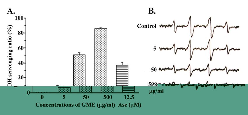
284                                                                                                   ScienceAsia 33 (2007)

total phenolic contents and an active constituent were       equivalents in milligram per gram of the extract, using
quantitated. Interestingly, this is the first report to      a standard curve generated with gallic acid. The
discover the cytoprotective activity of GME on human         concentrations of gallic acid (GA; mg/ml) in the reactions
ECV304 endothelial cells using the MTT assay to study        were analyzed from the following calibration curve
the survival rate of H2O2 treated cells and DCFH-DA          equation, determined by linear regression:
fluorescent probe to study the inhibition of intracellular      Absorbance = (21.51 x [GA]) + 0.1004, R2 = 0.9608
oxidative stress. We hope that our data will be the basis
to propose the preliminary mechanisms of radical             Reducing power
scavenging activities related to the cytoprotective              The Fe 3+ reducing power of the extract was
activity of G. mangostana extract.                           determined according to the method of Oyaizu8. The
                                                             extract (2 ml) was mixed with 0.2 M phosphate buffer,
MATERIALS AND METHODS                                        pH 6.6 (2 ml) and 1% potassium ferricyanide (2 ml). The
                                                             mixture was then incubated at 50°C for 20 min.
Plant material and extract preparation                       Afterwards, the mixture was stopped by adding 10%
    Garcinia mangostana hulls were collected from            trichloroacetic acid (2 ml) and then centrifuged at
Chantaburi Province, Thailand, during the summer             3,000 rpm for 10 min. The upper layer of supernatant
time in May 2004. Fruits were cleaned with running tap       (2 ml) was mixed with distilled water (2 ml) and 0.1%
water and fresh hulls were separated and chopped into        FeCl3 solution (0.5 ml). The absorbance was measured
pieces. They were dried under shade at room                  at 700 nm against a blank with a spectrophotometer,
temperature for 5 days and the air-dried hulls were          and ascorbic acid was used as a standard. Higher
then ground to powder for extraction.                        absorbance of the reaction mixture indicated greater
    An amount of 1 kg of powdered sample was                 reducing power. The percents of reducing power were
extracted with 5 L of methanol for a week with               presented as ascorbic acid equivalents using a
maceration at 37°C. The extract was then collected and       calibration curve between the absorbance of the
filtered through Whatman No.1 filter paper in a              reaction and the percent of the reducing power ability
Buchner funnel under vacuum. The filtrate was                of ascorbic acid:
concentrated by evaporation with a vacuum rotary                 OD = (0.0146 x [percent]) + 0.0016, R2 = 0.9999
evaporator at 45°C to yield 183.8 g of dried methanolic
extract. The dried extract was stored at 4°C for further     Ferrous ion chelating activity
uses. The methanolic extract was standardized by                 The chelating of ferrous ion by the extract was
quantifying the amount of α-mangostin, an active             estimated by the method of Gülçin9. Briefly, the extract
compound, using HPLC equipped with the BDS Hypersil          (2 ml) was added to a solution of 2 mM FeCl2 (0.1 ml).
C18 column (46 mm x 250 mm). The extract solution            The reaction was initiated by the addition of 5 mM
(5 ml) was injected in each run with 0.1% ortho-             ferrozine (0.2 ml) and the mixture was shaken
phosphoric acid and acetonitrile as mobile phase at 1        vigorously and left standing at room temperature for
ml/min, 25°C. The HPLC system was precalibrated with         5 min. Absorbance was measured at 562 nm and EDTA
α-mangostin standard (5-500 μg/ml) to evaluate the           was used as a positive control. The percents of inhibition
linear correlation curve. The amount of α-mangostin          of ferrozine-Fe2+ complex formation were given by the
was determined from the calibration curve of α-              following formula:
mangostin and the peak area of each concentration of         Ferrous ion chelating activity (%) = [1 - (A1 - A2) / A0] x 100%
the samples and calculated by the following linear               where A0 was the absorbance of the control (the
equation:                                                    mixture without the extract), A1 was the absorbance of
     Area = (11006 x [α-mangostin]) - 42616, R2 = 0.9998     the mixture in the presence of the extract and A2 was
                                                             the absorbance without ferrozine.
Total phenolic contents
    The extract was diluted with the same solvent used       DPPH scavenging activity
for extraction, to a suitable concentration for analysis.        The antioxidant activity of the extract was estimated
Total phenolic contents of GME were assessed                 on the basis of the radical scavenging effect of the
approximately by using the Folin-Ciocalteu phenol            stable DPPHi10. Various concentrations of the extract
reagent method7. The extract (1 ml) was added with           were added to a methanolic 0.4 mM DPPHi solution
10% Folin-Ciocalteu reagent and 0.5% sodium                  (0.1 ml) in a 96 well plate. The reaction mixture was
carbonate (0.5 ml). The contents were thoroughly             shaken vigorously and allowed to stand for 30 min at
mixed and allowed to stand for 30 min. Absorption at         37°C. The degree of DPPHi purple decolorization to
750 nm was measured in a spectrophotometer. The              DPPHH yellow indicated the scavenging efficiency of
total phenolic contents were expressed as gallic acid        the extract. The absorbance of the mixture was
ScienceAsia 33 (2007) 285
determined at 517 nm using UV-Vis microplate reader mixture containing 2 ml of the extract at different
and ascorbic acid was served as a positive control. concentrations and 50 mM SNP (0.5 ml) in 10 mM PBS
Lower absorbance of the reaction mixture indicated was incubated at 37°C for 60 min. An aliquot (0.5 ml)
higher free radical-scavenging activity. The scavenging of the incubation solution was pipetted out and diluted
activity against DPPHi was calculated using the following with 0.5 ml of Griess reagent (1% sulfanilamide in 5%
equation: H3PO 4 and 0.1% N-(1-naphthyl)ethylenediamine
Scavenging activity (%) = [1 - (A1 - A2) / A0] x 100% dihydrochloride (NED). The absorbance of the
where A0 was the absorbance of control (DPPHi chromophore that formed during diazotization of
solution without the extract), A1 was the absorbance of nitrite with sulfanilamide and subsequent coupling with
DPPHi solution in the presence of the extract and A2 NED was immediately recorded at 540 nm. The
was the absorbance without DPPH· solution. absorbance from various concentrations of sodium
nitrite salt treated the same way with Griess reagent
Hydroxyl radical scavenging activity was plotted for a standard curve. The amount of NO2¯
The iOH was measured by spin trapping iOH with (µM) was calculated from the following calibration
DMPO and the resultant DMPO-OH adduct was curve equation, determined by linear regression:
detected with an ESR spectrometry11. The ESR spectra Absorbance = (0.0276 x [NO2¯]) + 0.1162, R2 =
were measured at room temperature after mixing 0.02 0.9823
ml of 0.1 mM H2O2 with 0.01 ml of 2 M DMPO, 0.05 ml The capability to scavenge iNO radicals was
of the extract and 0.02 ml of 0.05 mM Fe2+ using the X- calculated using the following equation:
band ESR spectrometer (JES-RE1X, JEOL, Kyoto, Japan). Scavenging activity (%) = [1 - (A1 - A2) / A0] x 100%
ESR spectrometer setting parameters were as follows: where A0 was the absorbance of the control (the
external magnetic field 337.5 mT ± 5 mT, field reaction mixture without the extract), A 1 was the
modulation 0.1 mT of 100 kHz, microwave 10 mW of absorbance in the presence of the extract and A2 was
9.43 GHz. Ascorbic acid and ethanol were used as the absorbance without Griess reagent. α-Tocopherol
controls. The hydroxyl radical scavenging rate of the was used as a standard.
extracts was calculated by the following equation:
Scavenging rate = [(h0 - hx) / h0] x 100% Antioxidant activity in linoleic acid emulsion system
where hx and h0 were the ESR signal intensities of The degree of oxidation due to the formation of
reactions in the presence and absence of the extract, alkoxy radicals by the redox reaction with ferrous ions
respectively. (reducing agents) in a linoleic acid emulsion at
physiological pH was measured by the thiocyanate
Superoxide radical scavenging activity method13. The reaction mixture containing 0.5 ml of
In the ESR spin trapping, the generated superoxide the extract, 0.2 M sodium phosphate buffer, pH 7.0, (2
radicals were trapped by DMPO to form the spin ml) and linoleic emulsion (2.5 ml) was incubated at
adducts, DMPO-OOH using hypoxanthine-xanthine 37°C. Aliquots (1 ml) were taken at different intervals
oxidase system and detected with X-band ESR during incubation which was further diluted with 75%
spectrometry11. The mixture contained 0.01 ml of 4 ethanol (4.7 ml), 200 mM FeCl2 (0.1 ml) in 3.5% HCl and
mM hypoxanthine, 0.01 ml of 20 mM DTPA, 0.01 ml of 30% KSCN. The resulting red chromogen of the ferric
2 M DMPO, 0.05 ml of the extract and 0.02 ml of 0.4 (III)-thiocyanate complex could be measured at 500
U/ml xanthine oxidase. ESR spectra were measured at nm. The inhibition of lipid peroxidation (LPI) in percent
room temperature with ESR spectrometry under the was calculated by the following equation:
above conditions. Superoxide dismutase (SOD) was LPI (%) = [1 - (A1 - A2) / A0] x 100%
used as a control. The superoxide radical scavenging where A0 was the absorbance of control (the reaction
rate of the extract was calculated by the following mixture without the extract), A1 was the absorbance in
equation: the presence of the extract, and A2 was the absorbance
Scavenging rate = [(h0 - hx) / h0] x 100% without potassium thiocyanate solution. α-Tocopherol
where hx and h0 were the ESR signal intensities of was used as a standard.
reactions in the presence and absence of the extract,
respectively. Cell culture
Human umbilical vein endothelial cell line ECV304,
Nitric oxide radical scavenging assay obtained from the American Type Culture Collection
Sodium nitroprusside (SNP) in aqueous solution at (Rockville, MD), was grown in RPMI 1640 medium
physiological pH, spontaneously generated iNO which supplemented with fetal bovine serum, 100 units/ml of
interacted with oxygen to produce nitrite ion, which penicillin G and 0.1 mg/ml of streptomycin sulfate in a
was estimated using Griess reagent12. The reaction humidified atmosphere of a 5% CO2 at 37°C.286 ScienceAsia 33 (2007)
Cytoprotective activity Statistical analysis
Cell viability was determined by measuring the All the experiments were carried out in triplicate
metabolism of a tetrazolium substrate MTT6. Cells were and the results were expressed as mean ± SD. Statistical
seeded on 96-well plates at a density of 2x10⁴ cells/well analysis was performed using one-way analysis of
and incubated for 24 h. The medium was then aspirated variance (ANOVA) and the Student t - test. The values
and replaced with various concentrations of the extract of P < 0.01 and P < 0.05 were considered statistically
for 1 h. Afterwards, medium was removed and as significant. The amount of the effective concentration
incubated with 0.1 ml of H2O2 (1 mM) for 6 h. After of the extract needed to inhibit free radicals by 50%,
exposure to H2O2, the MTT solution (1 mg/ml) was IC50, was graphically estimated by interpolation from
incubated with cells for 60 min. MTT in solution is linear regression analysis between the scavenging
converted to a blue formazan crystal by mitochondrial activities (%) versus various concentrations of the
succinate dehydrogenase of living cells. The crystal extract.
was solubilized from cells with isopropanol and its
absorbance determined at 590 nm. The percents of cell RESULTS
viability were calculated with the following equation:
Cell viability (%) = (A1 / A0) x 100% Total phenolic contents
where A0 was the absorbance of the negative control It was well-known that plant phenolics are highly
(cells without the extract) and A1 was the absorbance effective free radical scavengers and antioxidants. G.
in the presence of the extract. mangostana contained high amounts of xanthones, a
class of phenolic compounds (Fig 1). In this study, the
Inhibition of intracellular reactive oxygen species contents of total phenolics in GME were determined
(ROS) spectrometrically according to the Folin-Ciocalteu
Intracellular ROS production was estimated using method and calculated as gallic acid equivalents (GAE).
fluorescent DCFH-DA probe14. Briefly, 2 x 10⁵ cells/ml It was shown that GME contained phenolic compounds
were pre-treated with various concentrations of the at 60.2 mg GAE. This result indicated that the potent
extract for 1 h prior to DCFH-DA (0.01 mM) exposure antioxidant activity of GME may be related to the
for 30 min. H2O2 (1 mM, 500 ml) was then added and phenolic compounds in the extract.
incubated at 37°C for 1 h. Intracellular ROS production
was evaluated by spectrofluorometer (Perkin-Elmer, Reducing power
MA). The percent of ROS production was calculated The direct correlation between antioxidant activity
according to the following equation: and reducing power of certain plant extracts were
Relative amount of intracellular ROS (%) = (FI1 / FI0) x 100% reported. The presence of reductants (antioxidants) in
where FI0 was the fluorescence intensity of the the extracts would result in the reduction of iron (III)
negative control (cells without the extract) and FI1 was to iron (II). The amount of iron (II)-ferricyanide complex
the fluorescence intensity in the presence of the extract could be determined by measuring the formation of
at an excitation wavelength of 485 nm and an emission Perl’s Prussian blue at 700 nm15. It was found that the
wavelength of 530 nm. reducing ability of GME against Fe3+ increased with
increasing concentration. GME showed 50% of activity
Fig 1. The important xanthones of G. mangostana: (1) nucleus of xanthone; (2) α-mangostin; (3) β-mangostin; (4) γ-mangostin;
(5) garcinone E; (6) 9-hydroxycalabaxanthone.ScienceAsia 33 (2007) 287
(IC50) at 84.35 μg/ml, while ascorbic acid had an IC50 of dependent manner with an IC50 value of 20.50 μg/ml
65.7 μg/ml. The result indicated that GME contained (Table 1).. Although the DPPHi radical-scavenging ability
electron donors and possessed the ability to reduce of GME was below that of ascorbic acid (IC50 value =
iron (III) to iron (II) in a concentration-dependent 13.57 μg/ml), it was evident that GME did show the high
fashion (Table 1). proton-donating ability on DPPHi to form stable
DPPHH molecules.
Ferrous ion chelating activity
Ferrozine could quantitatively form complexes with Hydroxyl radical scavenging activity
Fe2+. In the presence of chelating agent, the complex Hydroxyl radical (iOH) was produced by the Fenton
formation was disrupted with the result that the red reaction and trapped by DMPO. The typical 1:2:2:1 4-
color of the complex was decreased. Measurement of line ESR spectra of the DMPO-OH adduct were
color reduction, therefore, allowed the estimation of observed as shown in Fig 2. The correlation curve of
the chelating activity of the coexisting chelator. As scavenging ratio (%) and the concentrations of the
shown in Table 1, GME interfered the formation of extract was plotted to estimate its ability. When the
Fe2+- ferrozine complex and its activity showed an IC50 amount of iOH radicals was diminished, the peak high
value at 19.26 μg/ml. This indicated that GME captured of spectrum was reduced. GME scavenged i·· OH in a
Fe2+ before it formed the complex with ferrozine and dose-dependent manner with an IC50 value of 47 μg/ml.
the activity of GME was 3-times less than EDTA (IC50 In the other hand, GME showed strong activity to inhibit
value 6.49 μg/ml). the generation of iOH radicals from the Fenton reaction.
DPPH scavenging activity Superoxide radical scavenging activity
The DPPH assay was used to preliminarily screen In Fig 3, O2i·· ¯ scavenging activity was investigated
for antioxidant activity of the extract. The proton- with the hypoxanthine-xanthine oxidase system and
radical scavenging action is known as an important trapped by DMPO. The typical ESR signals of the DMPO-
mechanism of antioxidants. GME decolorized the OOH spin adduct were detected. The correlation curve
purple of DPPHi to the yellow of DPPHH in a dose- of the peak height and the concentration was calculated
Table 1. Antioxidant activities of methanolic extract from Garcinia mangostana (GME).
Method Sample Concentration(
Concentration(μμg/ml) Activity (%) IC50(μg/ml)
RP GME 5 4.30 ± 1.28 84.35
25 19.40 ± 1.06
100 56.35 ± 0.93
200 86.75 ± 2.11
Asc 100 77.42 ± 2.08 65.71
FC GME 5 40.51 ± 0.27 19.26
25 54.43 ± 0.01
100 78.12 ± 3.37
200 84.86 ± 2.90
EDTA 10 73.98 ± 4.20 6.49
DPPH GME 5 19.76 ± 3.05 20.50
25 64.36 ± 0.20
100 84.38 ± 4.12
200 84.13 ± 2.10
Asc 100 85.43 ± 2.50 16.00
NO GME 5 10.68 ± 2.14 55.61
25 34.49 ± 0.55
100 61.39 ± 1.64
200 67.95 ± 0.47
Toc 100 62.05 ± 1.60 24.98
LPO GME 5 22.75 ± 1.51 9.43
25 61.97 ± 1.80
100 71.74 ± 1.57
200 75.44 ± 1.67
Toc 100 75.74 ± 2.04 34.01
RP, reducing power presented as ascorbic acid equivalents; FC, ferrous ion chelating activity;
DPPH, DPPH scavenging activity; NO, nitric oxide scavenging activity; LPO, Inhibition of lipid peroxidation;
Asc, Ascorbic acid; Toc, α-Tocopherol; GME, Methanolic extract from G. mangostana hulls; IC50,
the concentration at which 50% of the maximum activity was produced.288 ScienceAsia 33 (2007)
Fig 2. The scavenging effect of GME on hydroxyl radicals was determined with the Fenton reaction and measured using ESR
spectroscopy. (A) The OHi scavenging activity of GME was shown in a dose-dependent manner and compared with
ascorbic acid (Asc). (B) ESR spectrum of spin adduct of DMPO-OH was demonstrated to study the effect of GME on OHi.
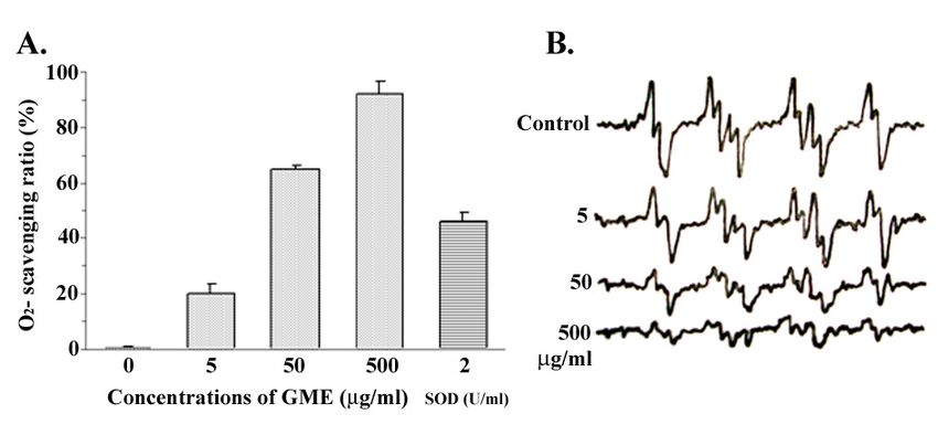
Fig 3. The scavenging effect of GME on superoxide radicals was determined with hypoxanthine-xanthine oxidase system and
measured using ESR spectroscopy. (A) The O2i¯ scavenging activity of GME was shown in a dose-dependent manner and
compared with superoxide dismutase (SOD). (B) The ESR spectrum of the spin adduct of DMPO-OOH was determined
to study the effect of GME on O2i¯.
to find the ability of the extract. GME possessed Inhibition on lipid peroxidation
powerful O2i¯ scavenging activity in a dose-dependent GME not only exhibited excellent ability for various
manner with an IC50 value of 25 μg/ml. We found that radical scavenging activities, but also suppressed lipid
GME acted as O2i¯ scavenger better than iOH scavenger. peroxidation. GME successfully inhibited the oxidation
of linoleic acid which was determined by a ferric
Nitric oxide scavenging activity thiocyanate (FTC) assay. The result demonstrated that
Formation of nitric oxide by SNP in aqueous GME inhibited lipid peroxidation in a concentration-
solution at physiological condition has been reported dependent manner with an IC50 value of 9.43 μg/ml,
to occur by spontaneous oxidation. iNO is a very while α-tocopherol showed IC50 value at 34.01 μg/ml
unstable species, under aerobic condition it reacts (Table 1).
with oxygen to produce, through intermediates such
as NO2, N2O4, N3O4, the stable products NO3¯and NO2¯. Cytoprotective activity
i
NO radicals were determined indirectly with Griess The cytoprotective activity of GME was studied on
reagent through the inhibition of nitrite production. As ECV304 cells after exposure with H2O2 as a cytotoxic
shown in Table 1, GME possessed iNO scavenging agent and oxidative stress inducer. As shown in Fig 4,
activity with an IC50 value of 55.61 μg/ml, while α- H2O2 decreased cell viability and its cytotoxic effect
tocopherol had an IC50 value of 24.98 μg/ml. was attenuated in the presence of GME. The resultScienceAsia 33 (2007) 289
Fig 4. Cytoprotective effect of GME on H2O2-induced ECV304 Fig 5. Effect of GME on ROS production in ECV304 cells as
cells was determined by the MTT assay. Cell controls determined using DCFH-DA fluorescent probe. Cell
without extract and H2O2 exposure had 100% of cell controls were cultured without extract and H2O2
viability ( ), whereas cells treated with H2O2 but exposure ( ), whereas the highest oxidative stress-
without extract showed the lowest percent of cell induced cells were grown in the presence of H2O2 (1
viability ( ). The pre-incubation of the extract on cells mM) without extract ( ). The inhibition of ROS
prior to H2O2 (1 mM) treatment was determined to be production in cells was conducted by pre-incubating
the cytoprotective ( ). The data were expressed as the extract with cells prior to H2O2 treatment and
mean ± SD of triplicate measurements. A P value < 0.05 detected by the DCFH-DA probe ( ). The relative
demonstrated significant cytoprotective activity. amount of ROS (%) is shown as mean ± SD of triplicate
measurements. A P value < 0.05 displayed significant
inhibitory activity.
demonstrated that GME at 2.5-25 μg/ml exhibited
significant cytoprotective effects on human ECV304 contained phenolic compounds at 60.2 mg GAE. It was
endothelial cells. The cell viability increased to 80.9% proposed here that the phenolic compounds of GME
at 25 μg/ml GME (P < 0.05), while the viability of H2O2- may play an important role in the observed antioxidant
treated cells without GME was 49.7%. activities through free radical quenching, electron
transfer, radical addition or radical recombination in
Inhibition on intracellular oxidative stress various tested systems. The majority of phenolic
Accumulation of intracellular ROS was detected compounds in the fruit hull of G. mangostana belong to
with DCFH-DA, which is freely permeable to cell the xanthone family, which may help to offset chronic
membranes. Once inside cells, DCFH-DA is hydrolyzed diseases related with ROS. Two major xanthones were
by the esterase activity to DCF, which can be trapped found in the fruit hull of this plant namely α-mangostin
intracellularly. DCF was then able to interact with and γ-mangostin17. In this study, α-mangostin, one of
peroxides and form the fluorescent 2′,7′- the major and interesting active compounds from G.
dichlorofluorescein, which was readily detected by mangostana was identified and quantitated with HPLC
spectrofluorometer14. H2O2 was used to produce the chromatography to standardize the quantity and quality
oxidative stress in human endothelial ECV304 cells. As of the extract. α-Mangostin was present in our GME at
shown in Fig 5, the intracellular ROS accumulation 25.19 ± 0.22 g/100 g of GME. Recently, the biological
resulting from H2O2 exposure was significantly reduced activities of the α-mangostin and γ-mangostin xanthones
in the presence of GME at 2.5-25 μg/ml (P < 0.05) in a were revealed. α-Mangostin showed antiproliferative
dose-dependent manner. activity against human leukemia HL60 cells through
apoptotic cell death18, while γ-mangostin inhibited the
DISCUSSION activity of cyclooxygenases (both COX-1 and COX-2)
and suppressed the conversion of arachidonic acid to
Phenolic compounds, such as benzoic acid prostaglandin E219. However, there has been no
derivatives, are commonly found in the plant kingdom previous report on the antioxidant activities of
and have been extensively exploited because of their xanthones from this plant against various kinds of
multiple biological activities, including antioxidant radical species to reveal the important mechanism.
effects. Since they contain at least one hydroxy- From this study, we assumed that xanthones in the
substituted aromatic ring system, they can form chelate extract may have an important role on antioxidant
complexes with metal ions and are easily oxidized, as activity.
well as serving as important units for donating To verify the radical scavenging ability of GME,
electrons16. In the current study, it was found that GME several methods were served to investigate its ability290 ScienceAsia 33 (2007)
compared with commercial antioxidants against radical H2O2 → iOH + OH- + Fe3+). Therefore, iOH scavenging
species by UV-Vis and ESR spectroscopy. Starting from activity could be influenced by electron-donating and
the study on reducing ability, the result showed that metal-chelating abilities of xanthones in GME. iOH is
GME expressed reducing ability as an electron donor widely implicated as the most potent oxidant and the
and possessed the ability to reduce iron (III) to iron (II) major damaging species in free radical pathology. It can
in a concentration-dependent fashion. The reducing initiate lipid peroxidation, cause DNA strand breaks,
power of GME might be due to the di- and and indiscriminatingly oxidize virtually any organic
monohydroxyl substitutions in the aromatic ring of molecule22. This result revealed that GME possessed
phenolic compounds, which possessed potent powerful iOH scavenging activity in Fig 2, while Fe2+
hydrogen donating abilities, and breaking of the free chelating activity was 3-times less than EDTA. It could
radical chain through donating a hydrogen atom20. be explained that the active compounds in GME might
Several antioxidants possess metal chelating activity have the main role to scavenge iOH radicals generated
to reduce the redox potential and stabilize the oxidized from Fenton reaction by donating electrons. Although
form of the metal ions, which related to the obstruction α-mangostin and β-mangostin as O-protected
on the peroxidative process and oxidative damage. xanthones might show low DPPH scavenging activity,
However, chelating activity might not be the main role these xanthones might unexpectedly possess powerful
i
of the extract, because its activity was 3-times less than OH radical scavenging activity due to their chelating
EDTA which was used as a standard (IC50 value = 6.49 effect with Fe2+ which prevents the generation of iOH
μg/ml). This result indicated that compounds in GME radicals in the system.
were not potent iron chelators; it could largely not O2i¯ is the first reactive oxygen radical produced by
obstruct the generation of iOH radicals from the Fenton one-electron reduction of molecular oxygen in
reaction. iOH radicals were still produced from the metabolism process and the source of other radicals.
reaction between iron (II) and H 2O 2. The more O2i¯ scavenging activity was investigated with the
important role of GME might be related to the hypoxanthine-xanthine oxidase system and trapping
scavenging of iOH after it was generated from the with DMPO10. The typical ESR signals of the DMPO-
Fenton reaction, whereby it would inhibit the reaction OOH spin adduct were detected, as shown in Fig 3.
of the radicals against biomolecules. Experimental data demonstrated that GME exhibited
DPPHi radicals have been used extensively as stable much stronger scavenging activity against O2i¯ radicals
radicals to preliminarily evaluate the antioxidant compared with iOH radicals. It was likely that GME
activities of plant extracts. GME did show the potent might well show an inhibition of oxidative damage in
proton-donating ability on DPPHi to produce DPPHH, the early stage to protect and/or balance the
which is an important mechanism of antioxidants. physiological system by concerting H-atom abstraction
Xanthones in the extract could transfer labile H-atoms (electron donation) and catalyzing the dismutation of
to radicals. Lee et al21 investigated the antioxidant O2i¯ (protonation effect).
activity of the xanthones of Cudrania tricuspidata. It was Abnormally high level of iNO has been implicated
found that a xanthones of C. tricuspidata (cudraxanthone in various inflammatory and degenerative diseases.
D), which contain a dihydroxyl group in the B-ring, Inflammatory cells, which were components of
exhibited strong radical scavenging activities. Such a atherosclerotic plaques, produced extracellular iNO
structure is found in γ-mangostin and garcinone E of G. which presumably interfered with the endothelial
mangostana. On the other hand, cudraxanthone C of C. proliferation 23. Thus, the pathophysiology of the
tricuspidata showed lower activity because one of its endothelium was modulated by iNO. Our indirect
hydroxyl groups in the vicinal diol was protected, as method to investigate iNO scavenging activity was based
found in α-mangostin, β-mangostin and 9- on the release of iNO from SNP in physiological solution
hydroxycalabaxanthone of G. mangostana. It could be and determined with Griess reagent through the
rationalized that O-protected xanthones could not inhibition of nitrite production. The decreased level of
transfer to quinone, while those with vicinal dihydroxyl nitrite in the reaction indicated an increased scavenging
groups could transfer to quinone easily by releasing effect against iNO since the amounts of nitrite were
two electrons in the molecule. On the other hand, such directly proportional to the amounts of iNO radicals in
a xanthone could easily donate electrons or protons to the reaction mixture. GME activity was weaker than
radical molecules to scavenge and quench the toxicity the standard α-tocopherol in the system. However, the
of the radicals. result indicated that some compounds in GME
In this study, the radical scavenging activity of GME scavenged iNO in the reaction, which might influence
against iOH and O2i¯ was determined by spin trapping the protection of cells, especially endothelial cells, by
technique and measuring with ESR spectrometry. iOH neutralizing the iNO produced in oxidative stress.
radicals were generated by the Fenton system (Fe2+ + Oxidation of unsaturated fatty acid in biologicalScienceAsia 33 (2007) 291
membranes leads to the formation and propagation of of Bio-function Science, Faculty of Pharmaceutical
lipid radicals (L i ), the uptake of oxygen, the Sciences, Kyushu University was greatly appreciated
rearrangement of the double bonds in unsaturated for his kind support and suggestions on this work.
lipids and the eventual destruction of membrane lipids
to produce breakdown products such as
malondialdehyde, which is known to be mutagenic and REFERENCES
carcinogenic24. Lipid peroxidation is a key process in
many pathological events and is one of the reactions 1. Yen GC, Lai HH and Chou HY (2001) Nitric oxide-scavenging
induced by oxidative stress. The GME prevented lipid and antioxidant effect of Uraria crinita root. Food Chem 74 74,
471-8.
peroxidation, which may explain its cytoprotective 2. Lee SE, Hwang HJ, Ha JS, Jeong HS and Kim JH (2003)
property on cell membrane damage caused by radicals Screening of medicinal plant extracts for antioxidative activity.
or toxic substances. Life Sci 73
73, 167-79.
To verify the cytoprotective activity of GME, the 3. Finkel T and Holbrook NJ (2002) Oxidants, oxidative stress
MTT assay was used to determine viability after and the biology of aging. Nature 408408, 239-47.
4. Suksamrarn S, Suwannapoch N, Phakhodee W, Thanuhiranlert
exposure of cells to H2O2 as a cytotoxic agent and J, Ratananukul P, Chimnoi N and Suksamrarn A (2003)
oxidative stress inducer. GME pretreatment was able Antimycobacterial activity of prenylated xanthones from the
to protect cells and increase the number of surviving fruits of Garcinia mangostana. Chem Pharm Bull 51 51, 857-9.
cells significantly. Interestingly, accumulation of 5. Farnsworth NR and Bunyapraphatsara N (1992) Thai medicinal
intracellular ROS using DCFH-DA fluorescent dye was plant: recommended for primary health care system. Mahidol
University, Bangkok.
decreased in GME-pretreated cells after exposure to 6. Moongkarndi P, Kosem N, Kaslungka S, Luanratana O,
H2O2. This could be explained as GME inhibition of Pongpan N and Neungton N (2004) Antiproliferation,
intracellular oxidative stress and protection of the antioxidation and induction of apoptosis by Garcinia
ECV304 cells from oxidative damage. Thus, GME- mangostana (mangosteen) on SKBR3 human breast cancer
pretreated cells survived after exposure with H2O2. cell line. J Ethnopharmacol 90
90, 161-6.
7. Singleton VL, Orthofer R and Lamuela-Raventós RM (1999)
In conclusion, the results obtained from the present Analysis of total phenols and other oxidation substrates and
study clearly revealed that GME possessed antioxidant antioxidants by means of Folin-Ciocalteu Reagent. Methods
and cytoprotective activities through its abilities to Enzymol 299299, 152-78.
scavenge various radicals produced in oxidative stress. 8. Oyaizu M (1986) Studies on products of browning reaction:
Interestingly, the uniqueness of GME was related to antioxidative activities of products of browning reaction
prepared from glucosamine. Japanese J Nutr 44 44, 307-15.
cytoprotective activity in the initial step of lipid .
9. Gülçin I, Beydemir S,, Alici HA, Elmastas, M and Büyükokuroglu
∪
peroxidation in the cell membranes of ECV304 ME (2004) In vitro antioxidant properties of morphine.
endothelial cells by the inhibition of superoxide radical Pharmacol Res 49 49, 59-66.
generation and the donation of hydrogen atoms or 10. Tepe B, Sokmen M and Akpulat HA (2005) In vitro antioxidant
electrons to other radical molecules such as hydroxyl activities of the methanol extract of five Allium species from
Turkey. Food Chem 92 92, 89-92.
radicals, nitric oxide radicals and lipid radicals. From 11. Rosen GM and Rauckman EJ (1984) Spin trapping of
the present study, it was found that GME contained superoxide and hydroxyl radicals. Methods Enzymol 105 105,
xanthones as the main phenolic compounds which 198-209.
may possess antioxidant and cytoprotective activities. 12. Geetha S, Ram MS, Singh V, Ilavazhagan G and Sawhney RC
G. mangostana extract is a good starting source for (2002) Effect of seabuckthorn on sodium nitroprusside-
induced cytotoxicity in murine macrophages. Biomed
further investigation on the exact constituents Pharmacother 56 56, 463-7.
responsible for these activities. Our preliminary study 13. Behera BC, Verma N, Sonone A and Makhija U (2006)
on isolation of methanolic extract compounds, Determination of antioxidative potential of lichen Usnea
including the chemical and biological identification, is ghattensis in vitro. LWT - Food Sci Tech 3939, 80-5.
now in process. We hope that the intensive study on the 14. Royall JA and Ischiropoulos H (1993) Evaluation of 2′,7′-
dichlorofluorescin and dihydrorhodamine123 as fluorescent
outcoming active constituents of G. mangostana will probes for intracellular H2O2 in cultured endothelial cells.
lead to the discovery of a novel botanical-drug for Arch Biochem Biophys 302 302, 348-55.
. .
chemoprevention. 15. Oktay M, Gülçin I and Küfreviolu I (2003) Determination of
in vitro antioxidant activity of fennel (Foeniculum vulgare)
ACKNOWLEDGEMENTS seed extracts. LWT - Food Sci Tech 36 36, 263-71.
16. Rice-Evans CA, Miller NJ, Paganga G (1997) Antioxidant
properties of phenolic compounds. Trends Plant Sci 2 , 152-
The authors would like to thank the Thailand 9.
Research Fund through the Royal Golden Jubilee Ph.D. 17. Chairungsrilerd N, Takeuchi K, Ohizumi Y, Nozoe S, Ohta T
Program for the financial support (Grant No. PHD/ (1996) Mangostanol, a prenyl xanthone from Garcinia
0011/2547). Professor Dr. Hideo Utsumi, Department mangostana. Phytochemistry 43 43, 1099-102.292 ScienceAsia 33 (2007)
18. Matsumoto K, Akao Y, Kobayashi E, Ohguchi K, Ito T, Tanaka
T, Iinuma M, et al (2003) Induction of apoptosis by xanthones
from mangosteen in human leukemia cell lines. J Nat Prod
66
66, 1124-7.
19. Nakatani K, Nakahata N, Arakawa T, Yasuda H, Ohizumi
(2002) Inhibition of cyclooxygenase and prostaglandin E2
synthesis by gamma-mangostin, a xanthone derivative in
mangosteen, in C6 rat glioma cells. Biochem Pharmacol 63 63,
73-9.
20. Shimada K, Fujikawa K, Yahara K and Nakamura T (1992)
Antioxidative properties of xanthones on the auto-oxidation
of soybean oil in cyclodextrin emulsion. J Agric Food Chem
40
40, 945-8.
21. Lee BW, Lee JW, Lee ST, Lee HS, Lee WS, Jeong TS, Park KH
(2005) Antioxidant and cytotoxic activities of xanthones
from Cudrania tricuspidata. Bioorg Med Chem Lett 15 15, 5548-
52.
22. Winterbourn CC (1987) The ability of scavengers to
distinguish OH· production in the iron-catalysed Haber-Weiss
reaction: comparison of four assays for OH·. Free Radic Biol
Med 3 , 33-9.
23. Marcocci L, Maguire JJ, Droy-Lefaix MT and Packer L (1994)
The nitric oxide-scavenging properties of Ginkgo biloba extract
EGb 761. Biochem Biophys Res Commun 201 201, 748-55.
24. Miyake T and Shibamoto T (1997) Antioxidant activities of
natural compounds found in plants. J Agric Food Chem 45 45,
1819-22.You can also read