SARS-COV-2 STRUCTURAL FEATURES MAY EXPLAIN LIMITED NEUTRALIZING-ANTIBODY RESPONSES - NATURE
←
→
Page content transcription
If your browser does not render page correctly, please read the page content below
www.nature.com/npjvaccines

                  COMMENT                      OPEN

                  SARS-CoV-2 structural features may explain limited
                  neutralizing-antibody responses
                  Martin F. Bachmann1,2,3 ✉, Mona O. Mohsen2,3, Lisha Zha1, Monique Vogel1 and Daniel E. Speiser                                     4✉

                      Neutralizing antibody responses of SARS-CoV-2-infected patients may be low and of short duration. We propose here that
                      coronaviruses employ a structural strategy to avoid strong and enduring antibody responses. Other viruses induce optimal and
                      long-lived neutralizing antibody responses, thanks to 20 or more repetitive, rigid antigenic epitopes, spaced by 5–10 nm, present on
                      the viral surface. Such arrays of repetitive and highly organized structures are recognized by the immune system as pathogen-
                      associated structural patterns (PASPs), which are characteristic for pathogen surfaces. In contrast, coronaviruses are large particles
                      with long spikes (S protein) embedded in a fluid membrane. Therefore, the neutralizing epitopes (which are on the S protein) are
                      loosely “floating” and widely spaced by an average of about 25 nm. Consequently, recruitment of complement is poor and
                      stimulation of B cells remains suboptimal, offering an explanation for the inefficient and short-lived neutralizing antibody
                      responses.
                      npj Vaccines (2021)6:2 ; https://doi.org/10.1038/s41541-020-00264-6
1234567890():,;

                  The immune response to severe acute respiratory syndrome                                  evolved strategies to mitigate induction of neutralizing antibodies
                  coronavirus 2 (SARS-CoV-2) infection is initiated by innate immune                        against this domain.
                  activation followed by antigen-specific B- and T-cell responses1.
                  An important mechanism protecting from viral disease is the
                  presence of virus-neutralizing antibodies, which is similar for                           DURATION AND QUALITY OF NEUTRALIZING ANTIBODY
                  almost all viruses causing acute disease followed by pathogen                             RESPONSES TO SARS-COV-2
                  clearance. In line with this, all currently available anti-viral vaccines                 As for infections with other viruses, COVID-19 patients produce
                  are primarily aiming at inducing virus-neutralizing antibodies.                           neutralizing antibodies at lower amounts than non-neutralizing ones.
                  Neutralizing antibodies generally block binding of the virus to                           There is disagreement about the stability of neutralizing antibodies in
                  cellular receptors. In some cases, neutralizing antibodies may                            COVID-19 patients, with several studies reporting stable persis-
                  prevent conformational changes necessary for fusion of the virus                          tence11,12, whereas others showing that neutralizing antibodies to
                  with the cell membrane or proteolytic cleavage. Neutralizing                              coronaviruses wane relatively rapidly, or appear late and remain at
                  antibodies against SARS-CoV-2 are directed against the spike (S)                          low titers13–15. Some patients may even lack long-lasting antibo-
                  protein, which contains multiple antigenic epitopes in the                                dies16. Indeed, there is increasing evidence that protection from
                  receptor-binding domain (RBD) and non-RBD epitopes2. A major                              disease can be short-lived: some patients experienced COVID-19
                  mechanism of neutralization is to block binding of RBD to                                 twice within months, proven by a virus-free interval17,18.
                  angiotensin-converting enzyme 2 (ACE2), the cellular receptor                                Antibody titers generally show an early decay after infection,
                  for the virus. The RBD is localized at the tip of the S protein (Fig. 1).                 because the first antibody wave is based on short-lived plasma
                  The receptor-binding motif (RBM) consists of about 70 aa within                           cells19. The second wave of antibodies is produced by more
                                                                                                            durable plasma cells20. Therefore, one cannot directly compare
                  the RBD and represents the actual amino acids directly interacting
                                                                                                            studies that differ in the time points at which antibodies
                  with ACE2.
                                                                                                            were measured. In addition, studies differ with respect to
                     Neutralizing antibodies are mostly directed against RBD and, in
                                                                                                            laboratory methods. The “gold-standard,” i.e., neutralization assays
                  particular, RBM3–5. Indeed, RBD-specific antibodies closely corre-
                                                                                                            that use live virus requires a safety level 3 laboratory, which is not
                  late with neutralization in convalescent sera3,6,7. Although S is
                                                                                                            always available. Although useful results are obtained by
                  heavily glycosylated, RBD only shows little glycosylation (and one                        alternative approaches (pseudotype neutralizing assays or
                  methylation) and the RBM is non-glycosylated, likely facilitating                         enzyme-linked immunosorbent assays designed to detect RBD-
                  protein–protein interactions with ACE2. This may also indicate that                       specific antibodies), they are less meaningful than the gold-
                  glycosylation of S is probably not the reason for induction of poor                       standard21.
                  neutralizing antibody responses. S is cleaved by furin and the                               The disagreement about the duration of the neutralizing
                  serine proteases TMPRSS2 and TMPRSS4, enabling fusion of viral                            antibody response is not surprising, given the lack of long-term
                  and cellular membranes, and consequent entry of viral RNA into                            follow up as SARS-CoV-2 has appeared less than one year ago.
                  the host cell8. This cleavage site may also be a target for                               Nevertheless, patients with minor or no symptoms often have only
                  neutralizing antibodies9,10. Overall, the RBD/RBM is the immuno-                          low and short-lived neutralizing antibody responses13,14. In
                  logical Achilles heel of the virus. Therefore, the virus may have                         addition, those patients are frequent in the current pandemic.

                  1
                   International Immunology Centre, Anhui Agricultural University, Hefei, China. 2Department of Rheumatology, Immunology and Allergology, University Hospital Bern, Bern,
                  Switzerland. 3Department of BioMedical Research, University of Bern, Bern, Switzerland. 4University Hospital and University of Lausanne, Lausanne, Switzerland. ✉email: martin.
                  bachmann@dbmr.unibe.ch; daniel.speiser@unil.ch

                  Published in partnership with the Sealy Institute for Vaccine Sciences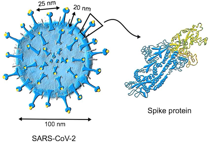
M.F. Bachmann et al.
                  2

                      Fig. 1 Structure of SARS-CoV-2. Coronaviruses have their names from the typical spikes which are made of the spike (S) protein that is
                      inserted in the lipid bilayer membrane of the virus. The receptor-binding domain (RBD) and its receptor-binding motif (RBM) enable
                      interaction with the cell surface receptor ACE2 mediating entry of the virus into host cells. This can be blocked by neutralizing antibodies.
                      Therefore, most neutralizing epitopes are located on RBD/RBM. Besides the S protein, SARS-CoV-2 has two further viral surface proteins (not
                      shown): envelope (E) and matrix (M).

                      Milder symptoms are observed when viral replication is restricted        STRUCTURE FUNCTION CONSIDERATIONS FOR SARS-COV-2
1234567890():,;

                      to the upper respiratory track. This occurs also in the usually mild     Most viruses have highly organized, repetitive and rigid surfaces37.
                      common cold infections caused by the endemic seasonal                    Typical RNA viruses cannot build up complex surfaces because of
                      coronaviruses, typically during winter and spring time20. For these      their limited genome of around 10,000 nucleotides (10 kb). Their
                      viruses, antibody responses that protect from disease are short-         capsid usually consists of multiple copies of only one or two
                      lived14, in the range of 1 year22 or less23, and infections occur        proteins, often arranged in icosahedral symmetry38, readily and
                      regularly, including re-infections with the same virus15,22,24.          efficiently inducing neutralizing antibody responses39. As the
                         Until last year, there existed only two coronaviruses that            vertebrate body is by and large devoid of such extracellular
                      frequently cause severe disease, SARS-CoV-1 and Middle East              repetitive and organized structures, the immune system has
                      Respiratory Syndrome Coronavirus (MERS-CoV). Neutralizing anti-          evolved to recognize such antigen organization as a pathogen-
                      body responses to SARS-CoV-1 can be measured in most                     associated structural pattern (PASP)40. In the 1970s, it was found
                      patients25,26 but may gradually disappear after recovery27. More         that optimal immune responses are induced by at least 12–16
                                                                                               epitopes spaced by 5–10 nm, called the immunon41. Figure 2A
                      is known about the MERS-CoV. This virus keeps on circulating in its
                                                                                               shows a typical RNA virus, with a diameter of 30 nm and 180
                      natural hosts, the dromedary populations, and animals may
                                                                                               copies of a single coat protein spaced by about 5 nm. Such viral
                      experience re-infections. The camels have a high seroprevalence          particles efficiently cross-link B-cell receptors42,43 and are recog-
                      (>90%), but virus transmission is not blocked by previous                nized by natural IgM, which induces the classical pathway of
                      infection28.                                                             complement activation. This facilitates binding of viral particles to
                         Different other reasons may hamper the immune response to             complement receptors followed by B-cell-mediated deposition on
                      coronaviruses, e.g., those concerning the innate immune system,          follicular dendritic cells causing efficient germinal center forma-
                      which is key for early activation of inflammatory cells and               tion44. Furthermore, complement-dependent stimulation of CD21
                      cytokines. SARS-CoV-2 may inhibit dendritic cells29 and interferon       on B cells facilitates induction of long-lived plasma cells, which is
                      (IFN)-I/III responses30,31. Relatively small percentages of patients     essential for durable antibody responses45. There is a vast
                      with severe COVID-19 bear various genetic variants that compro-          literature confirming these considerations for human vaccines,
                      mise innate immune mechanisms32, in particular, type I IFN               where repetitiveness is important for inducing long-lived antibody
                      pathways33, or have autoantibodies against type I IFNs34, which          responses37,46. Hence, repetitive, rigid structures spaced by
                      are likely aggravating diseases severity. Regarding T cells, several     5–10 nm are optimal for complement and B-cell activation,
                      studies showed impaired T-cell responses including CD4 helper            resulting in durable antibody responses.
                      and regulatory T cells29,35. This point could be important, as T cells      Figure 2A outlines the structure of SARS-CoV-2. It is immediately
                      may contribute to protection from disease, although this is not          evident that the structure of this coronavirus is quite different. The
                      proven36. It is also necessary to state that for neutralizing            virion has a relatively large body with a diameter of 100 nm (rather
                      antibodies, there is currently no proof that they indeed mediate         than 30–50 nm). Importantly, the S protein, which has a length of
                                                                                               about 20 nm, is present rather scarcely, floating in a sea of lipid
                      protection from COVID-19. For most current vaccines, neutralizing
                                                                                               bilayer. As mentioned above, RBD is sitting at the top of S;
                      antibodies are considered as correlate of protection from disease,
                                                                                               therefore, some 70 nm away from the center. The viral surface
                      although they do not necessarily equate to the only mechanism of         area, in which RBD is moving within a two-dimensional space,
                      protection. Finally, non-neutralizing antibodies such as those that      20 nm away from the lipid bilayer, can be calculated as 4 × πr2 =
                      fix complement on the viral surface or mediate antibody-                  4 × π70 nm2 = ca 62,000 nm2. Assuming an average number of ca.
                      dependent cellular cytotoxicity may also play a role, although           100 S per virion, each S covers a surface area of about 620 nm2.
                      this is not yet clear36.                                                 This leads to a grid-length of 25 nm, which indicates that S is
                         In the following, we propose that structural adaption of the          spaced by an average of 25 nm (Fig. 2C) rather than the 5–10 nm
                      virus family is co-responsible for the inefficiency of neutralizing       needed for optimal B-cell responses. Epitopes spaced by this large
                      antibody responses to the S protein of SARS-CoV-2, in particular         distance in a non-rigid manner are inefficient in cross-linking
                      RBD/RBM.                                                                 B-cell receptors or recruiting natural IgM antibodies, required for

                      npj Vaccines (2021) 2                                                        Published in partnership with the Sealy Institute for Vaccine Sciences
M.F. Bachmann et al.
3
A B C
D E
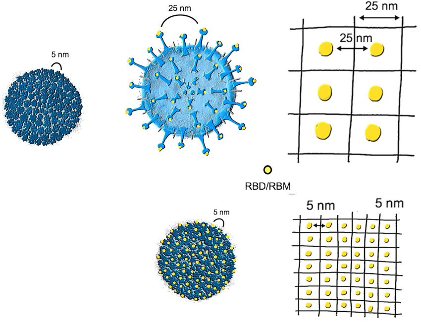
Fig. 2 Distances between neutralizing epitopes. A An example of a classical RNA virus with a capsule made of multiple copies of only
one protein that are rigidly structured, displaying highly immunogenic repetitive neutralizing epitopes spaced by 5–10 nm. This virus is
built with 180 monomers and has a total viral diameter of 30 nm. B, C A coronavirus with its S proteins, showing the distance of the
neutralizing epitopes of about 25 nm, which is large and unfavorable for triggering antibody responses. D, E A virus-like particle (VLP)
built by a viral protein into which the RBM of SARS-CoV-2 is genetically inserted. This VLP displays the neutralizing epitopes with an
optimized spacing of 5 nm.
complement activation and induction of long-live plasma cells. surfaces that allow evasion from immune recognition as a PASP
Hence, SARS-CoV-2 dilutes it’s Achilles heel, RBD on S, in a sea of and induction of enduring neutralizing antibody responses.
lipids and other proteins, avoiding potent neutralizing antibody Besides the non-enveloped viruses, also many enveloped
responses. viruses (Influenza, Rabies, Sindbis, and Vesicular Stomatitis
The S protein forms a trimer. Consequently, the RBD will display Virus) display highly immunogenic antigen arrays. Lytic viruses
three identical epitopes favorably spaced by about 3–5 nm. As generally induce potent antibody responses and produce
discussed above, three epitopes are, however, not enough to serotypes37. A notable exception were adenoviruses, which,
optimally activate B cells. On the contrary, epitopes occurring in similar to SARS-CoV-2, are lytic but also do not form serotypes.
low numbers inhibit, rather than activate, B-cell responses. Indeed, Parallel to coronaviruses, adenoviruses also dilute out the
Dintzis et al.47 concluded that increasing epitope density in a neutralizing epitopes on the surface of the virion thereby
molecular structure increases its immunogenicity if the threshold apparently avoiding stringent long-term neutralization (hence,
number of ∼20 is reached. In contrast, increasing the density in a no serotype formation). Similar to coronaviruses, adenoviruses
molecular structure below the threshold number increases its have a proofreading replication system, as they are DNA viruses49.
tolerogenicity47. Thus, trimeric RBD may reduce rather than Hence, the presence of proofreading may allow viruses to escape
increase neutralizing antibody responses. immune recognition as a PASP.
An additional consideration is the length of the viral genome, as We have previously discussed37 that viral structure predicts host
SARS-CoV-2 is encoded by 30,000 RNA nucleotides rather than the antibody responses and serotype formation. An interesting
usually about 10,000 nucleotides seen for most other RNA viruses. observation was that viruses with highly organized and rigid
Indeed, the longer genome of coronaviruses includes an RNA surfaces induce T-cell independent antibody responses and form
proofreading system, required for keeping the viral population serotypes, whereas viruses with a less rigid structure avoid potent
viable based on sufficient genome stability48. Hence, in contrast to antibody responses and do not form serotypes. However, the
other RNA viruses, coronaviruses can build up relatively complex “dilution” of the neutralizing epitopes performed by coronaviruses,
Published in partnership with the Sealy Institute for Vaccine Sciences npj Vaccines (2021) 2M.F. Bachmann et al.
4
as described in this study, is a unique strategy in the world of 11. Gudbjartsson, D. F. et al. Humoral immune response to SARS-CoV-2 in Iceland. N.
RNA viruses. Engl. J. Med. https://doi.org/10.1056/NEJMoa2026116 (2020).
12. Isho, B. et al. Persistence of serum and saliva antibody responses to SARS-CoV-2
spike antigens in COVID-19 patients. Sci. Immunol. 5, eabe5511 (2020).
IMPLICATIONS FOR VACCINE DESIGN 13. Tay, M. Z., Poh, C. M., Rénia, L., Macary, P. A. & Ng, L. F. P. The trinity of COVID-19:
immunity, inflammation and intervention. Nat. Rev. Immunol. 20, 363–374 (2020).
If the inefficient and short-lived neutralizing antibody responses 14. Sariol, A. & Perlman, S. Lessons for COVID-19 immunity from other coronavirus
induced by SARS-CoV-2 are indeed caused by the unusually infections. Immunity 53, 248–263 (2020).
large distance between neutralizing epitopes embedded in a 15. Jeyanathan, M. et al. Immunological considerations for COVID-19 vaccine stra-
fluid membrane, this has important implications for vaccine tegies. Nat. Rev. Immunol. 20, 615–632 (2020).
design. Specifically, by simple genetic extraction of RBD or RBM 16. CGTN. Recovered coronavirus patients are still prone to reinfection. Available at:
from SARS-CoV-2 followed by grafting onto highly repetitive and https://www.youtube.com/watch?v=GZ99J7mlaIQ. Accessed 10 Aug 2020.
immunogenic nanoparticles or virus-like particles (VLPs), one 17. Bentivegna, E. et al. New IgM seroconversion and positive RT-PCR test after
may render the poorly immunogenic RBD/RBM into a highly exposure to the virus in recovered COVID-19 patient. J. Med. Virol. https://doi.org/
10.1002/jmv.26160 (2020).
immunogenic version of it (Fig. 2D), with high numbers of
18. Ledford, H. COVID-19 reinfection: three questions scientists are asking. Nature
accessible epitopes at optimal distancing (Fig. 2E). Indeed, 585, 168–169 (2020).
chemical coupling or conjugation by the Spy-Catcher or similar 19. Alter, G. & Seder, R. The power of antibody-based surveillance. N. Engl. J. Med.
methods of RBD to VLPs results in highly immunogenic vaccine https://doi.org/10.1056/NEJMe2028079 (2020).
candidates that stimulate production of high levels of neutraliz- 20. Amanna, I. J., Carlson, N. E. & Slifka, M. K. Duration of humoral immunity to
ing antibodies in test animals50,51. An alternative strategy, which common viral and vaccine antigens. N. Engl. J. Med. 357, 1903–1915 (2007).
facilitates large scale production, is represented by genetic 21. Seow, J. et al. Longitudinal observation and decline of neutralizing antibody
fusion of RBD/RBM onto VLP-surfaces. Such approaches may responses in the three months following SARS-CoV-2 infection in humans. Nat.
represent attractive options that we and others are currently Microbiol. 5, 1598–1607 (2020).
22. Reed, S. E. The behaviour of recent isolates of human respiratory coronavirus
following50,52,53.
in vitro and in volunteers: evidence of heterogeneity among 229E-related strains.
J. Med. Virol. 13, 179–192 (1984).
23. Callow, K. A., Parry, H. F., Sergeant, M. & Tyrrell, D. A. The time course of the
CONCLUSION immune response to experimental coronavirus infection of man. Epidemiol. Infect.
SARS-CoV-2 induces inefficient neutralizing antibody responses 105, 435–446 (1990).
that are short-lived. In contrast to the other RNA virus families, 24. Galanti, M. & Shaman, J. Direct observation of repeated infections with endemic
which display arrays of neutralizing epitopes spaced by 5–10 nm coronaviruses. J. Infect. Dis. https://doi.org/10.1093/infdis/jiaa392 (2020).
in a rigid manner, SARS-CoV-2 displays a low number of 25. Temperton, N. J. et al. Longitudinally profiling neutralizing antibody response to
neutralizing epitopes spaced by 25 nm in a non-rigid manner, as SARS coronavirus with pseudotypes. Emerg. Infect. Dis. 11, 411–416 (2005).
26. Nie, Y. et al. Neutralizing antibodies in patients with severe acute respiratory
the S protein is embedded in a fluid membrane. Hence, SARS-
syndrome-associated coronavirus infection. J. Infect. Dis. 190, 1119–1126 (2004).
CoV-2 escapes an efficient neutralizing antibody response by 27. Cao, W.-C., Liu, W., Zhang, P.-H., Zhang, F. & Richardus, J. H. Disappearance of
structurally avoiding immunogenic display of its neutralizing antibodies to SARS-associated coronavirus after recovery. N. Engl. J. Med. 357,
epitopes. 1162–1163 (2007).
28. Hemida, M. G. et al. Longitudinal study of Middle East Respiratory Syndrome
coronavirus infection in dromedary camel herds in Saudi Arabia, 2014-2015.
Received: 16 October 2020; Accepted: 23 November 2020;
Emerg. Microbes Infect. 6, e56 (2017).
29. Zhou, R. et al. Acute SARS-CoV-2 infection impairs dendritic cell and T cell
responses. Immunity 53, 864–877.e5 (2020).
30. Blanco-Melo, D. et al. Imbalanced host response to SARS-CoV-2 drives develop-
ment of COVID-19. Cell 181, 1036–1045.e9 (2020).
REFERENCES 31. Sa Ribero, M., Jouvenet, N., Dreux, M. & Nisole, S. Interplay between SARS-CoV-2
1. Thevarajan, I. et al. Breadth of concomitant immune responses prior to and the type I interferon response. PLoS Pathog. 16, e1008737 (2020).
patient recovery: a case report of non-severe COVID-19. Nat. Med. 26, 32. Beck, D. B. & Aksentijevich, I. Susceptibility to severe COVID-19. Science 370,
453–455 (2020). 404–405 (2020).
2. Brouwer, P. J. M. et al. Potent neutralizing antibodies from COVID-19 patients 33. Zhang, Q. et al. Inborn errors of type I IFN immunity in patients with life-
define multiple targets of vulnerability. Science, https://doi.org/10.1126/science. threatening COVID-19. Science 370, eabd4570 (2020).
abc5902 (2020). 34. Bastard, P. et al. Auto-antibodies against type I IFNs in patients with life-
3. Berry, J. D. et al. Neutralizing epitopes of the SARS-CoV S-protein cluster inde- threatening COVID-19. Science, https://doi.org/10.1126/science.abd4585 (2020).
pendent of repertoire, antigen structure or mAb technology. MAbs 2, 53–66 35. Meckiff, B. J. et al. Imbalance of regulatory and cytotoxic SARS-CoV-2-reactive
(2010). CD4(+) T cells in COVID-19. Cell, https://doi.org/10.1016/j.cell.2020.10.001 (2020).
4. Shi, R. et al. A human neutralizing antibody targets the receptor-binding site of 36. Peiris, M. & Leung, G. M. What can we expect from first-generation COVID-19
SARS-CoV-2. Nature 584, 120–124 (2020). vaccines? Lancet, https://doi.org/10.1016/S0140-6736(20)31976-0 (2020).
5. Ju, B. et al. Human neutralizing antibodies elicited by SARS-CoV-2 infection. 37. Bachmann, M. F. & Zinkernagel, R. M. The influence of virus structure on antibody
Nature 584, 115–119 (2020). responses and virus serotype formation. Immunol. Today 17, 553–558 (1996).
6. Barnes, C. O. et al. Structures of human antibodies bound to SARS-CoV-2 spike 38. Mohsen, M. O., Augusto, G. & Bachmann, M. F. The 3Ds in virus‐like particle
reveal common epitopes and recurrent features of antibodies. Cell 182, 828–842. based‐vaccines: ‘Design, Delivery and Dynamics’. Immunol. Rev. 296, 155–168
e16 (2020). (2020).
7. Brigger, D. et al. Accuracy of serological testing for SARS-CoV-2 antibodies: first 39. Bachmann, M. F. & Zinkernagel, R. M. Neutralizing antiviral B cell responses. Annu.
results of a large mixed-method evaluation study. Allergy, https://doi.org/ Rev. Immunol. 15, 235–270 (1997).
10.1111/all.14608 (2020). 40. Bachmann, M. F. & Jennings, G. T. Vaccine delivery: a matter of size, geometry,
8. Zang, R. et al. TMPRSS2 and TMPRSS4 promote SARS-CoV-2 infection of human kinetics and molecular patterns. Nat. Rev. Immunol. 10, 787–796 (2010).
small intestinal enterocytes. Sci. Immunol. 5 (2020). 41. Dintzis, H. M., Dintzis, R. Z. & Vogelstein, B. Molecular determinants of immuno-
9. Sun, Z. et al. Mass spectrometry analysis of newly emerging coronavirus HCoV-19 genicity: the immunon model of immune response. Proc. Natl Acad. Sci. USA 73,
spike protein and human ACE2 reveals camouflaging glycans and unique post- 3671–3675 (1976).
translational modifications. Engineering. https://doi.org/10.1016/j.eng.2020.07.014 42. Bachmann, M. F. et al. The influence of antigen organization on B cell respon-
(2020). siveness. Science 262, 1448–1451 (1993).
10. Grant, O. C., Montgomery, D., Ito, K. & Woods, R. J. Analysis of the SARS-CoV-2 43. Chackerian, B., Lowy, D. R. & Schiller, J. T. Conjugation of a self-antigen to
spike protein glycan shield: implications for immune recognition. Sci. Rep. 10, papillomavirus-like particles allows for efficient induction of protective auto-
14991 (2020). antibodies. J. Clin. Invest. 108, 415–423 (2001).
npj Vaccines (2021) 2 Published in partnership with the Sealy Institute for Vaccine SciencesM.F. Bachmann et al.
5
44. Link, A. et al. Innate immunity mediates follicular transport of particulate but not AUTHOR CONTRIBUTIONS
soluble protein antigen. J. Immunol. 188, 3724–3733 (2012). M.F.B. designed the manuscript. M.F.B. and D.E.S. wrote the main parts. M.F.B., M.O.M.,
45. Gatto, D. et al. Complement receptors regulate differentiation of bone marrow and D.E.S. generated the figures. M.F.B., M.O.M., L.Z., M.V., and D.E.S. did literature
plasma cell precursors expressing transcription factors Blimp-1 and XBP-1. J. Exp. research, editing, and revisions.
Med. 201, 993–1005 (2005).
46. Slifka, M. K. & Amanna, I. J. Role of multivalency and antigenic threshold in
generating protective antibody responses. Front. Immunol. 10, 956 (2019). COMPETING INTERESTS
47. Dintzis, R. Z., Middleton, M. H. & Dintzis, H. M. Studies on the immunogenicity and
M.F.B. and M.O.M. own shares and/or receive salaries of Saiba GmbH, which is
tolerogenicity of T-independent antigens. J. Immunol. 131, 2196–2203 (1983).
involved in the development of a vaccine against COVID-19. L.Z., M.V., and D.E.S.
48. Sevajol, M., Subissi, L., Decroly, E., Canard, B. & Imbert, I. Insights into RNA
declare no competing interests.
synthesis, capping, and proofreading mechanisms of SARS-coronavirus. Virus Res.
194, 90–99 (2014).
49. Wang, D. & Hawley, D. K. Identification of a 3‘–>5’ exonuclease activity associated
with human RNA polymerase II. Proc. Natl Acad. Sci. USA 90, 843–847 (1993). ADDITIONAL INFORMATION
50. Zha, L. et al. Development of a COVID-19 vaccine based on the receptor binding Correspondence and requests for materials should be addressed to M.F.B. or D.E.S.
domain displayed on virus-like particles. Preprint at https://doi.org/10.1101/
2020.05.06.079830 (2020). Reprints and permission information is available at http://www.nature.com/
51. Fougeroux, C. et al. Capsid-like particles decorated with the SARS2-CoV-2 reprints
receptor-binding domain elicit strong virus neutralization activity. Res. Gate 1–23,
https://doi.org/10.21203/rs.3.rs-45062/v1 (2020). Publisher’s note Springer Nature remains neutral with regard to jurisdictional claims
52. Walls, A. C. et al. Elicitation of potent neutralizing antibody responses by in published maps and institutional affiliations.
designed protein nanoparticle vaccines for SARS-CoV-2. Cell 183, 1367–1382.e17
(2020).
53. Keech, C. et al. Phase 1-2 trial of a SARS-CoV-2 recombinant spike protein
nanoparticle vaccine. N. Engl. J. Med. https://doi.org/10.1056/NEJMoa2026920 Open Access This article is licensed under a Creative Commons
(2020). Attribution 4.0 International License, which permits use, sharing,
adaptation, distribution and reproduction in any medium or format, as long as you give
appropriate credit to the original author(s) and the source, provide a link to the Creative
Commons license, and indicate if changes were made. The images or other third party
ACKNOWLEDGEMENTS material in this article are included in the article’s Creative Commons license, unless
indicated otherwise in a credit line to the material. If material is not included in the
We thank Kaspars Tars for help with calculating viral dimensions and all members of
article’s Creative Commons license and your intended use is not permitted by statutory
the Bachmann lab for their numerous contributions. Worldwide, many scientists have
regulation or exceeds the permitted use, you will need to obtain permission directly
made seminal contributions to this field, which we could not cite due to space from the copyright holder. To view a copy of this license, visit http://creativecommons.
limitations. This work was supported by the Swiss National Science Foundation (SNF org/licenses/by/4.0/.
grants 31003A–149925 and 310030–179459), the Universities of Lausanne and Bern,
Switzerland, and the International Immunology Centre, Anhui Agricultural University,
Hefei, China. © The Author(s) 2021
Published in partnership with the Sealy Institute for Vaccine Sciences npj Vaccines (2021) 2You can also read


