Neutrophils and Neutrophil Extracellular Traps Drive Necroinflammation in COVID-19 - MDPI
←
→
Page content transcription
If your browser does not render page correctly, please read the page content below
cells
Perspective
Neutrophils and Neutrophil Extracellular Traps Drive
Necroinflammation in COVID-19
Bhawna Tomar 1 , Hans-Joachim Anders 2 , Jyaysi Desai 3 and Shrikant R. Mulay 1, *
 1    Division of Pharmacology, CSIR-Central Drug Research Institute, Lucknow 226031, India;
      bhawnatomar415@gmail.com
 2    Division of Nephrology, Department of Medicine IV University Hospital LMU, 80336 Munich, Germany;
      Hans-Joachim.Anders@med.uni-muenchen.de
 3    Department of Rheumatology, Leiden University Medical Center, 2333 ZA Leiden, The Netherlands;
      jyaysidesai@gmail.com
 *    Correspondence: shrikant.mulay@cdri.res.in

 Received: 6 May 2020; Accepted: 30 May 2020; Published: 2 June 2020

 Abstract: The COVID-19 pandemic is progressing worldwide with an alarming death toll. There is
 an urgent need for novel therapeutic strategies to combat potentially fatal complications. Distinctive
 clinical features of severe COVID-19 include acute respiratory distress syndrome, neutrophilia,
 and cytokine storm, along with severe inflammatory response syndrome or sepsis. Here, we propose
 the putative role of enhanced neutrophil infiltration and the release of neutrophil extracellular
 traps, complement activation and vascular thrombosis during necroinflammation in COVID-19.
 Furthermore, we discuss how neutrophilic inflammation contributes to the higher mortality of
 COVID-19 in patients with underlying co-morbidities such as diabetes and cardiovascular diseases.
 This perspective highlights neutrophils as a putative target for the immunopathologic complications of
 severely ill COVID-19 patients. Development of the novel therapeutic strategies targeting neutrophils
 may help reduce the overall disease fatality rate of COVID-19.

 Keywords: SARS-CoV-2; coronavirus; neutrophils; NETs; complement; thrombosis; MERS-CoV;
 necroinflammation

1. Introduction
      The novel severe acute respiratory syndrome coronavirus (SARS-CoV)-2 was first discovered in
Wuhan, China, and believed to have transmitted from bats to humans [1]. The SARS-CoV-2 has higher
human-to-human transmission capabilities compared to the SARS-CoV and Middle East respiratory
syndrome coronavirus (MERS-CoV) and has resulted in a pandemic. The World Health Organization
has named the disease COVID-19: coronavirus disease-2019; since it was first reported in December
2019. Although SARS-CoV-2 affects lungs at first, it can extend to many organs, including the heart,
kidneys, gut, blood vessels, and the brain [2].
      The SARS-CoV-2 is closely related to the SARS-CoV since they have 80% similarity in genome
sequence and seven conserved non-structural domains identified by protein sequence analysis [3,4].
Moreover, they both have a similar receptor-binding domain, and therefore both use the same cell
entry receptor, i.e. angiotensin-converting enzyme II (ACE2) [5]. Subsequently, viral replication
in combination with the subsequent antiviral immune response both contribute to the severity of
COVID-19, which in some patients involves cytokine storm followed by severe inflammatory response
syndrome (SIRS), sepsis, multi-organ failure, and death [6]. However, little is known about the immune
pathomechanisms that trigger the cytokine storm during COVID-19. We propose that as part of the
first line of the innate immune defense, neutrophils are critical for the exacerbation of the immune

Cells 2020, 9, 1383; doi:10.3390/cells9061383                                   www.mdpi.com/journal/cells
Cells 2020, 9, 1383 2 of 8
response, and that neutrophil extracellular traps (NETs)-related necroinflammation plays a central role
in the development of the cytokine storm, sepsis and multi-organ failure during COVID-19.
2. ACE2 and Neutrophils
ACE2, a homolog of ACE and central negative regulator of the renin-angiotensin system is a
type 1 integral membrane glycoprotein monocarboxypeptidase that converts angiotensin-II (AngII)
to Ang-(1–7) and is constitutively expressed by the epithelial cells of the lungs, kidney, heart,
and intestines on the outer surface [5,7]. Ang-(1–7) is a vasodilator that mediates anti-inflammatory,
anti-proliferative, and anti-fibrotic effects through the Mas receptor [8]. Using ACE2-mutant mice,
Imai, et al. demonstrated protective functions of ACE2 in acute respiratory distress syndrome (ARDS) [7].
They observed that ACE2 negatively regulates AngII, and thus, increases vascular permeability,
lung edema, and the infiltration of neutrophils, partially mediated by the angiotensin 1 receptor
(AT1R) [7]. Interestingly, SARS-CoV-infected mice or mice receiving injections of SARS-CoV spike
protein showed an aggravated phenotype compared to ACE2-mutant mice, suggesting the contribution
of ACE2 beyond being a mere receptor for SARS-CoV [9]. Similar to SARS-CoV, upon binding to ACE2,
SARS-CoV-2 enters cells along with ACE2 leading to reduced ACE2 expression on the cell surface [5].
Therefore, the loss of ACE2 might contribute to the severity of ARDS during COVID-19 by increasing
AngII- and AT1R-mediated vascular permeability, lung edema, and neutrophils infiltration [10].
How does ACE2 regulate the infiltration of neutrophils mechanistically? Sodhi et al. demonstrated
that attenuation of pulmonary ACE2 activity leads to activation of des-Arg9 bradykinin (DABK)/
bradykinin receptor B1 (BKB1R) axis, the release of pro-inflammatory chemokines e.g., C-X-C motif
chemokine ligand 5 (CXCL5), macrophage inflammatory protein-2 (MIP2), CXCL1, and tumor
necrosis factor (TNF)-α from airway epithelia, increased neutrophil infiltration, and exaggerated
endotoxin-induced lung inflammation and injury [11]. The dynamic variation of pulmonary ACE2
was found essential to control neutrophilic inflammation, i.e., a balanced reduction of ACE2 while
encountering a bacterial lung infection to recruit inflammatory neutrophils to combat the infection
and later its recovery to restrict neutrophil accumulation to alleviate the inflammation by limiting
interleukin (IL)-17 signaling by reducing STAT3 pathway activity [12]. Thus, ACE2 prevents the
infiltration of neutrophils at the injury or infection site.
3. SARS-CoV-2, Neutrophils, and Necroinflammation in COVID-19
An increased neutrophil-to-lymphocyte ratio predicts severe illness in the early stage of
SARS-CoV-2 infection, whereas neutrophilia frequently develops in COVID-19 patients in intensive
care units [6,13–16]. Being part of the first line of innate immune defense, neutrophils have been
thought to have protective roles during bacterial or fungal infections, where they kill bacteria or fungi
by phagocytosis as well as NET formation [16]. However, their role in viral infections remains unclear.
In murine SARS-CoV infection, neutrophils were dispensable for antibody-mediated clearance of
SARS-CoV from pulmonary cells as well as the survival of SARS-CoV-infected mice [17,18]. On the
other hand, continuous infiltration of neutrophils at the site of infection and their degranulation and
release of NETs in response to microbial stimuli to raise an immune response produces exaggerated
cytokines and chemokine that might result in the “cytokine storm” and contribute to the ARDS,
SIRS and sepsis development during COVID-19 [6,14,19]. Higher levels of interleukin (IL)-1β,
interferon-γ, CXCL10, monocyte chemoattractant protein-1, granulocyte colony-stimulating factor,
monocyte inhibitory protein-1, and TNF-α were observed in COVID-19 patients requiring ICU
admission [6,14]. A lung autopsy from a patient who succumbed to COVID-19 revealed an extensive
neutrophil infiltration in pulmonary capillaries with extravasation into the alveolar space displaying
acute capillaritis, as well as neutrophilic mucositis of the trachea indicating inflammation to the
entire airway [20]. Moreover, SARS-CoV-2 infection of endothelial cells and the accumulation of
inflammatory cells induced endothelitis in multiple organs, which may contribute to the systemicCells 2020, 9, 1383 3 of 8
Cells 2020, 9, x FOR PEER REVIEW 3 of 8
impaired microcirculatory function during COVID-19 [21] and to the phenomenon of the “happy
hypoxia”
may[22].
contribute to the systemic impaired microcirculatory function during COVID-19 [21] and to the
The SARS-CoV
phenomenon accessory
of the protein open
“happy hypoxia” [22]. reading frames SARS3a induced multimodal necrotic
cell death inTheepithelial
SARS-CoV cells [23]. Interestingly,
accessory SARS3aframes
protein open reading is conserved
SARS3a in SARS-CoV-2
induced multimodal[4], necrotic
suggesting
cell death inof
the engagement epithelial
similar cells [23]. Interestingly,
pathomechanisms SARS3a
during is conservedCellular
COVID-19. in SARS-CoV-2
necrosis[4],assuggesting
well as NET
the engagement
formation of similar
results in the releasepathomechanisms during COVID-19.
of several intracellular Cellular necrosis
danger-associated as well
molecular as NETthat
patterns
formation results in the release of several intracellular danger-associated molecular
activate the pattern recognition receptors on the surrounding immune and non-immune cells resulting patterns that
activate the pattern recognition receptors on the surrounding immune and non-immune cells
in more production of inflammatory cytokines and chemokines [24]. The release of NETs disperses
resulting in more production of inflammatory cytokines and chemokines [24]. The release of NETs
histones, DNA, and granule proteins, such as myeloperoxidase, neutrophil elastase, cathepsin G,
disperses histones, DNA, and granule proteins, such as myeloperoxidase, neutrophil elastase,
and proteinase
cathepsin 3,G,which
and results in severe
proteinase tissue
3, which destruction,
results in severesetting updestruction,
tissue the auto-amplification
setting up loopthe of
necroinflammation [24,25]
auto-amplification loop(Figure 1).
of necroinflammation [24,25] (Figure 1).
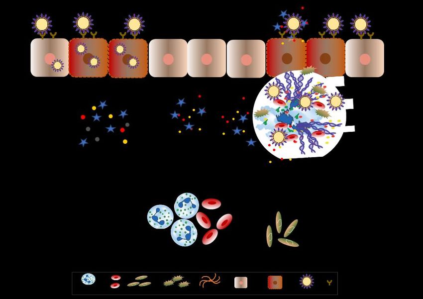
FigureFigure 1. Neutrophils
1. Neutrophils and andneutrophil
neutrophil extracellular
extracellulartraps drive
traps necroinflammation
drive in COVID-19.
necroinflammation The
in COVID-19.
severe acute respiratory syndrome coronavirus-2 (SARS-CoV-2) binds to ACE2 and enter epithelial
The severe acute respiratory syndrome coronavirus-2 (SARS-CoV-2) binds to ACE2 and enter epithelial
as well as endothelial cells along with it leading to reduced ACE2 expression that stimulates
as well as endothelial cells along with it leading to reduced ACE2 expression that stimulates
neutrophil recruitment. Subsequently, neutrophils undergo degranulation and NET formation
neutrophil recruitment. Subsequently, neutrophils undergo degranulation and NET formation releasing
releasing intracellular danger-associated molecular patterns, e.g., DNA, histones, neutrophil elastase
intracellular danger-associated
that activate molecular
the pattern recognition patterns,
receptors e.g., DNA,immune
on surrounding histones,andneutrophil
non-immune elastase
cells tothat
activate
induce cytokine secretion. The extracellular DNA released by NETs activates plateletsto and
the pattern recognition receptors on surrounding immune and non-immune cells induce
cytokine secretion. The extracellular DNA released by NETs activates platelets and
aggregated NETs provide a scaffold for binding of erythrocytes and activated platelets that promote aggregated
NETs provide
thrombusaformation.
scaffold forThebinding of erythrocytes
extracellular and activated
histones present on NETs platelets that promote
induce necrosis thrombus
in epithelial or
endothelial
formation. cells leading
The extracellular to thepresent
histones releaseonofNETs
associated
inducemolecular
necrosis inpatterns.
epithelialThis sets up ancells
or endothelial
leadingauto-amplification
to the release ofloop of necroinflammation
associated that aggravate
molecular patterns. Thisthe disease
sets up anseverity during COVID-19.
auto-amplification loop of
SARS-CoV-2 = severe acute respiratory syndrome coronavirus 2, ACE2 = angiotensin-converting
necroinflammation that aggravate the disease severity during COVID-19. SARS-CoV-2 = severe acute
enzyme 2, NET = neutrophil extracellular traps, DAMPs = danger-associated molecular patterns.
respiratory syndrome coronavirus 2, ACE2 = angiotensin-converting enzyme 2, NET = neutrophil
extracellular traps, DAMPs = danger-associated molecular patterns.
NETing neutrophils tend to form larger aggregates called “AggNETs” that drive the formation
of thrombi in blood vessels [26]. Interestingly, high incidences of venous thrombosis are reported in
NETing neutrophils tend to form larger aggregates called “AggNETs” that drive the formation
COVID-19 [27]. The extracellular DNA released by NETs activates the platelets, and the AggNETs
of thrombi in blood vessels [26]. Interestingly, high incidences of venous thrombosis are reported in
provide a scaffold for binding of the erythrocytes and activated platelets, which further promote the
COVID-19 [27]. Theand
NET formation extracellular DNAcycle
set up a vicious released by NETs
propagating activates
thrombus the platelets,
formation andalso
[26]. NETs theactivate
AggNETs
provide a scaffold for binding of the erythrocytes and activated platelets, which further promote the
NET formation and set up a vicious cycle propagating thrombus formation [26]. NETs also activateCells 2020, 9, 1383 4 of 8
the complement system. Myeloperoxidase, cathepsin G, and proteinase 3 activate properdin, factor B,
and C3, three components of the alternative pathway required to induce the complement cascade [28].
Activated neutrophils also express properdin, factor B, and C3, suggesting an important role of
neutrophils in complement activation. Of note, activation of the complement system has been reported
in the severe COVID-19 patients [27]. Together, neutrophils infiltration and NETs formation drive
necroinflammation during coronavirus infections (Table 1).
Table 1. Evidence for neutrophil-mediated necroinflammation in coronavirus infections.
Virus Evidence for Involvement of Neutrophils Reference
High levels of markers of NETs, e.g., cell free-DNA, myeloperoxidase-DNA,
[19]
and citrullinated histone 3 in sera from severely ill patients
SARS-CoV-2 High neutrophil-to-lymphocyte ratio cause ARDS in patients [13,15,29]
Neutrophil infiltration in pulmonary capillaries with extravasation into the alveolar space [20]
High neutrophil-to-lymphocyte ratio and D-dimer levels in patients [30]
C3 mediated neutrophil recruitment during disease progression in mice [31]
Neutrophils infiltration in lungs during the late phase of infection in mice [32]
SARS-CoV Neutrophils count correlate with the cytokine storm in patients [33]
Higher levels of neutrophil chemokine IL-8 found in patients [34]
Neutrophilia is associated with the severity of disease in patients [35]
Neutrophil-mediated innate inflammatory response in human DPP4 knock-in mice [36]
Increased neutrophils contribute to leukocytosis, an indicator of disease severity and
MERS-CoV [37]
fatality in patients
Increased release of ROS caused extensive pulmonary lesions and increased the disease
[38]
severity in marmosets
SARS-CoV = severe acute respiratory syndrome coronavirus, MERS-CoV = Middle East respiratory syndrome
coronavirus, NET = neutrophil extracellular trap, ARDS = acute respiratory distress syndrome, C3 = complement
factor 3, ROS = reactive oxygen species.
4. Diabetes, SARS-CoV-2, and Neutrophils
Many prevalent co-morbidities increase the severity and mortality of COVID-19 [14,27,39–41].
One of the most distinctive co-morbidities is diabetes mellitus [6]. Out of 1099 cases reported by
Guan et al., 16.2% of patients with severe disease had a higher prevalence of diabetes compared to
5.7% of patients with the non-severe disease [14]. Case fatality was higher in COVID patients with
diabetes [42]. This may be attributed to the dysfunctional innate immunity, as well as the exaggerated
pro-inflammatory cytokine response in patients with diabetes [43]. Furthermore, higher glucose levels
glycosylate and shed ACE2 [44] may contribute to the severity of ARDS during COVID-19 by increasing
vascular permeability, edema, and neutrophils infiltration in DM patients. On the other hand, it was
believed that patients with diabetes treated with ACE inhibitors and angiotensin-receptor blockers
may develop increased ACE2 expression, which could further facilitate the cell entry of SARS-CoV-2
and aggravate the infection [40]. However, a recent study reported no association with the likelihood
of COVID-19 positive test or severity of COVID-19 with renin-angiotensin system inhibitors [45].
Hyperglycemia in diabetes primes neutrophils to release NETs that might further contribute to the
cytokine storm, SIRS, and sepsis in COVID-19 [43]. Besides, sugar-activated neutrophils produce
S100 Calcium-binding proteins A8/A9 (S100A8/A9) that increased the production of thrombopoietin
in the liver and subsequent thrombocytosis [46], which might contribute to thrombus formation in
COVID-19. Th17-associated cytokine production promoted disease-predictive inflammation in DM [47].
Interestingly, a higher number of CCR6+ TH17 cells were found in the peripheral blood of COVID-19
patients, suggesting critical involvement of TH17 response [48]. Together, neutrophil-mediated cytokine
storm leads to sepsis and subsequent multi-organ failure to aggravate the severity of COVID-19 disease.Cells 2020, 9, 1383 5 of 8
5. Cardiovascular Diseases, SARS-CoV-2, and Neutrophils
Cardiovascular diseases, including coronary heart disease, cardiomyopathy, arrhythmias,
myocardial injury, and hypertension are other distinctive co-morbidities of COVID-19 that have
higher overall mortality rates [14,42,49]. Especially, the extent of myocardial injury correlated with
cardiac dysfunction, arrhythmias, and fatal outcome of COVID-19 [49]. ACE2 exerts vasodilatory effects
through Ang-(1–7) and the Mas receptor [8]. Therefore downregulation of ACE2 upon SARS-CoV-2 cell
entry induces vasoconstriction and subsequent hypertension. Subsequent ACE2-mediated neutrophil
infiltration, as well as NET formation, might be responsible for the exaggerated inflammatory
response, which in turn contributes to the development of cardiovascular diseases, e.g., thrombosis,
atherosclerosis, and endothelial injury, etc. One in five hospitalized COVID-19 patients showed
increased troponin, brain natriuretic peptide, lymphopenia, and inflammation markers, such as
c-reactive protein, IL-1β, and IL-6 in the early course of the disease suggesting cardiac injury [49,50].
Recently, NET-related endothelial cell injury was reported to contribute to vascular pathology in
pulmonary hypertension [39]. Moreover, IL-1β promoted the thrombus formation via NET-associated
tissue factor during atheroembolic events during cardiovascular diseases [51,52]. Furthermore, increased
neutrophil elastase activity was reported to contribute to obesity, insulin resistance, and related
inflammation [53]. Interestingly, the presence of obesity in metabolic associated fatty liver disease
increased the severity of COVID-19 six-fold [41]. All these reports indicate the involvement of
neutrophils and related necroinflammation in the pathology and severity of COVID-19.
6. Summary and Perspectives
To summarize, neutrophils play a central role in the immunopathology of COVID-19. SARS-CoV-2
infection, as well as downregulation of ACE2 upon the cell entry of SARS-CoV-2 triggers neutrophil
infiltration in the lungs. Necrotic cell death of alveolar epithelial cells, as well as NET formation,
releases damage-associated molecular patterns and alarmins in the surrounding extracellular space,
which induce production of pro-inflammatory cytokines and vice versa, setting up a loop of
necroinflammation that is responsible for the cytokine storm and sepsis. NETting neutrophils cause
endothelial injury and necroinflammation via complement activation, as well as promote the venous
thrombus formation during COVID-19. Underlying co-morbidities in COVID-19 patients, e.g., diabetes
and cardiovascular diseases enhance the neutrophilic inflammation and thereby severity of COVID-19.
Therefore, the development of novel therapeutic strategies targeted at neutrophils, e.g., inhibitors
of neutrophil recruitment or NET formation may help reduce the overall disease mortality rate
of COVID-19.
Author Contributions: S.R.M. conceived the idea. B.T., H.-J.A., J.D., S.R.M. wrote the manuscript. All authors
have read and agreed to the published version of the manuscript.
Funding: This research was funded by grants from the Department of Biotechnology, Government of India
(BT/RLF/Re-entry/01/2017) and Council of Scientific and Industrial Research (CSIR), India to S.R.M. and the
Deutsche Forschungsgemeinschaft (AN372/14-3, 23-1, and 24-1) to H.-J.A.
Acknowledgments: This manuscript has CDRI communication number 10074.
Conflicts of Interest: The authors declare no conflict of interest.
References
1. Zhou, P.; Yang, X.L.; Wang, X.G.; Hu, B.; Zhang, L.; Zhang, W.; Si, H.R.; Zhu, Y.; Li, B.; Huang, C.L.; et al.
A pneumonia outbreak associated with a new coronavirus of probable bat origin. Nature 2020, 579, 270–273.
[CrossRef] [PubMed]
2. Wadman, M.; Couzin-Frankel, J.; Kaiser, J.; Matacic, C. A rampage through the body. Science 2020, 368,
356–360. [PubMed]Cells 2020, 9, 1383 6 of 8
3. Lu, R.; Zhao, X.; Li, J.; Niu, P.; Yang, B.; Wu, H.; Wang, W.; Song, H.; Huang, B.; Zhu, N.; et al.
Genomic characterisation and epidemiology of 2019 novel coronavirus: Implications for virus origins
and receptor binding. Lancet 2020, 395, 565–574. [CrossRef]
4. Wrapp, D.; Wang, N.; Corbett, K.S.; Goldsmith, J.A.; Hsieh, C.L.; Abiona, O.; Graham, B.S.; McLellan, J.S.
Cryo-EM structure of the 2019-nCoV spike in the prefusion conformation. Science 2020, 367, 1260–1263.
[CrossRef] [PubMed]
5. Hoffmann, M.; Kleine-Weber, H.; Schroeder, S.; Kruger, N.; Herrler, T.; Erichsen, S.; Schiergens, T.S.; Herrler, G.;
Wu, N.H.; Nitsche, A.; et al. SARS-CoV-2 Cell Entry Depends on ACE2 and TMPRSS2 and Is Blocked by a
Clinically Proven Protease Inhibitor. Cell 2020, 181, 271–280.e278. [CrossRef] [PubMed]
6. Huang, C.; Wang, Y.; Li, X.; Ren, L.; Zhao, J.; Hu, Y.; Zhang, L.; Fan, G.; Xu, J.; Gu, X.; et al. Clinical features
of patients infected with 2019 novel coronavirus in Wuhan, China. Lancet 2020, 395, 497–506. [CrossRef]
7. Imai, Y.; Kuba, K.; Rao, S.; Huan, Y.; Guo, F.; Guan, B.; Yang, P.; Sarao, R.; Wada, T.; Leong-Poi, H.;
et al. Angiotensin-converting enzyme 2 protects from severe acute lung failure. Nature 2005, 436, 112–116.
[CrossRef]
8. Xu, J.; Fan, J.; Wu, F.; Huang, Q.; Guo, M.; Lv, Z.; Han, J.; Duan, L.; Hu, G.; Chen, L.; et al.
The ACE2/Angiotensin-(1-7)/Mas Receptor Axis: Pleiotropic Roles in Cancer. Front. Physiol. 2017, 8, 276.
[CrossRef]
9. Kuba, K.; Imai, Y.; Rao, S.; Gao, H.; Guo, F.; Guan, B.; Huan, Y.; Yang, P.; Zhang, Y.; Deng, W.; et al. A crucial
role of angiotensin converting enzyme 2 (ACE2) in SARS coronavirus-induced lung injury. Nat. Med. 2005,
11, 875–879. [CrossRef]
10. Eguchi, S.; Kawai, T.; Scalia, R.; Rizzo, V. Understanding Angiotensin II Type 1 Receptor Signaling in Vascular
Pathophysiology. Hypertension 2018, 71, 804–810. [CrossRef]
11. Sodhi, C.P.; Wohlford-Lenane, C.; Yamaguchi, Y.; Prindle, T.; Fulton, W.B.; Wang, S.; McCray, P.B., Jr.;
Chappell, M.; Hackam, D.J.; Jia, H. Attenuation of pulmonary ACE2 activity impairs inactivation of
des-Arg(9) bradykinin/BKB1R axis and facilitates LPS-induced neutrophil infiltration. Am. J. Physiol. Lung
Cell. Mol. Physiol. 2018, 314, L17–L31. [CrossRef] [PubMed]
12. Sodhi, C.P.; Nguyen, J.; Yamaguchi, Y.; Werts, A.D.; Lu, P.; Ladd, M.R.; Fulton, W.B.; Kovler, M.L.; Wang, S.;
Prindle, T., Jr.; et al. A Dynamic Variation of Pulmonary ACE2 Is Required to Modulate Neutrophilic
Inflammation in Response to Pseudomonas aeruginosa Lung Infection in Mice. J. Immunol. 2019, 203,
3000–3012. [CrossRef] [PubMed]
13. Liu, Y.; Du, X.; Chen, J.; Jin, Y.; Peng, L.; Wang, H.H.X.; Luo, M.; Chen, L.; Zhao, Y. Neutrophil-to-lymphocyte
ratio as an independent risk factor for mortality in hospitalized patients with COVID-19. J. Infect. 2020.
[CrossRef] [PubMed]
14. Guan, W.J.; Ni, Z.Y.; Hu, Y.; Liang, W.H.; Ou, C.Q.; He, J.X.; Liu, L.; Shan, H.; Lei, C.L.; Hui, D.S.C.; et al.
Clinical Characteristics of Coronavirus Disease 2019 in China. N. Engl. J. Med. 2020, 382, 1708–1720.
[CrossRef]
15. Lagunas-Rangel, F.A. Neutrophil-to-lymphocyte ratio and lymphocyte-to-C-reactive protein ratio in patients
with severe coronavirus disease 2019 (COVID-19): A meta-analysis. J. Med. Virol. 2020. [CrossRef]
16. Németh, T.; Sperandio, M.; Mócsai, A. Neutrophils as emerging therapeutic targets. Nat. Rev. Drug Discov.
2020, 19, 253–275. [CrossRef]
17. Yasui, F.; Kohara, M.; Kitabatake, M.; Nishiwaki, T.; Fujii, H.; Tateno, C.; Yoneda, M.; Morita, K.;
Matsushima, K.; Koyasu, S.; et al. Phagocytic cells contribute to the antibody-mediated elimination
of pulmonary-infected SARS coronavirus. Virology 2014, 454–455, 157–168. [CrossRef]
18. Channappanavar, R.; Fehr, A.R.; Vijay, R.; Mack, M.; Zhao, J.; Meyerholz, D.K.; Perlman, S. Dysregulated
Type I Interferon and Inflammatory Monocyte-Macrophage Responses Cause Lethal Pneumonia in
SARS-CoV-Infected Mice. Cell Host Microbe 2016, 19, 181–193. [CrossRef]
19. Zuo, Y.; Yalavarthi, S.; Shi, H.; Gockman, K.; Zuo, M.; Madison, J.A.; Blair, C.N.; Weber, A.; Barnes, B.J.;
Egeblad, M.; et al. Neutrophil extracellular traps in COVID-19. JCI Insight 2020. [CrossRef]
20. Barnes, B.J.; Adrover, J.M.; Baxter-Stoltzfus, A.; Borczuk, A.; Cools-Lartigue, J.; Crawford, J.M.;
Daßler-Plenker, J.; Guerci, P.; Huynh, C.; Knight, J.S.; et al. Targeting potential drivers of COVID-19:
Neutrophil extracellular traps. J. Exp. Med. 2020, 217, e20200652. [CrossRef]Cells 2020, 9, 1383 7 of 8
21. Varga, Z.; Flammer, A.J.; Steiger, P.; Haberecker, M.; Andermatt, R.; Zinkernagel, A.S.; Mehra, M.R.;
Schuepbach, R.A.; Ruschitzka, F.; Moch, H. Endothelial cell infection and endotheliitis in COVID-19. Lancet
2020, 395, 1417–1418. [CrossRef]
22. Couzin-Frankel, J. The mystery of the pandemic’s ‘happy hypoxia’. Science 2020, 368, 455–456. [CrossRef]
23. Yue, Y.; Nabar, N.R.; Shi, C.S.; Kamenyeva, O.; Xiao, X.; Hwang, I.Y.; Wang, M.; Kehrl, J.H. SARS-Coronavirus
Open Reading Frame-3a drives multimodal necrotic cell death. Cell Death Dis. 2018, 9, 904. [CrossRef]
24. Linkermann, A.; Stockwell, B.R.; Krautwald, S.; Anders, H.J. Regulated cell death and inflammation:
An auto-amplification loop causes organ failure. Nat. Rev. Immunol. 2014, 14, 759–767. [CrossRef]
25. Mulay, S.R.; Linkermann, A.; Anders, H.J. Necroinflammation in Kidney Disease. J. Am. Soc. Nephrol. 2016,
27, 27–39. [CrossRef]
26. Nakazawa, D.; Desai, J.; Steiger, S.; Müller, S.; Devarapu, S.K.; Mulay, S.R.; Iwakura, T.; Anders, H.J.
Activated platelets induce MLKL-driven neutrophil necroptosis and release of neutrophil extracellular traps
in venous thrombosis. Cell Death Discov. 2018, 4, 6. [CrossRef]
27. Llitjos, J.F.; Leclerc, M.; Chochois, C.; Monsallier, J.M.; Ramakers, M.; Auvray, M.; Merouani, K. High incidence
of venous thromboembolic events in anticoagulated severe COVID-19 patients. J. Thromb. Haemost. 2020.
[CrossRef]
28. de Bont, C.M.; Boelens, W.C.; Pruijn, G.J.M. NETosis, complement, and coagulation: A triangular relationship.
Cell. Mol. Immunol. 2019, 16, 19–27. [CrossRef]
29. Liu, J.; Liu, Y.; Xiang, P.; Pu, L.; Xiong, H.; Li, C.; Zhang, M.; Tan, J.; Xu, Y.; Song, R.; et al.
Neutrophil-to-lymphocyte ratio predicts critical illness patients with 2019 coronavirus disease in the
early stage. J. Transl. Med. 2020, 18, 206. [CrossRef]
30. Fu, J.; Kong, J.; Wang, W.; Wu, M.; Yao, L.; Wang, Z.; Jin, J.; Wu, D.; Yu, X. The clinical implication of
dynamic neutrophil to lymphocyte ratio and D-dimer in COVID-19: A retrospective study in Suzhou China.
Thromb. Res. 2020, 192, 3–8. [CrossRef]
31. Gralinski, L.E.; Sheahan, T.P.; Morrison, T.E.; Menachery, V.D.; Jensen, K.; Leist, S.R.; Whitmore, A.;
Heise, M.T.; Baric, R.S. Complement Activation Contributes to Severe Acute Respiratory Syndrome
Coronavirus Pathogenesis. mBio 2018, 9, e01753-18. [CrossRef] [PubMed]
32. Chen, J.; Lau, Y.F.; Lamirande, E.W.; Paddock, C.D.; Bartlett, J.H.; Zaki, S.R.; Subbarao, K. Cellular immune
responses to severe acute respiratory syndrome coronavirus (SARS-CoV) infection in senescent BALB/c
mice: CD4+ T cells are important in control of SARS-CoV infection. J. Virol. 2010, 84, 1289–1301. [CrossRef]
[PubMed]
33. Huang, K.J.; Su, I.J.; Theron, M.; Wu, Y.C.; Lai, S.K.; Liu, C.C.; Lei, H.Y. An interferon-gamma-related cytokine
storm in SARS patients. J. Med. Virol. 2005, 75, 185–194. [CrossRef] [PubMed]
34. Wong, C.K.; Lam, C.W.; Wu, A.K.; Ip, W.K.; Lee, N.L.; Chan, I.H.; Lit, L.C.; Hui, D.S.; Chan, M.H.; Chung, S.S.;
et al. Plasma inflammatory cytokines and chemokines in severe acute respiratory syndrome. Clin. Exp.
Immunol. 2004, 136, 95–103. [CrossRef]
35. Wong, R.S.; Wu, A.; To, K.F.; Lee, N.; Lam, C.W.; Wong, C.K.; Chan, P.K.; Ng, M.H.; Yu, L.M.; Hui, D.S.; et al.
Haematological manifestations in patients with severe acute respiratory syndrome: Retrospective analysis.
BMJ 2003, 326, 1358–1362. [CrossRef]
36. Li, K.; Wohlford-Lenane, C.L.; Channappanavar, R.; Park, J.E.; Earnest, J.T.; Bair, T.B.; Bates, A.M.;
Brogden, K.A.; Flaherty, H.A.; Gallagher, T.; et al. Mouse-adapted MERS coronavirus causes lethal
lung disease in human DPP4 knockin mice. Proc. Natl. Acad. Sci. USA 2017, 114, 3119–3128. [CrossRef]
37. Min, C.K.; Cheon, S.; Ha, N.Y.; Sohn, K.M.; Kim, Y.; Aigerim, A.; Shin, H.M.; Choi, J.Y.; Inn, K.S.; Kim, J.H.;
et al. Comparative and kinetic analysis of viral shedding and immunological responses in MERS patients
representing a broad spectrum of disease severity. Sci. Rep. 2016, 6, 25359. [CrossRef]
38. Baseler, L.J.; Falzarano, D.; Scott, D.P.; Rosenke, R.; Thomas, T.; Munster, V.J.; Feldmann, H.; de Wit, E.
An Acute Immune Response to Middle East Respiratory Syndrome Coronavirus Replication Contributes to
Viral Pathogenicity. Am. J. Pathol. 2016, 186, 630–638. [CrossRef]
39. Aldabbous, L.; Abdul-Salam, V.; McKinnon, T.; Duluc, L.; Pepke-Zaba, J.; Southwood, M.; Ainscough, A.J.;
Hadinnapola, C.; Wilkins, M.R.; Toshner, M.; et al. Neutrophil Extracellular Traps Promote Angiogenesis:
Evidence From Vascular Pathology in Pulmonary Hypertension. Arterioscler. Thromb. Vasc. Biol. 2016, 36,
2078–2087. [CrossRef]Cells 2020, 9, 1383 8 of 8
40. Fang, L.; Karakiulakis, G.; Roth, M. Are patients with hypertension and diabetes mellitus at increased risk
for COVID-19 infection? Lancet. Respir. Med. 2020, 8, e21. [CrossRef]
41. Zheng, K.I.; Gao, F.; Wang, X.B.; Sun, Q.F.; Pan, K.H.; Wang, T.Y.; Ma, H.L.; Liu, W.Y.; George, J.; Zheng, M.H.
Obesity as a risk factor for greater severity of COVID-19 in patients with metabolic associated fatty liver
disease. Metabolism 2020, 154244. [CrossRef] [PubMed]
42. Wu, Z.; McGoogan, J.M. Characteristics of and Important Lessons From the Coronavirus Disease 2019
(COVID-19) Outbreak in China: Summary of a Report of 72 314 Cases From the Chinese Center for Disease
Control and Prevention. Jama 2020. [CrossRef] [PubMed]
43. Wong, S.L.; Demers, M.; Martinod, K.; Gallant, M.; Wang, Y.; Goldfine, A.B.; Kahn, C.R.; Wagner, D.D.
Diabetes primes neutrophils to undergo NETosis, which impairs wound healing. Nat. Med. 2015, 21, 815–819.
[CrossRef]
44. Salem, E.S.; Grobe, N.; Elased, K.M. Insulin treatment attenuates renal ADAM17 and ACE2 shedding in
diabetic Akita mice. Am. J. Physiol. Renal. Physiol. 2014, 306, F629–F639. [CrossRef]
45. Reynolds, H.R.; Adhikari, S.; Pulgarin, C.; Troxel, A.B.; Iturrate, E.; Johnson, S.B.; Hausvater, A.; Newman, J.D.;
Berger, J.S.; Bangalore, S.; et al. Renin-Angiotensin-Aldosterone System Inhibitors and Risk of Covid-19.
N. Engl. J. Med. 2020. [CrossRef]
46. Kraakman, M.J.; Lee, M.K.; Al-Sharea, A.; Dragoljevic, D.; Barrett, T.J.; Montenont, E.; Basu, D.; Heywood, S.;
Kammoun, H.L.; Flynn, M.; et al. Neutrophil-derived S100 calcium-binding proteins A8/A9 promote
reticulated thrombocytosis and atherogenesis in diabetes. J. Clin. Investig. 2017, 127, 2133–2147. [CrossRef]
47. Nicholas, D.A.; Proctor, E.A.; Agrawal, M.; Belkina, A.C.; Van Nostrand, S.C.; Panneerseelan-Bharath, L.;
Jones, A.R.t.; Raval, F.; Ip, B.C.; Zhu, M.; et al. Fatty Acid Metabolites Combine with Reduced β Oxidation to
Activate Th17 Inflammation in Human Type 2 Diabetes. Cell Metab. 2019, 30, 447–461. [CrossRef]
48. Wu, D.; Yang, X.O. TH17 responses in cytokine storm of COVID-19: An emerging target of JAK2 inhibitor
Fedratinib. J. Microbiol. Immunol. Infect. 2020. [CrossRef]
49. Guo, T.; Fan, Y.; Chen, M.; Wu, X.; Zhang, L.; He, T.; Wang, H.; Wan, J.; Wang, X.; Lu, Z. Cardiovascular
Implications of Fatal Outcomes of Patients With Coronavirus Disease 2019 (COVID-19). JAMA Cardiol. 2020.
[CrossRef]
50. Chapman, A.R.; Bularga, A.; Mills, N.L. High-Sensitivity Cardiac Troponin Can Be an Ally in the Fight
Against COVID-19. Circulation 2020, 141, 1733–1735. [CrossRef]
51. Yadav, V.; Chi, L.; Zhao, R.; Tourdot, B.E.; Yalavarthi, S.; Jacobs, B.N.; Banka, A.; Liao, H.; Koonse, S.;
Anyanwu, A.C.; et al. Ectonucleotidase tri(di)phosphohydrolase-1 (ENTPD-1) disrupts inflammasome/
interleukin 1beta-driven venous thrombosis. J. Clin. Investig. 2019, 129, 2872–2877. [CrossRef]
52. Liberale, L.; Holy, E.W.; Akhmedov, A.; Bonetti, N.R.; Nietlispach, F.; Matter, C.M.; Mach, F.; Montecucco, F.;
Beer, J.H.; Paneni, F.; et al. Interleukin-1beta Mediates Arterial Thrombus Formation via NET-Associated
Tissue Factor. J. Clin. Med. 2019, 8, 2072. [CrossRef]
53. Mansuy-Aubert, V.; Zhou, Q.L.; Xie, X.; Gong, Z.; Huang, J.Y.; Khan, A.R.; Aubert, G.; Candelaria, K.;
Thomas, S.; Shin, D.J.; et al. Imbalance between neutrophil elastase and its inhibitor α1-antitrypsin in obesity
alters insulin sensitivity, inflammation, and energy expenditure. Cell Metab. 2013, 17, 534–548. [CrossRef]
© 2020 by the authors. Licensee MDPI, Basel, Switzerland. This article is an open access
article distributed under the terms and conditions of the Creative Commons Attribution
(CC BY) license (http://creativecommons.org/licenses/by/4.0/).You can also read


