Introducing V-Line as a New Strategy to Choose Surgical Corridor in Oblique Lumbar Interbody Fusion at the L5- S1 Segment - Hindawi.com
←
→
Page content transcription
If your browser does not render page correctly, please read the page content below
Hindawi
Disease Markers
Volume 2021, Article ID 5584372, 9 pages
https://doi.org/10.1155/2021/5584372

Research Article
Introducing V-Line as a New Strategy to Choose Surgical
Corridor in Oblique Lumbar Interbody Fusion at the L5-
S1 Segment

          Wei Zhang , Xing Du , Yong Zhu, Wei Luo, Ben Wang, Guanyin Jiang,
          and Yunsheng Ou
          Department of Orthopedics, The First Affiliated Hospital of Chongqing Medical University, YouYi Road 1#, YuZhong District,
          Chongqing 400016, China

          Correspondence should be addressed to Yunsheng Ou; ouyunsheng2001@163.com

          Received 7 January 2021; Revised 23 March 2021; Accepted 5 April 2021; Published 22 April 2021

          Academic Editor: Dong Pan

          Copyright © 2021 Wei Zhang et al. This is an open access article distributed under the Creative Commons Attribution License,
          which permits unrestricted use, distribution, and reproduction in any medium, provided the original work is properly cited.

          Purpose. A retrospective imaging study assessing the availability of oblique lumbar interbody fusion at the level of L5-S1 (OLIF51)
          and to choose ideal surgical corridor in OLIF51 by introducing V-line. Methods. The axial views through the center of L5-S1 disc
          were reviewed. We adopt 18 mm as the width of the simulated surgical corridor. The midline of the surgical corridor is at the center
          of L5-S1 disc. According to the traction distance of the left iliac vein (LCIV) and psoas major (PM), we defined all the subjects as V
          (+) (traction-difficultly LCIV), V (-) (traction-friendly LCIV), P (+) (traction-difficultly PM), and P (-) (traction-friendly PM). V-
          line was defined as a straight line dividing equally the simulated surgical corridor. All cases were divided into 2 groups: The V-line
          (+) group, more than half of the LCIV region, is located in the ventral part of V-line; the V-line (-) group, more than half of the
          LCIV region, is located in the dorsal part of V-line. Multiple variables regressive analysis was conducted to analyze the
          independent risk factors of V-line (+). Results. V-line (+) was found in 36 (38.7%) patients and V-line (-) in 57 (61.3%).
          Incidence of V (+) and P (+) was 35.4% (33/93) and 30.1% (28/93), respectively. 16.1% (15/93) subjects processed V (+) and P
          (+) at the same time. The independent risk factor of V-line (+) were gender of male (P = 0:034, OR: 12.152) and medial position
          of LCIV (P < 0:001, OR: 265.085). High iliac crest was a significant independent protective factor (P = 0:001, OR: 0.750).
          Conclusions. Most patients were suitable for OLIF51. V-line could assess the injury risk of LCIV. For patients who are V-line
          (+), mainly among males having the LCIV near the midline or the iliac crest relatively low, a surgical corridor external to the
          LCIV should be taken into consideration.

1. Introduction                                                          major and abdominal aorta, thus reducing the lumbar plexus
                                                                         injury during LLIF and abdominal large vessel injury during
The oblique interbody fusion (OLIF), as a new type of mini-              ALIF [3]. However, its application is often impeded for
mally invasive technique, has good applicability at L2-L5. It            severe canal stenosis, large disc herniation, or concomitant
has a wide range of indications including lumbar degenera-               ruptured disc herniation by its characteristics of indirect
tive disease, spinal deformities, trauma, infections, and neo-           decompression. Therefore, OLIF should be carefully chosen
plasms [1]. This procedure has many advantages in                        in clinical indications [4–6].
comparison with traditional spinal posterior approach sur-                   There were a few cases using OLIF of L5-S1 in recent lit-
gery. As with ALIF and LLIF, it also avoids iatrogenic injury            eratures; however, it is still quite difficult to perform OLIF51
to the paraspinal musculature and disruption of spinal canal             because of the risks associated with stretch of the iliac vessels
[2]. Besides, this surgical technique allows access to the               and the presence of the iliac wing disturbing insertion of the
anterolateral margin of the vertebral body between the psoas             cage. Silvestre et al. [7] reported 179 patients using miniopen
2 Disease Markers
anterior retroperitoneal lumbar interbody fusion concluding
that it is a safe approach for accessing the L2-L5 and advised
using another approach for the L5-S1 due to the danger of
injuring the iliac vessels.
A
Several studies have evaluated the size of OLIF51 surgical
corridor. Capellades et al. [8] confirmed the great anatomic
variability of vascular structures in the lumbosacral area,
P
D
and the window is really small in 18.05% of the study popu-
lation because of the venous structures overlapping the L5-S1
θ
V
D
disc. To our knowledge, there is no study to analyze the
anatomic structure at L5-S1 combined with OLIF surgical
corridor, as the oblique operation channel will change many
anatomical parameters.
The purpose of this study is to simulate the OLIF opera-
tion process at the L5-S1 level and then obtain anatomical
data through CT image analysis, thus providing a way to
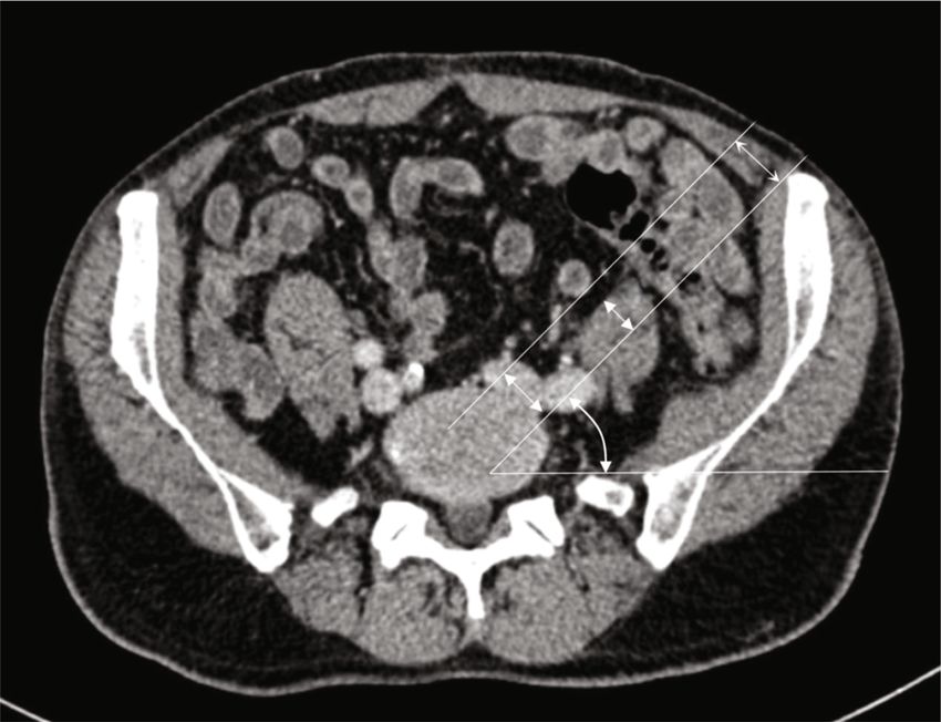
evaluate the feasibility of OLIF51 and to minimize vascular Figure 1: Illustration showing the simulated surgical corridor. The
complications in the preoperative planning stage. midline of surgical corridor is at the center of the L5-S1 disc and the
dorsal margin of surgical corridor is close to left iliac crest. Distance
A is the diameter of the surgical corridor (18 mm). DV is the traction
2. Methods and Methods distance of the left iliac vein. DP is the traction distance of the psoas
major. θ is the tilt angle of surgical corridor.
2.1. Subject Characteristics. 120 consecutive patients under-
going lumbar CT examination at our hospital’s Department
of Radiology from 1 October 2019 to 31 December 2019 were their right edge of the left iliac vein: lateral, intermediate, and
reviewed. All subjects had a clear thin-slice CT scan. Patients medial (Figure 2).
who had abdominal vascular abnormalities or diseases (i.e.,
abdominal aortic aneurysms, Budd-Chiari syndrome, 2.3.2. The Iliocaval Junction Position (JP). We used the classi-
abdominal aortic dissection, and iliac artery occlusion), spi- fication established by Capellades et al. [8]. A percentage of
nal deformity from any cause, lumbar spondylolisthesis, the distance between the inferior surface of the iliocaval junc-
transitional anatomy (i.e., sacralisation of L5, or lumbarisa- tion and the center of the L4-L5 disc as well as the distance
tion of S1), lumbar fracture, or a surgical history on lumbar between the center of the L4-L5 disc and the center of the
or retroperitoneum were excluded. The CT images were L5-S1 disc was calculated. Patients were classified into four
obtained via PACS (Picture Archiving and Communication groups according to their junction position: very high (ilioca-
System) with the patient in supine position. All the radiolog- val junction position less than -33.3%), high (iliocaval junc-
ical measurements were measured and recorded by two inde- tion position between -33.3% and 33.3%), low (iliocaval
pendent researchers, and the average value was taken as the junction position between 33.4% and 66.6%), or very low
final result. This study was approved by the institutional (iliocaval junction position greater than 66.7%) (Figure 3).
review board following the declaration of Helsinki principles, 2.3.3. The V-Line. In the present study, we proposed a new
and informed consent was obtained from all individual par- index that can evaluate the traction distance of iliac vessels.
ticipants. The whole research is being reported in line with We named this index the V-line. The “V” stands for “vessel”
the STROCSS criteria [9]. or “vein.” We defined V-line as a straight line dividing
equally the surgical corridor which we simulated before.
2.2. Surgical Simulation. The width of the OLIF cage is According to the V-line, all cases were divided into 2 groups:
18 mm (Medtronic, Inc, Minneapolis, Minnesota). As the V-line (+) group and V-line (-) group. In the V-line (+)
actual operative window for OLIF is not less than the width group, more than half of the left iliac vein region is located
of a cage, the author adopts 18 mm as the width of surgical in the ventral part of V-line, so the corridor external to the
corridor to simulate the operation process. The midline of left iliac vessels leads to less traction. In the V-line (-) group,
surgical corridor is at the center of the L5-S1 disc, and the more than half of the left iliac vein region is located in the
dorsal margin of surgical corridor is close to the left iliac dorsal part of V-line, which indicates that a surgical corridor
crest. We assumed that the cage is inserted obliquely with between the bifurcations of the iliac vessels is more optimal
the midline going through the center of the L5-S1 disc (Figure 4).
(Figure 1).
2.4. Quantitative Measurements. All the following parame-
2.3. Evaluation of Vascular Parameters ters were analyzed and recorded in an axial plane of the cen-
ter of the L5-S1 disc.
2.3.1. The Left Iliac Vein Position (VP). On the axial plane of
the L5-S1 disc, the area between the median line and the left 2.4.1. L5-S1 Disk Size. The anteroposterior diameter is
edge of the intervertebral disc was equally divided into three defined as the maximal distance of the anterior and posterior
zones. Patients were classified into three groups according to border of the intervertebral disk; the left-right diameter isDisease Markers 3
LCI
V
AP diameter
LR diameter
Medial Lateral
Intermediate
Figure 2: Diagram showing the anteroposterior diameter, the left-right diameter of L5-S1 disk, and the proposed classification for left
common iliac vein positions. As shown in the figure, the left common iliac vein is classified into intermediate group.
Very high
–33.3%
High
D 33.3%
Low
66.6%
Very low
100.0%
Figure 3: Diagram showing the proposed classification for iliocaval junction positions. D is the distance between the inferior surface of the
iliocaval junction and the center of the L4-L5 disc.
defined as the maximal distance of the left and right border of 2.4.4. Traction Distance of the Psoas Major. It is defined as the
the intervertebral disk (Figure 2). minimum distance to retract psoas major out of the surgical
corridor. In all cases, psoas major was retracted dorsally
2.4.2. Tilt Angle of Surgical Corridor. It is defined as the angle (Figure 1). The psoas was defined as P (+) (traction-difficultly
between the surgical corridor and the horizontal line. This PM) and P (-) (traction-friendly PM) in the same way as
parameter simulated the cage implantation angle when a LCIV.
patient was placed in the right lateral decubitus position
(Figure 1). 2.5. Statistical Analysis. Continuous variables were presented
as mean ± SD. χ2 analysis was used to find a statistical differ-
2.4.3. Traction Distance of the Left Iliac Vein. It is defined as ence in the left iliac vein position (VP) and the iliocaval junc-
the minimum distance to retract the iliac vein out of the sur- tion position (JP) between men and women. Univariate
gical corridor (Figure 1). The LCIV that needs to be stretched analysis for all risk factors of V-line (+) was conducted using
more than 9 mm were defined as V (+) (traction-difficultly the 2-tailed independent Student t-tests for continuous vari-
LCIV) and that less than 9 mm as V (-) (traction-friendly ables and χ2 or Fisher exact test for categorical variables. A
LCIV). multivariate logistic regression was conducted to find4 Disease Markers
LCIV LCIV
V-line
V-line
Figure 4: The diameter of the OLIF surgical corridor is 18 mm. The midline of the surgical corridor is at the center of the L5-S1 disc. Left, the
V-line (-) group, more than half of the left iliac vein region is located in the dorsal part of V-line. In this situation, we could retract the LCIV
dorsally. Right, the V-line (+) group, more than half of the left iliac vein region is located in ventral part of V-line. In this situation, we could
retract the LCIV ventrally.
independent risk factors for V-line (+). Risk factors for Chi-square analysis or Student t-tests was used to com-
V-line (+) with P < 0:15 by univariate analysis were pare gender, age, L5-S1 disk size, tilt angle of surgical corri-
included in the model. Using a forward (LR), variables with dor, sacral slope, the left iliac vein position (VP), and the
a P < 0:10 remained in the final model, with significant vari- iliocaval junction position (JP) in relation to the V-line.
ables having P < 0:05. Odds ratios (OR) and 95% confidence Differences were observed in tilt angle of surgical corridor,
intervals were calculated for all variables in the model. Statis- sacral slope, left iliac vein position (VP), and iliocaval junc-
tical analysis was performed using SPSS 20.0 (IBM Corpora- tion position (JP); they were all statistically significant (tilt
tion, Armonk, New York, USA). angle of surgical corridor, P = 0:001; sacral slope, P = 0:002;
left iliac vein position, P < 0:001; iliocaval junction position,
3. Results P < 0:001). The gender, age of the patient, and the L5-S1 disk
size will not significantly influence the V-line (Table 1).
We accessed 93 CT data in this paper, consisting of 49 men The multivariate analysis identified gender of male
and 44 women with a mean age of 55:80 ± 15:94 years. A (P = 0:034, odds ratio [OR]: 12.152) and medial position of
total of 27 subjects were excluded—among them 10 with LCIV (P < 0:001, odds ratio [OR]: 265.085) as significant
transitional anatomy, 7 with huge abdominal neoplasms, 5 independent risk factors for V-line (+), while high iliac crest
with spinal deformity, 4 with surgical history on lumbar or was a significant independent protective factor (P = 0:001,
retroperitoneum, and 1 with huge lumbosacral osteophyte. OR: 0.750) (Table 4).
The subjects’ other quantitative measurements are summa-
rized in Table 1. 4. Discussion
According to the VP classification, 35 patients (37.6%)
were grouped in the lateral group, 33 patients (35.5%) in 4.1. Application of OLIF51. OLIF51 is considered as mini-
the intermediate group, and 25 patients (26.9%) in the medial mally invasive ALIF through the oblique corridor in the lat-
group. Statistically significant difference was found between eral position [10]. It keeps the advantages of traditional
gender and the left iliac vein position. Males displayed a more ALIF with direct and extensive exposure of the intervertebral
medial position of LCIV. disc and avoidance of neural and muscular injury compared
According to the JP classification, 3 patients (3.2%) were with the posterior approach. As a new surgical technique,
grouped in the very high group, 39 patients (41.9%) in the OLIF51 could be described as laterally positioned ALIF, but
high group, 38 patients (40.9%) in the low group, and 13 the latter was superior to the former in many aspects [3, 11,
patients (14.0%) in the very low group. No statistically signif- 12]. First, it can be extended to upper levels in a single posi-
icant differences were found between gender and the iliocaval tion with less mobilization of the great vessels, especially for
junction position (Table 2). ALIF at L4-5. Secondly, it can avoid rectus abdominis muscle
To evaluate precisely the ease of surgical exposure of injury, as well as minimize the mobilization of the peritoneal
OLIF51, the study population was classified into 4 configura- content [3]. Moreover, OLIF51 is advantageous in obese
tions by combining traction distance of LCIV and psoas patients because gravity pulls the visceral fat away from the
major. According to the four-configuration classification, spine [13]. Even so, OLIF at L5-S1 is still difficult because
47 patients (50.5%) were included in P (-) V (-) group, 18 of the risks associated with mobilization of the vessels and
(19.4%) in P (-) V (+) group, 13 (14.0%) in P (+) V (-) group, the presence of the iliac wing.
and 15 (16.1%) in P (+) V (+) group. The P (+) V (+) group is Many literatures have proved the practicability of
considered not suitable for OLIF51 due to hard exposure. OLIF51 [3, 10, 14–18]. Silvestre et al. [7] first reported
There were 61.3% (57/93) of the subjects that were defined OLIF51 through a retroperitoneal approach was performed
as V-line (-) and 38.7% (36/93) as V-line (+) (Table 3). successfully in 6 patients, but one patient had to be abortedDisease Markers 5
Table 1: Univariate analysis of risk factors for V-line (+) during OLIF51.
Risk factors Patients with V-line (+), n = 36 Patients with V-line (-), n = 57 χ2 /t P
Sex
Female 14 30
1.672 0.196
Male 22 27
Age 57:39 ± 14:07 54:79 ± 17:06 -0.764 0.447
L5-S1 disk AP diameter 37:66 ± 3:44 37:48 ± 3:80 -0.229 0.820
L5-S1 disk left-right diameter 55:95 ± 6:49 54:04 ± 5:21 -1.564 0.121
Tilt angle of surgical corridor 27:15 ± 10:20 34:05 ± 6:72 3.595 0.001∗
Sacral slope 35:42 ± 5:34 39:44 ± 6:02 3.269 0.002∗
#
Low iliac crest 22 21
5.228 0.022 ∗
High iliac crest 14 36
The left iliac vein position (VP)
Medial 22 3
Intermediate 13 20 44.5706 Disease Markers
Table 4: Multivariate analysis of risk factors for V-line (+) during OLIF51.
Risk factor Odds ratio 95% confidence interval P
Sex 12.152 1.208-122.276 0.034
The left iliac vein position (VP) 265.085 16.629-4225.839Disease Markers 7
comprehensively studied. Previous work on the morphologi- ination is important to minimize the vascular injury. For
cal characteristics of the LCIV cannot solve the problem patients who are V-line (+), mainly among males having
properly. In this thesis, a new concept, V-line, is proposed the LCIV near the midline or the iliac crest relatively low, a
for assessing the mobilization risk of these two approaches. surgical corridor external to the LCIV should be taken into
By introducing the concept of V-line, we could qualitatively consideration, and vice versa. Finally, it is worth noting
evaluate the extent of vascular traction of two approaches that all the subjects in P (+) V (+) group were divided
to guide surgical treatment. into V-line (-) taking almost a quarter of the V-line (-) group,
In our work, there were 61.3% (57/93) of the subjects indicating potential difficulties in operating procedure.
were defined as V-line (-). In this group, a surgical corridor
between the bifurcations of the iliac vessels is more favorable
4.5. Limitations. The present research has several limitations.
which is exactly the mainstream approach in the world. One
First, the study object of this article is patients undergoing
important concern with this approach is the injury to the
lumbar CT examination, whose regional anatomical charac-
SHP (superior hypogastric plexus), which overlies the L5-S1
ter in the lumbosacral area are different from those with lum-
disk between the bifurcations and supplies the sympathetic
bar degeneration disease. In addition, we did not include the
function for the urogenital system [25]. Consequently, dam-
disc height in the measurement which might change in dif-
age to the SHP could result in retrograde ejaculation in male
ferent pathologies and impact the result of the iliocaval junc-
patients [26]. Careful unilateral blunt dissection of the SHP
tion position. The second limitation concerns the patient’s
and avoidance of monopolar coagulation is recommended
position, as CT images are obtained in the supine position,
[3, 17]. Besides, middle sacral vessels are also important
whereas OLIF is performed in the right lateral decubitus
structures during this central approach; we could simply
position, the location and configuration of LCIV and psoas
divide these vessels by the application of bipolar cautery or
major may vary [28]. Third, all the subjects in our study were
vascular clips.
Chinese; the positive rate of V-line might be different in other
Naturally, the rest of the subjects (36/93, 38.7%) were
ethnic populations. Lastly, a high-quality prospective study is
defined as V-line (+), in which the corridor external to the
much needed to confirm the validity of this retrospective
left iliac vessels is superior for less stretch of the LCIV. On
anatomical imaging study.
this condition, particular attention should be paid to the
identification and handling of the iliolumbar vein (ILV)
[13]. The ILV travels laterally approximately 3-4 cm below 5. Conclusions
the bifurcation and then traverses medially along the L5 ver-
tebral body, coursing between the obturator nerve and lum- This study demonstrated that the majority of the patients
bar trunks [27]. In exposing the L5-S1 level external to the were suitable for OLIF51 without excessive traction of the
LCIV, the ILV is easily avulsed due to medial traction of LCIV and the psoas major. There was a relatively high inci-
LCIV. Zairi et al. had gained access to the L5-S1 level by find- dence of V-line (+) in the Asian population. Among male
ing and clipping the ILV before retracting the iliac artery and patients having the LCIV near the midline or the iliac crest
vein anteriorly. This study considered this a safer and feasible relatively low, a surgical corridor external to the LCIV should
approach [14]. Coagulation or ligation is recommended in be taken into consideration to minimize the risk of vascular
case the ILV is identified. We could also take gentle dissec- injury.
tion of the fat around the L5-S1 level lateral to the iliac vessels
in order to obtain better recognition of the ILV.
Recently, many studies analyzing the venous anatomy in 6. Trial Registry Number
the lumbosacral area have been reported. Whether they are
useful for predicting the surgical approach of OLIF51? Mul- The study had been registered in chictr.org.cn (UIN =
tiple variables regressive analysis demonstrated that gender ChiCTR2000038598). Full detail can be accessed via http://
of male, medial position of LCIV, and high iliac crest were www.chictr.org.cn/showprojen.aspx?proj=61903.
predictive factors of V-line, while age of the patient, L5-S1
disk size, sacral slope, and iliocaval junction position (JP)
were not. As to the left iliac vein position (VP), it is found Abbreviations
that almost all subjects of the medial group were classified LCIV: Left common iliac vein
into the V-line (+) group and they accounted for nearly 2/3 OLIF: Oblique lumbar interbody fusion
of the V-line (+) group. In addition, the height of iliac crest ALIF: Anterior lumbar interbody fusion
is also crucial for the preoperative evaluation, there were LLIF: Lateral lumbar interbody fusion
more than half of low iliac crest group could choose an exter- VP: Left iliac vein position
nal corridor to decrease the stretch of the LCIV. However, the JP: Iliocaval junction position.
iliocaval junction position (JP) was not a crucial factor. A
possible explanation for this is that although lower junction
positions have more medial LCIV, it is not the only factor, Data Availability
the junction angle will affect the distribution pattern of iliac
vein as well. Therefore, in the clinical practice of OLIF51, a The datasets used to support the findings of this study are
comprehensive, detailed analysis and conventional CT exam- available from the corresponding author upon request.8 Disease Markers
Ethical Approval junction and left iliac vein positions related to L5-S1 disc,”
Spine (Phila Pa 1976), vol. 25, no. 13, pp. 1695–1700, 2000.
This study was approved by the institutional review board [9] R. Agha, A. Abdall-Razak, E. Crossley, N. Dowlut, C. Iosifidis,
following the declaration of Helsinki principles. and G. Mathew, “STROCSS 2019 Guideline: strengthening
the reporting of cohort studies in surgery,” International
Consent journal of surgery (London, England), vol. 72, pp. 156–165,
2019.
Informed consent was obtained from all individual partici- [10] N. Anand, A. Alayan, A. Agrawal, S. Kahwaty, E. Nomoto, and
pants. The whole research is being reported in line with the B. Khandehroo, “Analysis of spino-pelvic parameters and seg-
STROCSS criteria. mental lordosis with L5-S1 oblique lateral interbody fusion at
the bottom of a long construct in circumferential minimally
invasive surgical correction of adult spinal deformity,” World
Conflicts of Interest Neurosurgery, vol. 130, pp. e1077–e1083, 2019.
The authors declare that they have no competing interests. [11] R. C. Sasso, J. Kenneth Burkus, and J. C. LeHuec, “Retrograde
ejaculation after anterior lumbar interbody fusion: transperito-
neal versus retroperitoneal exposure,” Spine (Phila Pa 1976),
Acknowledgments vol. 28, no. 10, pp. 1023–1026, 2003.
This article has been presented as preprint according to the [12] B. K. Weiner, M. Walker, and R. D. Fraser, “Vascular anatomy
following link: https://www.researchsquare.com/article/rs- anterior to lumbosacral transitional vertebrae and implications
118245/v1. This work was supported by the Basic Research for anterior lumbar interbody fusion,” The Spine Journal,
vol. 1, no. 6, pp. 442–444, 2001.
and Frontiers Exploration Project of Yuzhong District,
Chongqing (2018114) and the Science and Technology Inno- [13] N. S. Chung, H. D. Lee, and C. H. Jeon, “Vascular anatomy and
surgical approach in oblique lateral interbody fusion at lumbo-
vation Project for Postgraduate of Chongqing Municipal
sacral transitional vertebrae,” Journal of orthopaedic science :
Education Commission (CYS18200). All authors gratefully official journal of the Japanese Orthopaedic Association, 2020.
acknowledge the support of the subjects who participated in
[14] F. Zairi, T. P. Sunna, H. J. Westwick et al., “Mini-open oblique
the present study.
lumbar interbody fusion (OLIF) approach for multi-level dis-
cectomy and fusion involving L5-S1: preliminary experience,”
References Orthopaedics & traumatology, surgery & research : OTSR.,
vol. 103, no. 2, pp. 295–299, 2017.
[1] K. Phan, M. Maharaj, Y. Assem, and R. J. Mobbs, “Review of [15] N. S. Chung, C. H. Jeon, and H. D. Lee, “Use of an alternative
early clinical results and complications associated with oblique surgical corridor in oblique lateral interbody fusion at the L5-
lumbar interbody fusion (OLIF),” Journal of clinical neurosci- S1 segment: a technical report,” Clinical Spine Surgery: A Spine
ence : official journal of the Neurosurgical Society of Austral- Publication, vol. 31, no. 7, pp. 293–296, 2018.
asia, vol. 31, pp. 23–29, 2016.
[16] K. Kanno, S. Ohtori, S. Orita et al., “Miniopen oblique lateral
[2] J. Allain and T. Dufour, “Anterior lumbar fusion techniques:
L5-s1 interbody fusion: a report of 2 cases,” Case reports in
ALIF, OLIF, DLIF, LLIF, IXLIF,” Orthopaedics & Traumatol-
orthopedics, vol. 2014, Article ID 603531, 5 pages, 2014.
ogy: Surgery & Research, vol. 106, no. 1, pp. S149–S157, 2020.
[17] J. S. Kim and S. B. Sharma, “How I do it? Oblique lumbar inter-
[3] K. R. Woods, J. B. Billys, and R. A. Hynes, “Technical descrip-
body fusion at L5S1(OLIF51),” Acta Neurochirurgica, vol. 161,
tion of oblique lateral interbody fusion at L1-L5 (OLIF25) and
no. 6, pp. 1079–1083, 2019.
at L5-S1 (OLIF51) and evaluation of complication and fusion
rates,” The Spine Journal, vol. 17, no. 4, pp. 545–553, 2017. [18] N. S. Chung, C. H. Jeon, H. D. Lee, and H. J. Kweon, “Preop-
[4] D. H. Heo and J. S. Kim, “Clinical and radiological outcomes of erative evaluation of left common iliac vein in oblique lateral
spinal endoscopic discectomy-assisted oblique lumbar inter- interbody fusion at L5-S1,” European Spine Journal, vol. 26,
body fusion: preliminary results,” Neurosurgical Focus, no. 11, pp. 2797–2803, 2017.
vol. 43, no. 2, article E13, 2017. [19] H. Y. Mun, M. J. Ko, Y. B. Kim, and S. W. Park, “Usefulness of
[5] J. Quillo-Olvera, G. X. Lin, H. J. Jo, and J. S. Kim, “Complica- oblique lateral interbody fusion at L5-S1 level compared to
tions on minimally invasive oblique lumbar interbody fusion transforaminal lumbar interbody fusion,” Journal of Korean
at L2-L5 levels: a review of the literature and surgical strate- Neurosurgical Society, vol. 63, no. 6, pp. 723–729, 2020.
gies,” Annals of Translational Medicine, vol. 6, no. 6, p. 101, [20] C. B. Tribus and T. Belanger, “The vascular anatomy anterior
2018. to the L5-S1 disk space,” Spine (Phila Pa 1976), vol. 26,
[6] D. S. Xu, C. T. Walker, J. Godzik, J. D. Turner, W. Smith, and no. 11, pp. 1205–1208, 2001.
J. S. Uribe, “Minimally invasive anterior, lateral, and oblique [21] T. T. Davis, R. A. Hynes, D. A. Fung et al., “Retroperitoneal
lumbar interbody fusion: a literature review,” Annals of Trans- oblique corridor to the L2-S1 intervertebral discs in the lateral
lational Medicine, vol. 6, no. 6, p. 104, 2018. position: an anatomic study,” Journal of Neurosurgery. Spine,
[7] C. Silvestre, J. M. Mac-Thiong, R. Hilmi, and P. Roussouly, vol. 21, no. 5, pp. 785–793, 2014.
“Complications and morbidities of mini-open anterior retro- [22] D. M. Molinares, T. T. Davis, and D. A. Fung, “Retroperitoneal
peritoneal lumbar interbody fusion: oblique lumbar interbody oblique corridor to the L2-S1 intervertebral discs: an MRI
fusion in 179 patients,” Asian spine journal, vol. 6, no. 2, study,” Journal of Neurosurgery. Spine, vol. 24, no. 2,
pp. 89–97, 2012. pp. 248–255, 2016.
[8] J. Capellades, F. Pellisé, A. Rovira, E. Grivé, S. Pedraza, and [23] N. A. Quraishi, M. Konig, S. J. Booker et al., “Access related
C. Villanueva, “Magnetic resonance anatomic study of iliocava complications in anterior lumbar surgery performed by spinalDisease Markers 9
surgeons,” European Spine Journal, vol. 22, no. S1, pp. 16–20,
2013.
[24] J. Garg, K. Woo, J. Hirsch, J. D. Bruffey, and R. B. Dilley, “Vas-
cular complications of exposure for anterior lumbar interbody
fusion,” Journal of vascular surgery., vol. 51, no. 4, pp. 946–950,
2010.
[25] S. Eid, J. Iwanaga, J. R. Chapman, R. J. Oskouian, M. Loukas,
and R. S. Tubbs, “Superior hypogastric plexus and its surgical
implications during spine surgery: a review,” World Neurosur-
gery, vol. 120, pp. 163–167, 2018.
[26] H. Tiusanen, S. Seitsalo, K. Osterman, and J. Soini, “Retro-
grade ejaculation after anterior interbody lumbar fusion,”
European Spine Journal, vol. 4, no. 6, pp. 339–342, 1995.
[27] M. Davis, S. Jenkins, S. Bordes et al., “Iliolumbar vein: anatomy
and surgical importance during lateral transpsoas and oblique
approaches to lumbar spine,” World Neurosurgery, vol. 128,
pp. e768–e772, 2019.
[28] F. Zhang, H. Xu, B. Yin et al., “Does right lateral decubitus
position change retroperitoneal oblique corridor? A radio-
graphic evaluation from L1 to L5,” European Spine Journal,
vol. 26, no. 3, pp. 646–650, 2017.You can also read