Prevalence of elevated intracranial pressure in patients with classical trigeminal neuralgia with overweight and obesity
←
→
Page content transcription
If your browser does not render page correctly, please read the page content below
AN              Archivos de Neurociencias (Mex) INNN
                                                                                                    ARTÍCULO ORIGINAL

Prevalence of elevated intracranial
pressure in patients with classical
trigeminal neuralgia with overweight
and obesity
Oseguera-Zavala Betzaida Saraía, Munguía-Rodríguez Aarón Giovannia, Carranza-Rentería Octavioa,
Flores-Solís María Doloresb, Segura-Lozano Mauro Albertoa
a
  Unidad de Investigación de Neurología Segura, Hospital Angeles Morelia, Morelia, México
b
  Servicio de Nutrición Enteral y Parenteral, Hospital General de Morelia “Dr. Miguel Silva”, Morelia, México
Correspondence: Mauro Alberto Segura Lozano                                         Recibido         23 de junio de 2020
MD, PhD. Av. Montaña Monarca, Norte 331 B-607,
Desarrollo Montaña Monarca, 58350 Morelia                                           Aceptado 14 de septiembre de 2020
Michoacán, México.                                                                  Publicado 14 de octubre de 2020
E-mail: info@neurologiasegura.net

        Abstract
       Background: There is a clear association between obesity and Idiopathic Intracranial
       Hypertension (IIH), a syndrome characterized by increased Intracranial Pressure (ICP).
       The clinical manifestations of IHH include headache and visual/oculomotor disorders due
       to the involvement of abducens nerve. Thus far, it has not been widely studied whether
       affectations by ICP elevation could involve other cranial nerves such as the trigeminal nerve.
       Objective: The aim of this study is to analyze the prevalence of elevated ICP in
       patients with BMI ≥ 25 that suffer vascular compression of the trigeminal nerve.
       Methods: A case series including 19 patients evaluated during a period of 8
       months with BMI ≥ 25 and a clinical diagnosis of classic trigeminal neuralgia (TN)
       who underwent Microvascular Decompression (MVD) surgery is reported. Patients
       with TN presenting another cause of intracranial hypertension were excluded.
       The ICP was determined just before MVD surgery by introducing an enteral tube
       through a 2 mm incision in the dura and measuring the level reached by the CSF.
       Results: In our series, 42.1% of patients suffered overweight (n = 8), 47.3% grade I obesity
       (n = 9) and 10.5% grade II obesity (n = 2). The ICP was elevated in 47.4% of patients.
       Conclusion: IHH is an obesity-related disorder. Patients with BMI ≥ 25 and TN show a high
       prevalence of ICP. It is important to consider that an obese patient may present high ICP
       during and after MVD surgery.
       Keywords: body mass index, idiopathic intracranial hypertension, trigeminal neuralgia,
       pseudotumor cerebri, obesity

                                              2020, Oseguera-Zavala, B.S., et al.. Este es un artículo de acceso abierto
                                              distribuido bajo los términos de la Creative Commons Attribution License CC BY
                                              4.0 International NC, que permite el uso, la distribución y la reproducción sin
                                              restricciones en cualquier medio, siempre que se acredite el autor original y la fuente.

Vol 25 • Num 3 • 2020 • 6                                                                        ☞ http://archivosdeneurociencias.com
Archivos de Neurociencias (Mex) INNN AN
Introduction Table 1. Modified Dandy Diagnostic Criteria for
Idiopathic Intracranial Hypertension
The global prevalence of overweight and obesity
has increased dramatically in recent decades. Modified Dandy Criteria
The current obesity and overweight epidemic is
1.-Sings and symptoms of intracranial hypertension
associated with lifestyle, as well as genetic and
(headache, nausea, vomiting, transient vision loss,
environmental aspects(1). Obesity rates increase
papillae edema).
regardless of age, sex, geographical locality,
2.-Absence of sings of neurological focality, except for
ethnicity or socioeconomic status. Nearly a third
unilateral of bilateral paralysis of VIth cranial nerve.
of the world's population is now classified as
3.-Incrased CSF pressure without chemical or
overweight or obese(2). As a complex multifactorial
cytological abnormalities.
disease, obesity is directly related to the
4.-Neuroimaging studies do not reveal alternative
development and evolution of a wide spectrum of
causes or intracranial hypertension.
co-morbidities, including type 2 diabetes mellitus,
5.-Conscious and alert patient.
dyslipidemias, non-alcoholic fatty liver disease,
respiratory abnormalities, osteoarticular diseases, An oculomotor affection of the abducens nerve
psychiatric conditions, reproductive dysfunction, is a common finding during IIH classically causing
certain types of cancer, cardiovascular disease and binocular horizontal diplopia that worsens with
hypertension(3,4). long-distance viewing. The gold standard for
diagnosing elevated ICP is either a lumbar puncture
Idiopathic Intracranial Hypertension (IIH) also or a direct intracranial measurement through a
known as Pseudotumor cerebri, is a disorder craniotomy. A normal ICP is considered between
characterized by increased Intracranial Pressure 7 - 15 mmHg, while 20 - 25 mmHg is postulated
(ICP) with no apparent cause. It was first described as the upper limit of normal and sometimes may
by Quincke at the end of the 19th century naming require therapeutic intervention(11,12).
it “Serous Meningitis”, referring to the presence The causes of IIH are still debated although obesity
of Intracranial Hypertension (ICH) without and weight gain are clearly established as risk
hydrocephalus or spaceoccupying lesion(5). The factors. Initially, the most accepted hypothesis
symptoms of IIH include: headaches, transient or proposed that the central fat increased intra
persistent vision loss, pulsatile tinnitus, photopsia, abdominal pressure, generating an elevation in
and/or diplopia; this in the context of a normal the central venous pressure and subsequently
composition of cerebrospinal fluid (CSF), in addition intracranial venous pressure. This hypothesis was
to the absence of other causes of ICH evident in refuted by Kesler, since most of the patients with
neuroimaging or clinical evaluations and drugs IIH had a higher proportion of fat in the lower body
that can cause the syndrome(6,7). Some of the risk than central obesity (13). Another theory unifies
factors for developing IIH include: female gender, various effects on the mineralocorticoid receptor
BMI ≥ 25 kg/m2, rapid and considerable weight (MR) to explain a possible mechanism that triggers
gain, endocrine or nutritional disorders and age an increased production of CSF and consequently
(20 to 50 years)(8,9). Generally, the modified Dandy the ICP during IIH(14). The MR is abundantly found
criteria are used for the diagnosis of IIH (Table 1)(10). in the choroid plexus epithelium, regulating the
☞ http://archivosdeneurociencias.com Vol 25 • Num 3 • 2020 • 7Archivos de Neurociencias (Mex) INNN IIH IN OBESE PATIENTS WITH TN
production of CSF. Activation of MRs or their the possibly of a pressure-related phenomenon
downstream pathways can stimulate the Na+/ that induces neurovascular conflict(22,23,24).
K+-ATPase to transport sodium ions in the
apical membrane of the choroid plexus towards Table 2. Main mechanisms involved in IIH pathogenesis.
cerebral ventricle creating an osmotic gradient to IIH Pathogenesis
enhance CSF secretion and therefore increasing
1. Elevation of intracranial venous pressure due to stenosis
ICP(15). Cortisol levels in CSF are regulated by the
of the venous sinuses.
11-ß-hydroxysteroid dehydrogenase, abundant
2. Increase in CSF production and increased resistance in
in the choroid plexus that converts inactive its absorption.
cortisone into cortisol, which shows great affinity 3. Increased venous pressure abdominal and intracranial
to MR(16). The corticosteroid axis disorders, through in obesity.
exogenous or endogenous stimuli, can lead to 4. Alternation in the mechanisms of water and sodium
development of IIH through this mechanism. retention.
5. Conscious and alert patient.
Human fat, an active endocrine tissue, secretes
mineralocorticoidreleasing factors, providing TN is a disease characterized by sudden, severe,
another possible link for ICP elevation in obese periodic, stabbing, lancinating and electric shock-like
patients with IIH(16,17,18). Another hypothesis about pain attacks that are usually one-sided and occurs
IIH pathophysiology focuses on the insufficiency of specifically on the trigeminal nerve distribution
jugular vein valves and its potential to facilitate the of the face. The paroxysms of severe pain can
pressure transmission contributing to intracranial be associated to one or more branches of the
hypertension(19). Moreover, during obstructive sleep nerve with periods of remission and exacerbation
apnea that is often associated with obesity, it has of pain(25). TN has a prevalence of 1-2 per 10,000
been described that hypoxia and hypercapnia result habitants and an incidence that varies from 4-5
in cerebral vasodilation that causes an increase cases per 100,000/year reaching 20 per 100,000/
in ICP that can be maintained if there is sufficient year after the age of 60. It can be developed at any
compression of the venous sinus(20,21). In summary, age and occurs more frequently in women than
conditions associated with obesity can be factors to in men with a ratio of 3:226. According to a recent
consider for the development of IIH. Nevertheless, classification, TN is divided into classical, secondary
the precise pathogenesis of IIH is not known exactly, and idiopathic(27). For the purposes of this research,
multiple coexisting mechanisms are needed to we focused on studying only the classical form.
consider the presence of this syndrome (Table 2).
Classical TN occurs in multiple episodes
The association between IIH and Trigeminal throughout the day of short duration. However,
Neuralgia (TN) has been slightly reported. Some attacks may occur more frequently, becoming
cases where IIH was accompanied by a clinical more intense and the characteristics of the pain
presentation of TN and were solved by lumbar change, indicating a progressive nature of the
puncture and pharmacological treatment with disease. The etiology of classical TN is due to
acetazolamide and/or gabapentin. The relation chronic compression of the trigeminal nerve at the
between the reduction of CSF pressure and the root entry zone; this compression may be caused
improvement of TN symptoms and signs, suggests by tumors or more frequently by blood vessels.
Vol 25 • Num 3 • 2020 • 8 ☞ http://archivosdeneurociencias.comOSEGUERA-ZAVALA BETZAIDA SARAÍ, ET. AL. Archivos de Neurociencias (Mex) INNN
The most often implicated vessels include: superior danazol, tamoxifen, corticosteroids, anabolics
cerebellar artery, anterior inferior cerebellar and growth hormone); those which had some
artery and superior petrosal vein complex with disease related to secondary IIH (hypothyroidism,
multiple tributaries(28). The diagnosis of the disease hypoparathyroidism, Cushing's syndrome, deficiency
can be supported with a MRI. However, certain anemias, chronic renal failure, Addison's disease);
studies have shown that the MRI has a sensitivity patients with any disease that could manifest CSF
of 52% for diagnosing neurovascular contact abnormalities or with CSF leakage prior to sampling or
of the trigeminal nerve(29). The initial treatment during sample collection due to technical incidents.
of TN is pharmacological, carbamazepine or We also excluded patients with abnormal imaging
oxcarbazepine are first line therapy. Other drugs studies (ventriculomegaly or intracranial tumors)
such as gabapentin, phenytoin, pregabalin, and patients older than 65 years as they could have
lamotrigine, baclofen and botulinum toxin-A cerebral atrophy which favors an increase in ICP.
are alternative treatments. Surgical options are
available if medications are no longer effective After considering our inclusion and exclusion
or tolerated. Percutaneous Rhizotomies (PR), criteria, our series comprised 19 cases. ICP was
Stereotactic Radiosurgery (SRS) and Microvascular determined for all patients under general anesthesia
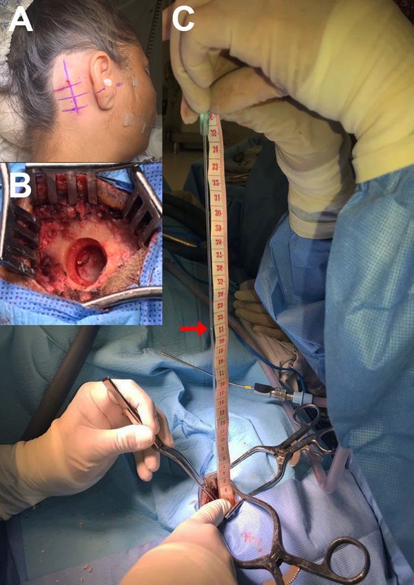
Decompression (MVD) are the most promising before MVD surgery. For the measurement, the
surgical alternatives(30). The MVD surgery procedure patient was placed in a lateral decubitus position,
requires the establishment of a microsurgical trichotomy and antisepsis were performed at
site that involves a craniotomy and the opening the retroauricular region followed by a minimally
of the dura; procedure that allows the CSF to be invasive retrosigmoid craniectomy (Figure 1A
obtained or manipulated to determine some of its and 1B). A 3 mm incision in the dura was then
parameters. The aim of this study is to analyze the performed where approximately 5 mm of a 5 Fr
prevalence of high ICP in patients with BMI ≥ 25 that enteral feeding tube was gently inserted at an angle
suffer vascular compression of the trigeminal nerve. of 45° to position it between the dorsal surface of
the cerebellum and the internal face of the adjacent
Methods dura and directed towards the cistern of the
We selected 97 patients with diagnosis of TN bulbopontine sulcus. The CSF flows up through the
treated at our medical center between December probe placed in a totally vertical position and when
2017 and August 2018 who underwent MVD the CSF movement stabilized, the level reached
surgery. All of them were diagnosed with TN based was determined using a sterilized measuring
on clinical criteria and vascular compression of the tape (Figure 1C). Subsequently, a CSF sample was
trigeminal nerve was confirmed by MRI 3D-FIESTA collected for biochemical and cytological analyses.
sequence (Signa; GE Medical Systems, Milwaukee, This sample was obtained by placing a 10 cc syringe
WI, USA). We excluded patients with BMI < 25; with on the aforementioned probe and aspirating
idiopathic and secondary TN (epidermoid cyst, approximately 5 cc of CSF. Afterwards, the
post-herpetic, meningioma, multiple sclerosis), neurosurgeon proceeded with the MVD surgery.
patients under medication that could condition
IIH (hypervitaminosis, tetracyclines, nalidixic acid, The following values for ICP were used as
nitrofurantoin, sulfonamides, retinoids, cimetidine, reference: < 7 - 15 cmH2O = Normal; 20-25
cyclosporine, diphenylhydantoin, lithium carbonate, cmH2O = Inconclusive; and ≥ 25 cmH2O = High.
☞ http://archivosdeneurociencias.com Vol 25 • Num 3 • 2020 • 9Archivos de Neurociencias (Mex) INNN IIH IN OBESE PATIENTS WITH TN
The weight and height of each patient were measured The mean ICP was 14.4 cmH2O for normal value,
with a mechanical scale and a RGZ-160 stadimeter. 20.0 cmH2O for inconclusive and 27.7 cmH2O in the
The patients were followed up in the immediate case of high ICP. Graph 1 demonstrates a certain
postoperative period and with consultations at one tendency of the ICP to rise according to the increase
month, three months and one year after surgery. in BMI; Pearson's correlation coefficient was +0.122
Statistical analyses were performed with SPSS 22.0 (p = 0.618). On the other hand, the analysis of
software (IBM Corporation, Armonk, New York, USA). CSF samples showed positive for the presence of
Central tendency measures were stablished and a countless erythrocytes in all the samples, probably
Pearson correlation coefficient between BMI and secondary to a traumatic puncture and the implicit
ICP was obtained. surgical procedure. Furthermore, all the samples
showed elevated proteins, glucose and DHL. VDRL
and Gram staining in CSF were negative in all cases.
Graph 1. Correlation between intracranial pressure and body
max index
Figure 1. (A) Patient preparation for MVD surgery. (B)
Minimally invasive retrosigmoid craniectomy before incision
in the dura. (C) Enteral feeding tube colocation for ICP During MVD surgery, we found that the
determination. Red arrow indicates de level reached by the neurovascular contact was arterial in 4 cases,
CSF. venous in 8 and mixed in 7. The most commonly
involved artery was the superior cerebellar artery
Results (n = 7), followed by the vertebrobasilar artery
Of the 19 patients, 89.5% were women and 10.5% (n = 4) and the anterior inferior cerebellar artery
men, with an age range between 26-59 years (n = 2), while the most commonly involved veins
(M = 47). It was determined that 42.1% of these were the pontine vein (n = 6), an innominate vein
patients were overweight (n = 8), 47.4% grade I (n = 5), ponto-trigeminal (n = 3), superior petrosal
obesity (n = 9) and 10.5% with grade II obesity (n = 2). venous complex (n = 2) and bridging vein (n = 1).
After ICP analysis, 37.8% of our patients had a
normal ICP (n = 7), 15.8% were inconclusive ICP After the follow-up period, 78.9% of patients
(n = 3) and 47.4% showed high ICP (n = 9). (n = 15) had a complete remission of pain and 21.1%
Vol 25 • Num 3 • 2020 • 10 ☞ http://archivosdeneurociencias.comOSEGUERA-ZAVALA BETZAIDA SARAÍ, ET. AL. Archivos de Neurociencias (Mex) INNN
(n = 4) presented recurrence of pain during the of the patients with IIH presented its characteristic
first post-surgical year, however all the recurrences signs or symptoms, however, it is possible that these
were treatable with a pharmacological approach. were masked by the trigeminal pain syndrome.
Moreover, it is necessary to demonstrate the
Conclusion absence of CSF alterations for IIH diagnosis. All
Our study suggests that a high percentage of patients in our series showed an increase in CSF cell
patients with BMI ≥ 25 and TN suffer of elevated count although we assume that this anomaly may
ICP without apparent cause. Although the ICP be secondary to the technique required to obtain
raised according to the increase in BMI and a the sample. CSF analysis should be an important
positive Pearson's coefficient, the correlation was point to consider for following studies in our group.
non-statistically significant (p = 0.618). Some of
the limitations of the study include the relatively As mentioned, due to the limitations of the
low number of patients and the absence of observational nature of our study, we believe that
patients without overweight/obesity as control. cohort or control-cases studies are needed to
clearly determine the association between IHH
A positive feedback loop is often proposed for ICP and TN. If this association could be proven, the
where constriction in transverse sinuses raises prevention and management of obesity may be
venous pressure, decreasing CSF resorption and considered in the treatment of patients with TN
subsequently elevating the ICP. We observed and could support the hypothesis that IIH could
that some patients with IIH showed a reduction be a factor involved in the pathophysiology of TN.
in the volume of the cerebellopontine angle
cistern where the trigeminal nerve and adjacent Founding
vascular structures are located, this reduction The authors received no financial support for the
could be favoring the neurovascular contact, research, authorship, and/or publication of this article.
being the superior cerebellar artery and pontine
vein the vessels more frequently involved. Conflict of interest
Therefore, we think that cistern volume reduction The authors declare having no conflicts of interest.
could be also a consequence of an increase
of global retrograde venous pressure in brain Acknowledgment
tissues in a phenomenon analogous to venous We thank to the members of Neurología Segura
stasis of the lower limbs in obese patients. for their support during the development of this
Another important point to mention is that none investigation.
References
1. Barquera Cervera, S, Rojas R, Rivera J. Obesity in Mexico: epidemiology and health policies for its control and
prevention. Gac Med Mex. 2010; 146:397-407.
2. Chooi YC, Ding C, Magkos F. The epidemiology of obesity. Metabolism. 2019; 92:6-10. DOI: 10.1016/j.
metabol.2018.09.005
3. Kopelman PG. Obesity as a medical problem. Nature. 2000; 404(6778):65-643. DOI: 10.1038/35007508
☞ http://archivosdeneurociencias.com Vol 25 • Num 3 • 2020 • 11Archivos de Neurociencias (Mex) INNN IIH IN OBESE PATIENTS WITH TN
4. Knight, Joseph. Diseases and Disorders Associated with Excess Body Weight. Ann Clin Lab Sci. 2011;41(2):107-21.
5. Friedman D, Diger K, Liu G. Revised diagnostic criteria for the pseudotumorcerebri syndrome in adults and
children. Neurology. 2013;81(24):1159-1163. DOI: 10.1212/WNL.0b013e3182a55f17
6. Uddin AB. Drug-induced pseudotumor cerebri. Clin Neuropharmacol. 2003; 26(5):236-238. DOI:
10.1097/00002826-200309000-00007
7. Mondragon J, Klovenski V. Pseudotumor Cerebri. In: StatPearls. StatPearls Publishing, Treasure Island (FL); 2019.
8. Chen J, Wall M. Epidemiology and risk factors for idiopathic intracranial hypertension. Int Ophthalmol Clin.
2014;54(1):1-11. DOI: 10.1097/IIO.0b013e3182aabf11
9. Daniels AB, Liu GT, Volpe NJ, Galetta SL, Moster ML, Newman NJ, et al. Profiles of obesity, weight gain, and quality
of life in idiopathic intracranial hypertension (pseudotumor cerebri). Am J Ophthalmol. 2007; 143(4):635-41. DOI:
10.1016/j.ajo.2006.12.040
10. Smith JL. Whence pseudotumor cerebri? J Clin Neuroophthalmol. 1985;5(1):55-56.
11. Gaye A. Pseudotumor Cerebral. Rev Urug Med Int. 2016;1(3):5261.
12. Ghajar J. Traumatic brain injury. Lancet. 2000;356(9233):923929. DOI: 10.1016/S0140-6736(00)02689-1
13. Kesler A, Kliper E, Shenkerman G, Stern N. Idiopathic intracranial hypertension is associated with lower body
adiposity. Ophthalmology. 2010; 117(1):169-174. DOI: 10.1016/j.ophtha.2009.06.030
14. Salpietro V, Polizzi A, Berte LF, Chimenz R, Chirico V, Manti S, Ferraù V, Salpietro A, Arrigo T, Ruggieri M. Idiopathic
intracranial hypertension: a unifying neuroendocrine hypothesis through the adrenal-brain axis. Neuro Endocrinol
Lett. 2012; 33(6):569–573.
15. Speake T, Whitwell C, Kajita H, Majid A, Brown PD. Mechanisms of CSF secretion by the choroid plexus. Microsc Res
Tech. 2001; 52:49–59. DOI: 10.1002/1097-0029(20010101)52:13.0.CO;2-C
16. Sinclair AJ, Walker EA, Burdon MA, van Beek AP, Kema IP, Hughes BA, Murray PI, Nightingale PG, Stewart PM,
Rauz S, Tomlinson JW. Cerebrospinal fluid corticosteroid levels and cortisol metabolism in patients with idiopathic
intracranial hypertension: a link between 11ß-HSD1 and intracranial pressure regulation? J Clin Endocrinol Metab.
2010; 95:5348–5356. DOI: 10.1210/jc.2010-0729
17. Andrews LE, Liu GT, Ko MW. Idiopathic intracranial hypertension and obesity. Horm Res Paediatr. 2014;81(4):217-
225. DOI: 10.1159/000357730
18. Salpietro V, Polizzi A, Berte LF, Chimenz R, Chirico V, Manti S, et al. Idiopathic intracranial hypertension: a unifying
neuroendocrine hypothesis through the adrenal-brain axis. Neuroendocrinol Lett. 2012;33(6):569-573.
19. Nedelmann M, Kaps M, Mueller-Forell W. Venous obstruction and jugular valve insufficiency in idiopathic
intracranial hypertension. J Neurol. 2009; 256(6):964-969. DOI: 10.1007/s00415-009-5056-z
20. Rabbani CC, Saltagi MZ, Nelson RF. The role of obesity, sleep apnea, and elevated intracranial pressure in
spontaneous cerebrospinal fluid leaks. Curr Opin Otolaryngol Head Neck Surg. 2019;27(5):349-355. DOI: 10.1097/
MOO.0000000000000562
21. Thurtell MJ, Trotti LM, Bixler EO, Rye DB, Bliwise DL, Newman NJ, Biousse V, Bruce BB. Obstructive sleep apnea in
idiopathic intracranial hypertension: comparison with matched population data. J Neurol. 2013;260(7):1748-1751.
DOI: 10.1007/s00415-013-6858-6
22. Davenport RJ, Will RG, Galloway PJ. Isolated intracranial hypertension presenting with trigeminal neuropathy. J
Neurol Neurosurg Psychiatry. 1994; 57(3):381 DOI: 10.1136/jnnp.57.3.381
23. Hussein A, Saleh B, Tahir HO. Idiopathic Intracranial Hypertension. Atypical Presentation. Saudi Med J.
2007;28(5):762765.
Vol 25 • Num 3 • 2020 • 12 ☞ http://archivosdeneurociencias.comOSEGUERA-ZAVALA BETZAIDA SARAÍ, ET. AL. Archivos de Neurociencias (Mex) INNN
24. Iftikhar PM, Munawar M, Pour MA, Nasir S, Inayat A. (2020). Atypical Presentation of Trigeminal Neuralgia
Induced by Intracranial Hypertension Mimicking Sinusitis. Arch Clin Med Case Rep. 2020; 4(2):285-291.
25. Rasmussen P. Facial pain II. A prospective survey of 1052 patients with a view of: Character of the attacks, onset,
course, and character of pain. Acta Neurochir. 1990;107(3-4):121–128. https://doi.org/10.1007/BF01405790
26. Manzoni GC, Torelli P. Epidemiology of typical and atypical craniofacial neuralgias. Neurol Sci. 2005;26 Suppl2:
s65-67. DOI: 10.1007/s10072-005-0410-0
27. Cruccu G, Finnerup NB, Jensen TS, Scholz J, Sindou M, Svensson P, et al. Trigeminal neuralgia: New
classification and diagnostic grading for practice and research. Neurology. 2016;87(2):220-228. DOI: 10.1212/
WNL.0000000000002840
28. Thomas KL, Vilensky JA. The anatomy of vascular compression in trigeminal neuralgia. Clin Anat. 2014;27(1):89-
93. DOI: 10.1002/ca.22157
29. Antonini G, Di Pasquale A, Cruccu G, Truini A, Morino S, Saltelli G, et al. Magnetic resonance imaging contribution
for diagnosing symptomatic neurovascular contact in classical trigeminal neuralgia: a blinded case-control study
and metaanalysis. Pain 2014;155(8):1464-1471. DOI: 10.1016/j.pain.2014.04.020
30. Al-Quliti KW. Update on neuropathic pain treatment for trigeminal neuralgia. The pharmacological and surgical
options. Neurosciences (Riyadh) 2015;20(2):107-114.
Artículo sin conflicto de interés
© Archivos de Neurociencias
☞ http://archivosdeneurociencias.com Vol 25 • Num 3 • 2020 • 13You can also read