Detection of Crimean-Congo hemorrhagic fever virus (CCHFV) in Hyalomma ticks collected from Mauritanian livestock
←
→
Page content transcription
If your browser does not render page correctly, please read the page content below
Detection of Crimean-Congo hemorrhagic fever
virus (CCHFV) in Hyalomma ticks collected from
Mauritanian livestock
Ansgar Schulz
Friedrich-Loe er-Institut Bundesforschungsinstitut fur Tiergesundheit
Yahya Barry
ONARDEL
Franziska Stoek
Friedrich-Loe er-Institut Bundesforschungsinstitut fur Tiergesundheit
Matthew J. Pickin
Friedrich-Loe er-Institut Bundesforschungsinstitut fur Tiergesundheit
Aliou Ba
ONARDEL
Lidia Chitimia-Dobler
Institut fur Mikrobiologie der Bundeswehr
Mohamed L. Haki
Ministère du Développement Rural
Baba A. Doumbia
Ministère du Développement Rural
Albert Eisenbarth
Friedrich-Loe er-Institut Bundesforschungsinstitut fur Tiergesundheit
Abdellahi Diambar
ONARDEL
Mohamend Y. Bah
Ministère du Développement Rural
Martin Eiden
Friedrich-Loe er-Institut Bundesforschungsinstitut fur Tiergesundheit
Martin H. Groschup ( martin.groschup@ i.de )
Friedrich-Loe er-Institut Bundesforschungsinstitut fur Tiergesundheit https://orcid.org/0000-0003-
0215-185X
Research
Keywords: CCHFV, Hyalomma spp., livestock, epidemiology, Mauritania
Page 1/20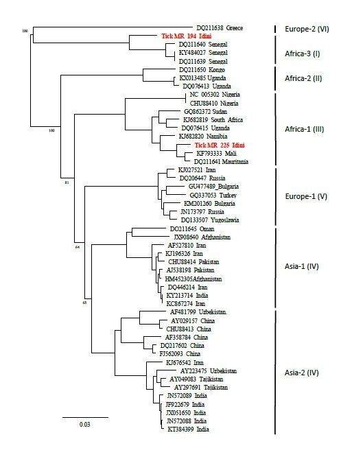
DOI: https://doi.org/10.21203/rs.3.rs-45301/v1
License: This work is licensed under a Creative Commons Attribution 4.0 International License.
Read Full License
Page 2/20
Abstract
Background: Crimean-Congo hemorrhagic fever virus (CCHFV) belongs to the Nairovididae family in the
Orthonairovirus genus and is an emerging tick-borne virus. It is endemic in most parts of Africa, Asia, as
well as southern Europe, and can cause severe hemorrhagic symptoms in humans with high fatality rates
(5-30 %).

Methods: Hyalomma ticks were collected from four different livestock herds (cattle and camel) from
Mauritania in 2018. The tick species was determined morphologically and con rmed on a molecular level
by using cytochrome oxidase 1 gene marker (CO1). For the detection of CCHFV, ticks were tested
individually with a one-step multiplex real-time RT-qPCR. Subsequently, the S-segment of all positive
samples were sequenced to determine the CCHFV genotype.

Results: Overall, 39 of 1,523 ticks (2.56 %) collected from 63 cattle and 28 camels were tested positive for
CCHFV. Three Hyalomma (H.) species were identi ed. The highest prevalence of CCHFV was found in
Hyalomma ru pes (5.67 %; 16/282), followed by H. dromedarii (1.89 %; 23/1,214) and H. impeltatum (0
%; 0/21). Positive ticks were found on only 6 out of 91 host animals. Sequence analysis of the positive
samples revealed the presence of two different CCHFV lineages (Africa I and Africa III).

Conclusions: This study reveals a CCHFV prevalence of 2.56 % in Hyalomma ticks collected from camels
and cattle in Mauritania. The true prevalence of unfed ticks may however be lower since a considerable
number of ticks may have been passively infected during the ingestion of the blood meal by co-feeding or
viremia of the host. The study shows that tick control measures should be implemented, especially in the
examined areas.

Background
Crimean-Congo hemorrhagic fever virus (CCHFV) belongs to the Nairovididae family in the
Orthonairovirus genus and is an emerging zoonotic arthropod-borne virus that causes the Crimean-Congo
hemorrhagic fever in humans (CCHF). CCHFV is present in most parts of Africa, southern Asia as well as
southern Europe [1, 2]. The virus is characterized by a high genetic diversity [3]. Hard ticks of the genus
Hyalomma are considered as main vectors and reservoirs of the virus [4]. Most, if not all of the various
feeding hosts of Hyalomma (ranging from wildlife species to domesticated animals) can be infected,
although they seem to develop only a short-term viremia without clinical symptoms [5]. In contrast,
CCHFV infections in humans can lead to severe hemorrhagic symptoms with a high lethality rate of up to
30 % [4]. Seroepidemiological studies of different susceptible livestock and wildlife species can provide a
 rst indication whether CCHFV is circulating in a certain region. Hence it became a widely used
epidemiological instrument to de ne potential endemic risk areas [5, 6]. However, serological surveys
cannot provide information on the true virus prevalence, CCHFV strain genetics and host-vector
dynamics. Such data can only be obtained by complex experimental studies of tick-host transmission,
which have to be conducted according to stringent guidelines as described by Gargili et al. [7]. To

                                                 Page 3/20
circumvent these highly elaborate experiments, ticks from less complicated eld studies are often
screened for the presence of the virus, although these ndings must be interpreted cautiously. The
detection of CCHFV in engorged ticks is only an evidence for the virus presence, but it does not imply the
vector competence of a tick, since passive contamination from blood of the host cannot be excluded [7].
According to World Health Organization (WHO) data, West African countries, including Mauritania, are
considered highly endemic with a large annual incidence of human CCHF cases. Traditional husbandry is
common in Mauritania, meaning close contact between farmers and their livestock. The rst evidence of
a human CCHFV infection in Mauritania was reported in 1983 [8]. Moreover, several serological studies on
livestock revealed a high seroprevalence of 15 % in small ruminants [9, 10] and up to 67 % in cattle [11],
underlining the high endemic status of the country. Despite those high seroprevalences, CCHFV
prevalences in ticks have not been systematically studied in Mauritania. The rst comprehensive survey
was conducted in 1985 and included 2,539 ticks collected from cattle, sheep, goats, camels and horses
[12]. The samples were pooled and analyzed using complement- xation test, resulting in 12/172 (6-9 %)
CCHFV-positive tick pools. Of the four analyzed tick species (Hyalomma ru pes, Hyalomma marginatum,
Hyalomma impeltatum and Hyalomma dromedarii), only H. ru pes pools were positive. In a second
study, 378 engorged and non-engorged ticks of two different genera (Hyalomma and Rhipicephalus) were
tested for the presence of CCHFV. Only in four Rhipicephalus evertsi evertsi collected from sheep CCHFV
was detected, whereas all of the Hyalomma ticks showed negative results [10].
The aim of this study was to obtain recent data on the circulation of CCHFV in livestock and Hyalomma
ticks in Mauritania. Special emphasis was laid on an accurate tick species identi cation [7, 13] and on
recording sample histories to improve our understanding of the host-vector dynamics in the sampled
herds. Moreover, CCHFV genotypes were determined. The results contribute to a CCHFV exposure risk
analysis for local farmers, butchers and other exposed groups in close contact with livestock.
Materials And Methods
1.1. Collection sites
Mauritania is a large country in West Africa with mostly desert-covered landscape of the Sahara, and a
low population density. Samples were collected in the surrounding region of the capital Nouakchott and
the town of Rosso. Camels were sampled at a livestock market with an associated slaughterhouse on the
outskirts of Nouakchott. In addition, one cattle herd was sampled at a dairy farm 60 km east of
Nouakchott near the small village Idini (Trarza). Two other sampling sites for cattle and camels,
respectively, were located in the surrounding area of Rosso (Trarza) in southwestern Mauritania. The
distance between Nouakchott and Rosso is about 160 km (Fig.).
1.2. Collection of samples
Up to 30 Hyalomma ticks were collected per animal from a total of 28 camels and 63 cattle from the four
aforementioned herds. Moreover, blood samples were taken from most of these animals (Idini, cattle: n=
49; Nouakchott slaughterhouse, camels: n= 13; Rosso, camels: n=15). No blood samples were available
Page 4/20from the cattle herd in Rosso. In total, 77 blood samples were collected among the different herds. Since
Hyalomma ticks are considered the main vector and reservoir for CCHFV, ticks from other genera were
excluded from this study. The collected ticks were stored at -80°C at the O ce National de Recherche et
de Développement de l'Elevage (ONARDEL) in Nouakchott. Ethanol (90 %) was added to the samples
prior to their shipment to the Friedrich-Loe er-Institut (FLI), Germany.
1.3. Morphological and molecular tick species identi cation
All ticks were morphologically identi ed using the identi cation keys of Apanaskevich et al. [14-16].
Individual ticks were homogenized in AVL buffer (Qiagen, Hilden, Germany) using a Tissuelyser II (Qiagen,
Hilden, Germany) machine. The homogenates were cleared by centrifugation and supernatants were used
for nucleic acid extraction. DNA/RNA was extracted using a a KingFisher Flex instrument (ThermoFisher,
Waltham, USA) with the NucleoMag® VET kit (Macherey-Nagel, Düren, Germany) according to the
manufacturer`s protocol. A selected number of ticks that were hard to determine as well as CCHFV-
positive specimens were identi ed using partial cytochrome oxidase 1 (CO1) gene Sanger sequencing
and restriction fragment length polymorphism method (RFLP) [17].
1.4. CCHFV genome detection
RNA extracted using KingFisher instrument (ThermoFisher Scientifc, Waltham, USA) alongside the
NucleoMag Vet kit (Machery-Nagel, Düren, Germany) from individual ticks and serum samples was used
to screen for CCHFV. The screening was performed using a one-step real-time reverse-transcriptase PCR
assay (RT-qPCR) as described previously [18]. The assay targets a conserved region within the S-
Segment. Samples were considered positive in case of ct-values below 35 and weak-positive if between
35 and 40.
1.5. Molecular and phylogenetic analyses of CCHFV genotypes
In order to get a rst insight into the detected CCHFV genotypes, amplicons (127 bp) of the RT-qPCR
products of all positively tested samples were sequenced by Sanger sequencing (Euro ns, Luxembourg,
Luxembourg) and aligned with GeneBank entries by using the BLAST tool (NCBI, Bethesda, USA). For this
purpose, the PCR protocol was performed using only one primer pair speci c for the African CCHFV
lineage III. Due to the small length of the PCR product, it was necessary to amplify a larger segment from
the S-segment, which allows a more meaningful phylogenetic analysis. Therefore, complimentary primers
close to the terminal regions were selected based on the most related sequence in the BLAST results. The
primers to amplify African linage 1 were: “for 5’- AACACGTGCCGCTTACGC” and “rev 5’ –
TATCGTTGCCGCACAGCC”; and for African linage 3, “for 5’ –
ATGGAAAACAAAATCGAGGTGAATAACAAAGAT” and “rev 5’ – TTAGATAATGTTAGCACTGGTGGCATT”.
Both the reverse transcription using SuperScript IV Reverse Transcriptase (ThermoFisher, Waltham, USA)
with the reverse primer as well as the PCR using the KAPA HiFi HotStart ReadyMix PCR Kit (Roche, Basel,
Switzerland) were performed according to the manufacturer's instructions. The ampli ed fragment was
Page 5/20sequenced by Sanger sequencing (Euro ns, Luxembourg, Luxembourg) and used to create a phylogenetic
tree.
1.6. Statistical analysis
Statistical analyses included 95 % con dence intervals (CI), Fisher's exact test and Chi-square test.
Therefore, R software and R-Studio (an integrated development interface for R) were used for the
calculation [19]. Those ticks that could not be species identi ed were excluded for the Fisher's exact test
and Chi-square test.
Results
1.1. Tick species and their distribution on hosts
Overall, 1,523 blood-fed Hyalomma ticks were collected from 63 cattle and 28 camels. Morphological
identi cation revealed the presence of three different tick species (H. dromedarii, H. ru pes and H.
impeltatum) in the surveyed areas. Due to the large morphological diversity of Hyalomma ticks and the
similarity of H. dromedarii and H. impeltatum, 219 specimens were identi ed genetically by the RFLP
approach. Furthermore, the CO1 gene amplicon used for restriction digest was sequenced from 47 ticks
to unambiguously identify their species. Due to the very poor condition of six ticks, neither a
morphological nor a molecular identi cation of these specimens was possible. Therefore, those ticks
were only determined up to genus level. Results of species identi cation and distribution among the
different collection sites and hosts are summarized in Table 1. H. dromedarii represented the by far
largest population both in cattle and camels (79.58 %), followed by H. ru pes (18.52 %) and H.
impeltatum (1.51 %). Despite the predominance of H. dromedarii, a great variation was observed
regarding the regional distribution of tick species in their hosts. In both camel herds, the percentage of H.
dromedarii was very high (97.01 – 98.58 %), while ticks of the other species were found only sporadically.
In contrast, the distribution of tick species among the two cattle herds was considerably more
heterogeneous. A large number of H. dromedarii individuals (88.76 %) were identi ed on the dairy farm in
Idini, although H. ru pes (6.94 %) and H. impeltatum (3.31 %) were also recorded. On the other hand,
85.34 % of the collected ticks from the cattle herd in Rosso were identi ed as H. ru pes and only 14.66 %
as H. dromedarii. The distribution of sex showed nearly a 70:30 ratio of males to females across all four
collection sites. In the cattle herd from Rosso, the proportion of male ticks was slightly higher compared
to the other sampling sites.
1.2. CCHFV prevalence in ticks and sera
Of the 1,523 individual Hyalomma ticks analyzed, 39 tested positive for CCHFV by RT-qPCR and 23 were
considered weak-positive. A comprehensive summary is given in Table 1. The CCHFV prevalence between
species and sampling sites showed signi cant differences. Overall, 23 out of 1,214 H. dromedarii (1.89
%) and 16 out of 282 H. ru pes (5.67 %) were found to be positive for CCHFV, respectively. All 21
examined H. impeltatum ticks were negative. Only three H. dromedarii collected from camels at the
Page 6/20Tenweich slaughterhouse tested positive (0.87 %). In contrast, all ticks from the camel herd nearby Rosso
(Trarza) tested negative, whereas 7 out of 266 ticks (all H. ru pes) originating from cattle of the same
region were positive (2.63 %). The highest prevalence (4.79 %) was found in the cattle herd from Idini
(Trarza), where 29 positive ticks were identi ed out of 605 collected ticks. Overall, 20 of 537 H. dromedarii
(3.72 %) and 9 of 42 H. ru pes (21.43 %) were CCHFV-positive, while all H. impeltatum ticks (n = 26) were
negative. In total, 2.28 % of the female and 2.67 % male sampled ticks were CCHFV-positive. No
signi cant sex differences were observed in terms of CCHFV prevalence. All 77 collected serum samples
of camels and cattle were negative for CCHFV.
1.3. Distribution of CCHFV-positive ticks per sampled livestock
The distribution of all CCHFV-positive Hyalomma spp. on cattle and camels is summarized in Table 2.
CCHFV-positive ticks were collected on 6 out of 91 (6.59 %) sampled animals ( ve cattle: “no. 1 to no. 5“
and one camel: “no. 1”). In one case (“cattle no. 1”), all 22 collected ticks were CCHFV-positive. In four
cattle, negatively tested Hyalomma spp. were associated with a variable number of positively and weak-
positively tested ticks. In each of these four cattle, one of the collected ticks was highly-positive with
much higher levels of S-segment RNA (lower ct-value) compared to the other ticks collected from the
same cattle. In three of the four cases, these highly positive ticks were identi ed as H. ru pes, and in one
case identi ed to be H. dromedarii. In "cattle no. 5“, only a single H. ru pes was CCHFV-positive, while the
remaining ticks were negative. There was only one camel ("camel no. 1") found with CCHFV-positive ticks
(n = 3) which were identi ed as H. dromedarii. Viral RNA concentrations of the S-segment were almost
identical in all three samples, and no “weak-positive” ticks were found.
1.4. Phylogeny of CCHFV genotypes
To determine the CCHFV genotypes, the 127 bp long RT-qPCR amplicons were sequenced. Consistent
sequence data were obtained for 28 of the 39 CCHFV-positive ticks and compared by using the BLAST
tool (NCBI). Essentially, two different CCHFV genotypes – Africa I and Africa III – were detected (Tables 2
and 3). Ticks originating from the same animal always carried the same CCHFV genotype and haplotype.
However, two different lineages (Africa I and III) circulated simultaneously in tick vectors of cattle from
the Idini region (Table 2). The CCHFV lineages also differed between the collection sites and/or host
species (Table 3). The Africa I lineage detected in ticks from cattle (Idini) and camel (Nouakchott
slaughterhouse) showed nucleotide differences of 3.15 %. The Africa III genotypes found in ticks feeding
on cattle from Idini and from Rosso varied by 2.17 %. The overall genetic distance between the Africa I
and Africa III lineages ranged from 10.04 % between the cattle herds from Idini (I) and Rosso (III) up to a
maximum of 13.19 % between camels from Nouakchott slaughterhouse (I) and cattle from Rossi/Idini
(III). To con rm that the short amplicon (127 bp) was representative for the whole segment, the complete
coding region of the S-segment from two CCHFV-positive ticks was RT-PCR ampli ed and sequenced.
Alignments with GenBank database sequences showed a nucleotide homology of 97.5 % and 99.4 % for
the consensus sequences of Africa I and Africa III strains, respectively. Construction of a phylogenetic tree
Page 7/20(Fig. 2) showed that both sequences clustered well with the reference strains of Africa I (Senegal) and III
(Mauritania/Mali).
Discussion
Previous serological studies have shown a high CCHFV antibody seroprevalence in the livestock
population of Mauritania. Several severe CCHF cases have been reported in humans. Hence, Mauritania
is considered as a high-endemic country for CCHFV [8–11, 20]. CCHF cases were also reported in Senegal
which borders Mauritana to the south [21, 22]. In Mali, eastwards of Mauritania, no human cases of CCHF
have been reported, but serological data [23] and virus detection in ticks [24] have also proven a CCHFV
circulation in Mali. However, CCHFV monitoring in ticks (especially of the genus Hyalomma) has not yet
been conducted systematically in this West African region. Existing datasets are either small-scaled,
outdated or generated by the analysis of tick pools with focus on virus detection thus only allowing
limited conclusions on vector species distribution and competence [10, 12, 24, 25]. Therefore, this study
was carried out to provide a better understanding of host-vector dynamics and the current
epidemiological situation in Mauritanian livestock herds.
The presence of the three tick species H. ru pes, H. dromedarii, and H. impeltatum in Mauritania is
consistent with previous reports from the region [14–16]. The primary host of H. dromedarii are camels
[16], which explains the high proportion of specimens (97.01–98.58%) found on camels in Rosso and
Nouakchott slaughterhouse (Table 1). Due to the important role camels play as agricultural livestock for
milk and meat production in Mauritania, there is a relatively high camel density in the country [26].
Moreover, cattle and other ungulates can also be infested by adult stages of H. dromedarii [16], especially
if camels and cattle are held in close contact. Thus, the 88.71% of H. dromedarii ticks found on cattle
from Idini were not an exceptional nding. The higher rate of male ticks across all four sampling sites is
probably caused by the absence of female ticks from the host during oviposition, which is taking place in
the environment and not on the host itself [27].
In total, 39/1,523 (2.56%) of the blood-fed ticks collected from cattle and camels were CCHFV-positive
(Table 1). The highest prevalence was found in both cattle herds from Idini (4.79%) and Rosso (2.63%),
followed by the camels from the Nouakchott slaughterhouse (0.85%). No CCHFV-positive ticks were
found on camels from Rosso. The reasons for different CCHFV prevalences across collection sites and
host species are various and require careful interpretation. One explanation could be a difference in
susceptibility of cattle and camels for the virus and/or a better capability of cattle to support a longer
lasting viremia. So far, only a small number of experimental CCHFV infection studies have been
conducted with different livestock species (cattle, sheep and horses), which showed that all species
develop a short-term viremia of similar durations [6]. However, no experimental data of CCHFV infections
of camelids are available to date to prove this assumption. There remains a need for further infection
experiments comparing the susceptibility for CCHFV in different host species.
Page 8/20The high CCHFV prevalence of ticks from cattle in Idini may be related to the geographically isolated
location of the sampled farm. It is assumed that fragmented CCHFV foci consisting of susceptible hosts
and competent vector ticks may induce stable virus ampli cation, leading to a high prevalence in these
isolated geographical clusters [28]. This dairy farm is located far away from the next village Idini in a
desert-like region. The remote location results in limited contact with new naive hosts (wildlife, livestock),
which might negatively affect the CCHFV prevalence [29]. In contrast, the fertile lands around Rosso lead
to a higher density of livestock as well as human population and thus to an increased movement and
interaction between the animals. A similar situation exists at the livestock market in Nouakchott, where a
large number of cattle, sheep, goats and camels from various regions of Mauritania are sold or
slaughtered every day (Fig. 3).
The tick species themselves may also have an impact on the CCHFV prevalence. Ticks of the genus
Hyalomma are considered as main vector and reservoir of CCHFV [7], but it is still unknown whether all of
the currently recognized 27 Hyalomma spp. [30] can function e ciently as virus reservoir and/or vector.
Despite the considerably higher occurrence of H. dromedarii (79.71%) ticks in the study region (Table 1),
signi cantly more H. ru pes (5.67%) than H. dromedarii (1.89%) ticks were CCHFV-positive. This
difference was most obvious among the cattle herd from Idini, where 21.43% of H. ru pes and only 3.72%
of H. dromedarii were CCHFV-positive. Nevertheless, since our data were derived from blood-fed ticks,
speculations on vector competence have to be interpreted cautiously. Furthermore, feeding on viremic
hosts and/or co-feeding transmission [31–34] may also have contributed to the concentrated occurrence
of some of the 39 positively tested ticks collected from 6 (of 91) animals (Table 2). Interestingly, one of
the CCHFV-positive ticks collected from four bovines in Idini and Rosso (no. 1- no. 4) each had much
higher levels of S-segment RNA than the co-infesting ticks of the same animal. It is also noteworthy that
three of four highly positive ticks in Idini (total occurrence: 88.76% H. dromedarii vs. 6.94% H. ru pes)
were identi ed as H. ru pes (Table 2), which is suggestive for a better vector competence of H. ru pes for
CCHFV. The genomic data proving a 100% sequence identity of CCHFV for all infected ticks from a given
bovine host supports this assumption (Tables 2 and 3). Therefore, the true CCHFV prevalence in the tick
population and thus the absolute risk of exposure for local farmers may actually be considerably lower
than the measured 2.56%. Nevertheless, it is recommended to enforce tick control strategies and
encourage public awareness of tick bite prevention in the examined areas.
In addition, at least two different CCHFV genotypes (Africa I and III) were found in ticks in Mauritania,
either alone or even circulating side by side simultaneously as observed in one cattle herd from Idini
(Tables 2 and 3). Signi cant genetic variability also occurred within the genotypes. The underlying
mechanisms of the high genetic diversity are still not fully understood and require further research on the
driving factors.
Conclusion
This study reveals a high CCHFV prevalence (2.56%) in Hyalomma ticks collected from camels and cattle
in Mauritania. H. ru pes showed signi cantly higher CCHFV infection rates compared to H. dromedarii
Page 9/20and H. impeltatum. However, it must be considered the data was obtained from engorged ticks thus
skewing statements on true vector competence. Two different CCHFV genotypes (Africa I and III) were
found in the ticks. The absolute risk of exposure for local farmers is probably lower than this determined
prevalence would suggest, because a considerable number of ticks may have been passively infected
during ingestion of the blood meal by co-feeding with infected ticks or feeding on a viremic host. It is
recommended to enforce tick control strategies and encourage public awareness of tick bite prevention in
these areas.
Declarations
Ethics approval and consent to participate
The sample collection was carried out by the Mauritanian State Veterinary Laboratory “O ce National de
Recherches et de Développement de l'Elevage“ (ONARDEL) following all relevant national as well as
international regulations and according to fundamental ethical principles for diagnostic purposes in the
framework of a governmental program for the animal health surveillance.
Consent for publication
Not applicable.
Availability of data and materials
All data generated or analysed during this study are included in this published article.
Competing interests
There were no competing interests among the authors.
M.J. Pickin
molecular assay methodology, practical implementation, writing (review & editing)
Aliou Ba
on-site methodology implementation, data curation
Lidia Chitimia-Dobler
entomological expertise and supervision, writing (review & editing)
Mohamed L. Haki
local supervision of project administration
Baba A. Doumbia
Page 10/20resources, project administration
Abdellahi Diambar
local project implementation
Mohamend Y. Bah
resources, project administration
M.H. Groschup
design of research strategy and overall supervision of its implementation, data interpretation, funding
acquisition and professional project administration, manuscript writing ( nal review & editing)
Funding
This study was co-funded by the German O ce for Foreign Affairs (German Partnership Program for
Biosecurity), by the Deutsche Forschungsgemeinschaft (grant number: GR 980/4 − 1) as well as by the
European Union (LEAP-AGRI LEARN project (ID 222)).
Authors contribution
Acknowledgement
We thank Kristin Vorpahl for the technical support and Jana Schulz for creating the phylogenetic tree and
the map. Moreover, we especially thank all Mauritanian collaboration partners for their help collecting the
tick samples in Mauritania.
References
1. Bente DA, Forrester NL, Watts DM, McAuley AJ, Whitehouse CA, Bray M. Crimean-Congo hemorrhagic
fever: history, epidemiology, pathogenesis, clinical syndrome and genetic diversity. Antiviral research.
2013;100(1):159–89.
2. Hoogstraal H. The epidemiology of tick-borne Crimean-Congo hemorrhagic fever in Asia, Europe, and
Africa. J Med Entomol. 1979;15(4):307–417.
3. Deyde VM, Khristova ML, Rollin PE, Ksiazek TG, Nichol ST. Crimean-Congo hemorrhagic fever virus
genomics and global diversity. J Virol. 2006;80(17):8834–42.
4. Whitehouse CA. Crimean-Congo hemorrhagic fever. Antiviral research. 2004;64(3):145–60.
5. Spengler JR, Bergeron E, Rollin PE. Seroepidemiological Studies of Crimean-Congo Hemorrhagic
Fever Virus in Domestic and Wild Animals. PLoS Negl Trop Dis. 2016;10(1):e0004210.
6. Spengler JR, Estrada-Pena A, Garrison AR, Schmaljohn C, Spiropoulou CF, Bergeron E, et al. A
chronological review of experimental infection studies of the role of wild animals and livestock in the
Page 11/20maintenance and transmission of Crimean-Congo hemorrhagic fever virus. Antiviral research.
2016;135:31–47.
7. Gargili A, Estrada-Pena A, Spengler JR, Lukashev A, Nuttall PA, Bente DA. The role of ticks in the
maintenance and transmission of Crimean-Congo hemorrhagic fever virus: A review of published
eld and laboratory studies. Antiviral research. 2017;144:93–119.
8. Saluzzo JF, Aubry P, McCormick J, Digoutte JP. Haemorrhagic fever caused by Crimean Congo
haemorrhagic fever virus in Mauritania. Trans R Soc Trop Med Hyg. 1985;79(2):268.
9. Gonzalez JP, LeGuenno B, Guillaud M, Wilson ML. A fatal case of Crimean-Congo haemorrhagic fever
in Mauritania: virological and serological evidence suggesting epidemic transmission. Trans R Soc
Trop Med Hyg. 1990;84(4):573–6.
10. Nabeth P, Cheikh DO, Lo B, Faye O, Vall IO, Niang M, et al. Crimean-Congo hemorrhagic fever,
Mauritania. Emerg Infect Dis. 2004;10(12):2143–9.
11. Sas MA, Mertens M, Isselmou E, Reimer N, El Mamy BO, Doumbia B, et al. Crimean-Congo
Hemorrhagic Fever Virus-Speci c Antibody Detection in Cattle in Mauritania. Vector borne zoonotic
diseases. 2017;17(8):582–7.
12. Saluzzo JF, Digoutte JP, Camicas JL, Chauvancy G. Crimean-Congo haemorrhagic fever and Rift
Valley fever in south-eastern Mauritania. Lancet. 1985;1(8420):116.
13. Estrada-Pena A, Gray JS, Kahl O, Lane RS, Nijhof AM. Research on the ecology of ticks and tick-borne
pathogens–methodological principles and caveats. Front Cell Infect Microbiol. 2013;3:29.
14. Apanaskevich DA, Horak IG. The genus Hyalomma Koch, 1844: V. re-evaluation of the taxonomic
rank of taxa comprising the H. (Euhyalomma) marginatum Koch complex of species (Acari:
Ixodidae) with redescription of all parasitic stages and notes on biology. Int J Acarol. 2008;34(1):13–
42.
15. Apanaskevich DA, Horak IG. The genus Hyalomma Koch, 1844. IX. Redescription of all parasitic
stages of H. (Euhyalomma) impeltatum Schulze & Schlottke, 1930 and H. (E.) somalicum Tonelli
Rondelli, 1935 (Acari: Ixodidae). Systematic parasitology. 2009;73(3):199–218.
16. Apanaskevich DA, Schuster AL, Horak IG. The genus Hyalomma: VII. Redescription of all parasitic
stages of H. (Euhyalomma) dromedarii and H. (E.) schulzei (Acari: Ixodidae). J Med Entomol.
2008;45(5):817–31.
17. Schulz A, Karger A, Bettin B, Eisenbarth A, Sas MA, Silaghi C, et al. Molecular discrimination of
Hyalomma tick species serving as reservoirs and vectors for Crimean-Congo hemorrhagic fever virus
in sub-Saharan Africa. Ticks and tick-borne diseases. 2020:101382.
18. Kasi KK, von Arnim F, Schulz A, Rehman A, Chudhary A, Oneeb M, et al. Crimean-Congo haemorrhagic
fever virus in ticks collected from livestock in Balochistan, Pakistan. Transbound Emerg Dis. 2020.
19. R Core Team. R: A language and environment for statistical computing. Vienna: R Foundation for
Statistical Computing; 2019.
20. Kleib AS, Salihy SM, Ghaber SM, Sidiel BW, Sidiya KC, Bettar ES. Crimean-Congo Hemorrhagic Fever
with Acute Subdural Hematoma, Mauritania, 2012. Emerg Infect Dis. 2016;22(7):1305–6.
Page 12/2021. Nabeth P, Thior M, Faye O, Simon F. Human Crimean-Congo hemorrhagic fever, Senegal. Emerg Infect
Dis. 2004;10(10):1881–2.
22. Tall A, Sall AA, Faye O, Diatta B, Sylla R, Faye J, et al. [Two cases of Crimean-Congo haemorrhagic
fever (CCHF) in two tourists in Senegal in 2004]. Bull Soc Pathol Exot. 2009;102(3):159–61.
23. Maiga O, Sas MA, Rosenke K, Kamissoko B, Mertens M, Sogoba N, et al. Serosurvey of Crimean-
Congo Hemorrhagic Fever Virus in Cattle, Mali, West Africa. Am J Trop Med Hyg. 2017;96(6):1341–5.
24. Zivcec M, Maiga O, Kelly A, Feldmann F, Sogoba N, Schwan TG, et al. Unique strain of Crimean-Congo
hemorrhagic fever virus, Mali. Emerg Infect Dis. 2014;20(5):911–3.
25. Zeller HG, Cornet JP, Diop A, Camicas JL. Crimean-Congo hemorrhagic fever in ticks (Acari: Ixodidae)
and ruminants: eld observations of an epizootic in Bandia, Senegal (1989–1992). J Med Entomol.
1997;34(5):511–6.
26. Mint Mohamed Lemine A, Ould Lemrabott MA, Hasni Ebou M, Mint Lekweiry K, Ould Ahmedou Salem
MS, Ould Brahim K, et al. Mosquitoes (Diptera: Culicidae) in Mauritania: a review of their biodiversity,
distribution and medical importance. Parasites vectors. 2017;10(1):35.
27. Estrada-Pena A, de la Fuente J. The ecology of ticks and epidemiology of tick-borne viral diseases.
Antiviral research. 2014;108:104–28.
28. Estrada-Pena A, Vatansever Z, Gargili A, Ergonul O. The trend towards habitat fragmentation is the
key factor driving the spread of Crimean-Congo haemorrhagic fever. Epidemiol Infect.
2010;138(8):1194–203.
29. Estrada-Pena A, Jameson L, Medlock J, Vatansever Z, Tishkova F. Unraveling the ecological
complexities of tick-associated Crimean-Congo hemorrhagic fever virus transmission: a gap analysis
for the western Palearctic. Vector borne zoonotic diseases. 2012;12(9):743–52.
30. Horak IG, Camicas JL, Keirans JE. The Argasidae, Ixodidae and Nuttalliellidae (Acari: Ixodida): a
world list of valid tick names. Exp Appl Acarol. 2002;28(1–4):27–54.
31. Gonzalez JP, Camicas JL, Cornet JP, Faye O, Wilson ML. Sexual and transovarian transmission of
Crimean-Congo haemorrhagic fever virus in Hyalomma truncatum ticks. Res Virol. 1992;143(1):23–
8.
32. Gordon SW, Linthicum KJ, Moulton JR. Transmission of Crimean-Congo hemorrhagic fever virus in
two species of Hyalomma ticks from infected adults to cofeeding immature forms. Am J Trop Med
Hyg. 1993;48(4):576–80.
33. Jones LD, Gaunt M, Hails RS, Laurenson K, Hudson PJ, Reid H, et al. Transmission of louping ill virus
between infected and uninfected ticks co-feeding on mountain hares. Med Vet Entomol.
1997;11(2):172–6.
34. Logan TM, Linthicum KJ, Bailey CL, Watts DM, Dohm DJ, Moulton JR. Replication of Crimean-Congo
hemorrhagic fever virus in four species of ixodid ticks (Acari) infected experimentally. J Med
Entomol. 1990;27(4):537–42.
35. Legends.
Page 13/20Tables
Table 1. CCHFV prevalence rates in ticks at the different sampling sites
Overview showing results of all four sampled herds, including the collected number of different tick species,
CCHFV-positive ticks and the analysis of the correlation between the three identified species and CCHFV status
by chi-square test (p < 0.05).
Page 14/20Location, Tick species Tick number % Number of CCHFV Prev./total Prev./tick
Number of collected positive ticks species
sampled animals
Idini Hyalomma 537 88.76 20 3.31% 3.72% (2.29-
43 cattle dromedarii % (2.03-5.06 5.69 %)
%)
Hyalomma 20 3.31 0 0% (0-0.06 0% (0-16.84
impeltatum % %) %)
Hyalomma 42 6.94 9 1.49% 21.43%
rufipes % (0.06-2.8 (10.3-
%) 36.81%)
Hyalomma spp. 6 0.99 0 0% (0-0.06 0% (0-16.84
% %) %)
Total 605 29 4.79 % p-value:
(3.23-6.81Prev. = CCHFV prevalence; Hyalomma spp. = not identified species; 95 % confidence interval (CI %) in brackets;
*= no p-value available, because of the absence of H. impeltatum at this sampling site
Table 2. Distribution of positive ticks over the sampled animals
All sampled animals on which CCHFV-positive ticks were found (6 out of 91), including weak-positive tested ticks
on those individuals and the species of the most positive sample. The CCHFV genotype was determined by
comparing the RT-qPCR products (127 bp) using the BLAST Tool (NCBI).
Location Host n p (+) Tick sample with the lowest CCHFV ct-value CCHFV
Lowest ct- Tick species Second- Tick species genotype
value (lowest ct- lowest (sec.- lowest ct-
value) ct- value value)
Idini Cattle 22 22 0 18.26 H. rufipes 28.62 H. dromedarii Africa I
no. 1
Idini Cattle 30 1 8 19.44 H. dromedarii 35.22 H. dromedarii Africa I
no. 2
Idini Cattle 22 5 9 19.68 H. rufipes 34.44 H. dromedarii Africa I
no. 3
Rosso Cattle 21 7 3 28.8 H. rufipes 31.52 H. rufipes Africa III
no. 4
Idini Cattle 8 1 0 26.66 H. rufipes - - Africa III
no. 5
Nouakchott Camel 24 3 0 28.67 H. (29.89; - Africa I
no. 1 dromedarii (3x) 29.56)
Total 127 39 20
n= number of ticks; p= positive ticks; (+) = weak-positive ticks
Table 3. Genetic distances (%) between the CCHFV genotypes
Genetic distances (%) between the CCHFV lineages found in the respective positive ticks on cattle and camels
by comparing the RT-qPCR amplicons (127 bp). In all positive ticks originated from the same host animal, both
the genotype and the detected gene sequence were identical.
Page 16/20Host/location/genotype Cattle Cattle Cattle Cattle Cattle Camel No.1
No.1 No.2 No.3 No.4 No.5
Idini Idini Idini Rosso Idini Nouakchott
Africa I Africa I Africa I Africa III Africa III Africa I
Cattle Idini Africa I - 100 % 100 % 89.96 % 89.76 % 96.85 %
No.1
Cattle Idini Africa I 100 % - 100 % 89.96 % 89.76 % 96.85 %
No.2
Cattle Idini Africa I 100 % 100 % - 89.96 % 89.76 % 96.85 %
No.3
Cattle Rosso Africa 89.96 % 89.96 % 89.96 % - 97.83 % 86.81 %
No.4 III
Cattle Idini Africa 89.76 % 89.76 % 89.76 % 97.83 % - 86.81 %
No.5 III
Camel Nouakchott Africa I 96.85 % 96.85 % 96.85 % 86.81 % 86.81 % -
No.1
Figures
Page 17/20Figure 1
Map of Mauritania and the neighboring countries showing the different sampling sites of cattle and
camels.
Page 18/20Figure 2
Phylogenetic tree showing the genetic distances of the small (S-) segment of the consensus strains and
selected CCHFV-positive samples from Mauritania (1384 bp). The tree was generated using Neighbor-
Joining algorithm and Jukes-Cantor distance model in Geneious version 2019.2 (Biomatters, available
from https://www.geneious.com). The tree was midpoint-rooted using FigTree v1.4.4 (available from
https://github.com/rambaut/ gtree/releases). Values at branches represent support in 1,000 bootstrap
replicates. Bootstrap values are only shown at major branches.
Page 19/20Figure 3
Different habitats of the livestock herds (cattle and camels) selected for sampling Cattle (a) and camel
herds (c) grazing nearby Rosso (Trarza) close to the Senegal River (southern Sahel). The favourable
climatic conditions permit the growth of larger trees in the savanna landscape and also the simple
cultivating of eld crops. In contrast, a modern dairy farm in Idini (b) is fully surrounded by arid desert.
Picture d) depicts camels that are gathered for being sold at the highly-frequented Nouakchott livestock
market/slaughterhouse.
Page 20/20You can also read