Redescription of Anopheles Nyssorhynchus antunesi Galvão & Amaral and description of a new species of the Myzorhynchella Section Diptera: ...
←
→
Page content transcription
If your browser does not render page correctly, please read the page content below
278      Mem Inst Oswaldo Cruz, Rio de Janeiro, Vol. 105(3): 278-285, May 2010

        Redescription of Anopheles (Nyssorhynchus) antunesi Galvão
      & Amaral and description of a new species of the Myzorhynchella
        Section (Diptera: Culicidae) from Serra da Mantiqueira, Brazil
         Sandra Sayuri Nagaki1, Monique de Albuquerque Motta2, Maria Anice Mureb Sallum1/+
 Departamento de Epidemiologia, Faculdade de Saúde Pública, Universidade de São Paulo, Av. Dr. Arnaldo 715, 01246-904, São Paulo,
 1

        SP, Brasil 2Laboratório de Transmissores de Hematozoários, Instituto Oswaldo Cruz-Fiocruz, Rio de Janeiro, RJ, Brasil

   Anopheles (Nyssorhynchus) pristinus Nagaki & Sallum, n. sp. of the Myzorhynchella Section is described based on
morphological characters of adult females, males, fourth-instar larvae, pupae and male genitalia. Anopheles (Nys-
sorhynchus) antunesi Galvão & Amaral is characterized to fix its identity and distinguish it from An. pristinus. The
eggs of An. antunesi are described for the first time. Molecular characterization employing sequences of the COI mi-
tochondrial gene and the ITS2 region of ribosomal DNA are provided for each species. An. antunesi and An. pristinus
are compared with morphologically similar species of the Myzorhynchella Section. The results of the present study
suggest that the new species has been misidentified as both An. antunesi and Anopheles lutzii Cruz. An. antunesi and
An. pristinus are sympatric, occurring at high altitudes in Serra da Mantiqueira, southeastern Brazil.

                               Key words: COI - ITS2 - morphology - Serra da Mantiqueira - new species

     The genus Anopheles Meigen includes 458 species,               reserve in the Serra do Mar within the Mata Atlântica,
which are subdivided into seven subgenera (Harbach                  in municipality of Salesópolis, state of São Paulo (SP).
2008). The subgenus Nyssorhynchus Blanchard includes                More recently, Forattini et al. (1997) described the eggs
some of the most important vector species of human ma-              of An. antunesi from specimens collected in Campos do
laria parasites in Central and South America (Sallum &              Jordão, Serra da Mantiqueira.
Wilkerson 1997) and is divided into three sections, the                 The type specimen of An. antunesi was deposited in
Albimanus, Argyritarsis and Myzorhynchella Sections                 the Faculdade de Medicina, Universidade de São Paulo
(Harbach 2008). The Albimanus Section includes 19 for-              (FMUSP) (Galvão & Amaral 1940). The FMUSP collec-
mally recognized species (Faran 1980), the Argyritarsis             tion was transferred to the Faculdade de Saúde Pública
11 species (Linthicum 1988, Rosa-Freitas 1989, Mo-                  (FSP) of USP in the late 1960s; however, except for one
toki et al. 2009) and the Myzorhynchella four species,              adult female paratype (E-2034) (Forattini et al. 1970), no
i.e., Anopheles lutzii Cruz, Anopheles parvus (Chagas),             other type specimen of An. antunesi was found among
Anopheles nigritarsis (Chagas) and Anopheles antunesi               the individuals in the FMUSP collection. It is likely that
Galvão & Amaral (Galvão 1941). These four taxa were                 the holotype of An. antunesi is either lost or deposited in
described from specimens collected in Brazil (Belkin et             an unknown collection.
al. 1971) and there is no epidemiological evidence sup-                 While examining specimens of An. antunesi from
porting the involvement of any Myzorhynchella species               Serra da Mantiqueira, we observed that they could be sep-
as vectors of pathogens.                                            arated into two morphological forms based on the pattern
     Galvão & Amaral (1940) described An. antunesi                  of pale and dark wing spots and characters of the larvae
based on morphological characters of all life stages, in-           and male genitalia. One morphological form corresponds
cluding eggs. Specimens were from Vila Emilio Ribas,                to An. antunesi, whereas the second form belongs to an
municipality of Campos do Jordão, Serra da Mantiquei-               undescribed species. The objective of this paper is to de-
ra, southeastern Brazil. Since the description of An. an-           scribe the new species of the Myzorhynchella Section and
tunesi, few studies have been conducted for species of              to characterize An. antunesi employing both morphologi-
the Myzorhynchella Section. Galvão (1941) described                 cal characters and DNA sequence for the ITS2 region of
and illustrated the eggs and the male genitalia of speci-           ribosomal DNA and the COI mitochondrial gene.
mens of An. antunesi collected in Casa Grande, a forest                           Materials and Methods
                                                                         Females were collected in a Shannon trap in the
                                                                    vicinities of the Parque Nacional do Itatiaia, munici-
                                                                    pality of Itatiaia, state of Rio de Janeiro (RJ), Brazil
                                                                    (-22.41630556S 44.62213889W, Datum SAD69) and in
Financial support: FAPESP (2005/53973-0 to MAMS, Master fellow-     Pico do Itapeva, municipality of Pindamonhangaba, SP,
ship 07/01870-8 to SSN), CNPq (BPP 300351/2008-9 to MAMS)
                                                                    Brazil (-22.75847222S -45.51527778W, Datum SAD69).
+ Corresponding author: masallum@usp.br
Received 17 December 2009                                           Eggs were collected from two females of each local-
Accepted 1 April 2010                                               ity. Females were blood-fed and one wing was removed

online | memorias.ioc.fiocruz.br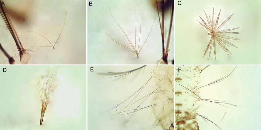
Species of the Myzorhynchella Section • Sandra Sayuri Nagaki et al. 279
48 h later to induce oviposition. Eggs were fixed in for PCR. Sequencing reactions were purified in Sephadex
Bouin’s solution 36 h after oviposition and prepared for G50® columns (GE Healthcare). Sequences were analyzed
scanning electron microscopy (SEM) following the pro- on an ABI Prism 3100 - Avant Genetic Analyzer (Applied
tocol described by Forattini & Marucci (1993). External Biosystems, Foster City, CA, USA).
morphology of the eggs was examined in a JEOL-P15 Sequences were edited using Sequencher ® for Win-
scanning electron microscope (JEOL, Japan). dows version 4.9 (Gene Codes Corporation, Ann Arbor,
Morphological characters of the adult females, males, USA), aligned in CLUSTAL X 1.8 (Thompson et al.
fourth-instar larvae, pupae and male genitalia were ex- 1997) and optimized manually in MacClade version 4.08
amined. Abbreviations for the life stages are: F: adult (Maddison & Maddison 2000). Similarity of the ITS2
female; M: adult male; G: male genitalia; L: larva; P: sequences generated in this study with those previously
pupa; Le: larval exuviae; Pe: pupal exuviae; E: eggs. Ter- available in GenBank was assessed using FASTA search
minology for morphological characters follows Harbach (http://www.ncbi.nlm.nih.gov/BLAST/) and document-
& Knight (1980). The nomenclature of the wing veins ed using PAUP* 4.0 software (Swofford 2003).
follows Belkin (1962), and the terminology for wing Larval and pupal exuviae, male genitalia, wings
spots follows Wilkerson & Peyton (1990). Specimens and legs of the specimens used for DNA extraction are
used in this study are deposited in the Entomological deposited in the FSP-USP as vouchers. Template DNA
Collection of the FSP-USP. from this study is retained at -70ºC in the FSP-USP for
Sequences of the ITS2 of ribosomal DNA and COI future reference.
barcode subunit of the mitochondrial genome were gen-
erated for six specimens of An. antunesi and 12 speci- An. (Nyssorhynchus) antunesi Galvão & Amaral
mens of Anopheles pristinus. Genomic DNA was extract- (Figs 1-4)
ed from individual mosquitoes following the insect DNA An. (Nyssorhynchus) antunesi of Galvão & Amaral,
extraction protocol provided by the QIAgen DNeasy® 1940: 150. Galvão
�������������������������������������������
1941: 552 (taxonomy, bionomics); Ro-
Blood and Tissue Kit (QIAgen Ltd, Crawley, UK). For one driguez & Varela 1962: 246 (first record in Uruguay);
specimen of An. antunesi, only the abdomen was used to Gorham et al. 1967: 42 (distribution, illustrated key); Fo-
obtain sequences. The extraction protocol for this speci- rattini 1962: 432 (distribution, taxonomy, identification
men was the same used for whole specimens except that key); Belkin et al. 1971: four (type information, bionom-
the DNA was eluted in 50 µl of buffer AE. Considering ics); Knight & Stone 1977: 61 (distribution); Forattini
that the chance of cross contamination was high, DNA 2002: 213 (identification key).
was extracted in a flow microbiological safety cabinet.
The ITS2 was amplified using the 5.8SF (5’-ATC ACT Morphological characterization - An. antunesi can
CGG CTC GTG GAT CG - 3’) and 28SR (5’-ATG CTT be recognized by the following characteristics of the
AAA TTT AGG GGG TAG TC - 3’) primers (Djadid et male genitalia: ventral claspette without spicules, apex
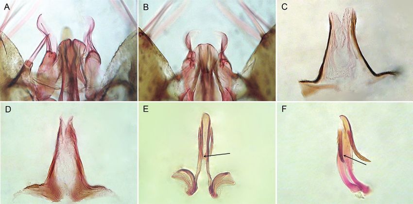
al. 2007). Polymerase chain reactions (PCR) were carried narrow (Fig. 1A), ventral surface with distinctly swol-
out in a 25 μl reaction mix containing 1 µl of DNA of len, striated lobes; dorsal claspette with three long se-
the first elution; 10 mM Tris-HCl, pH 8.3; 50 mM KCl; tae, most dorsal seta narrow, arising subapically, two
1.5 mM MgCl2; 2.5 μl DMSO; 5 pM of each primer; large, contorted, flattened ventral setae arising apically;
200 μM each dNTPs; 2.5 U New England Biolabs® Taq aedeagus long, slender, strongly sclerotized laterally,
polymerase (New England Biolabs, Ispwich, MA); the with a pair of subapical leaflets, leaflets well developed,
remaining volume of ultrapure H2O up to 25 μl. Am- straight, parallel to the longitudinal axis, strongly sclero-
plification consisted of a 3 min denaturation at 94ºC, 34 tized, serrated on dorsal and lateral surfaces; apex of ae-
cycles at 94ºC, 60ºC and 72ºC for 30 s each, followed by deagus somewhat rounded (Fig. 1E); proctiger membra-
a 10-min extension at 72ºC. nous, without spicules (Fig. 1C). The adult female can
The primers LCO1490 (5’GGT CAA CAA ATC ATA be recognized by having abdominal terga II-VII without
AAG ATA TTG G 3’) and HCO2198 (5’TAA ACT TCA scales; variable pattern of pale and dark scales on vein
GGG TGA CCA AAA AAT CA 3’) (Folmer et al. 1994) R4+5, mostly pale-scaled, with pale scales medially, with
were used to amplify 658 bp of the COI gene. PCR reac- small dark spots at proximal and distal ends; vein CuA2
tions (25 μl) contained 1 μl DNA; 10 mM Tris-HCl, pH pale-scaled on proximal 0.5, dark-scaled distally (Fig.
8.3; 50 mM KCl; 1.5 mM MgCl2; 2.5 μL DMSO; 5 pM of 2A); foretarsomeres 1-3 mostly dark-scaled with narrow
each primer; 200 μM each dNTPs; 2.5 U New England apical pale rings, foretarsomeres 4,5 entirely dark-scaled;
Biolabs® Taq polymerase (New England Biolabs, Ispwich, midtarsomere 1 with few pale scales dorsally at apex,
MA); the remaining volume of ultrapure H2O up to 25 μl. midtarsomeres 2-5 dark-scaled; hindtarsomere 1 mostly
Amplification consisted of a 3-min denaturation at 94oC dark-scaled, with an apical white ring, hindtarsomere
and 35 cycles at 94oC, 55oC and 72oC for 1 min each, fol- 2 dark-scaled on approximately basal 0.5, white-scaled
lowed by a 7-min extension at 72oC. COI and ITS2 PCR on apical 0.5, hindtarsomeres 3-5 entirely white-scaled.
products were purified using PEG precipitation (20% Based on fourth-instar larvae, An. antunesi can be rec-
polyethylene glycol 8000/2.5 M NaCl). Sequencing reac- ognized by the following combination of characters: seta
tions were carried out in both directions using the ABI 2-C weakly aciculate, seta 4-C single, long, aciculate;
Big Dye Terminator Kit version 3.1 (PE Applied Biosys- seta 14-P usually with three long, slender branches (Fig.
tems, Warrington, England) with the same primers used 3A); seta 1-II-VII palmate, with short pedicel, leaflets ta-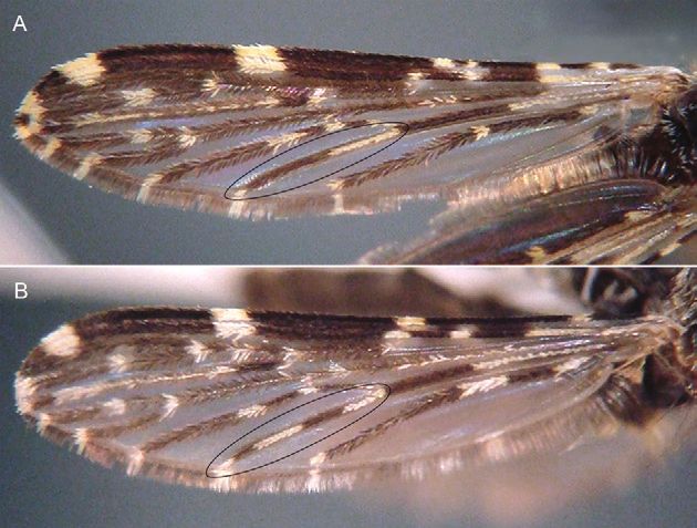
280     Mem Inst Oswaldo Cruz, Rio de Janeiro, Vol. 105(3), May 2010

Fig. 1: male genitalia of Anopheles antunesi (A, C, E) and Anopheles pristinus, n. sp. (B, D, F) from southern Brazil. A, B: ventral claspette
(dorsal aspect); C, D: proctiger; E, F: dissected aedeagus showing the position of the ventromesal subtriangular projection.

                                                                             Molecular characterization - The amplicon length of
                                                                         the ITS2 sequence was consistent at 472 bp (without the
                                                                         flanking 5.8S and 28S). Six sequences revealed a single
                                                                         haplotype comprised of 19.1% T, 19.3% A, 28.8% C and
                                                                         32.8% G. A FASTA search using the Database: nucle-
                                                                         otide collection - Optimize for: Somewhat Similar (http://
                                                                         www.ncbi.nlm.nih.gov/BLAST/) revealed that the ITS2
                                                                         sequence of An. antunesi shares 77% similarity with
                                                                         Anopheles pictipennis (Philippi) within a query cover-
                                                                         age of 84%. Two unique COI haplotypes were detected
                                                                         in the 658 bp (without the primer regions) fragment.
                                                                         Haplotype 1 is comprised of 39.2% T, 28.7% A, 15.8%
                                                                         C and 16.3% G, whereas haplotype 2 is comprised of
                                                                         39.5% T, 28.6% A, 15.5% C and 16.4% G. The ITS2 and
                                                                         COI sequences generated during this study are avail-
                                                                         able in GenBank under the following accessions: ITS2
Fig. 2: dorsal surface of the wings of Anopheles antunesi (A) and        (GU989324 - GU989329), COI (GU989342 - GU989347,
Anopheles pristinus, n. sp. (B) from southern Brazil, showing details
of the vein CuA 2.
                                                                         GU989349).
                                                                             Distribution - An. antunesi was recorded in Brazil
                                                                         (Galvão & Amaral 1940, Galvão 1940, Forattini 1962,
                                                                         2002, Gorham et al. 1967), Argentina (Darsie 1985,
pering to apex, ending in a pointed apex (Fig. 3C); 6-IV-                Gorham et al. 1967) and Uruguay (Rodriguez & Varela
VI long, bifid (Fig. 3E). Pupae have the trumpet darkly                  1962, Gorham et al. 1967). In Brazil, the species was
pigmented at midlength, paddle obovate, unpigmented or                   reported in SP, municipality of Campos do Jordão (Gal-
very weakly pigmented, lighter than posterior abdominal                  vão & Amaral 1940, Galvão 1940, Forattini et al. 1997),
segments, outer edge of paddle distal to external buttress               state of Maranhão (MA), northeast (Rebêlo et al. 2007),
with spicules. Eggs with floats lateral in position, long,               state of Rio Grande do Sul (RS) (Cardoso et al. 2004),
covering part of dorsal surface, ribs weakly divided into                municipality of Guaíba (Deane & Neto 1969) and state
lobes (Fig. 4A, D); deck narrow, uniformly covered with                  of Paraná (PR) (Rachou & Ricciardi 1951), municipality
irregularly shaped tubercles (Fig. 4C); ventral and lat-                 of Foz do Iguaçu (Consolim et al. 1993). However, in
eral plastron with somewhat circular pores (Fig. 4B).                    considering the difficulties of identifying species of the
Species of the Myzorhynchella Section • Sandra Sayuri Nagaki et al. 281
Fig. 3: larval characters of Anopheles antunesi (A, C, E) and Anopheles pristinus, n. sp. (B, D, F) from southern Brazil. A, B: seta 14-P; C, D:
palmate seta 1-II-VII; E, F: seta 6-IV-VI.
Myzorhynchella Section based on morphological charac- streams. The water was fresh, stagnant, cool, well oxy-
teristics of the adult females, it is likely that the records genated, pH approximately 5, with decomposing leaves
of An. antunesi in Argentina (Darsie 1985, Gorham et and little vegetation in partial shade. An. antunesi was
al. 1967), Uruguay (Rodriguez & Varela 1962, Gorham considered to be zoophilic by Galvão & Amaral (1940)
et al. 1967) and Brazil, in MA (Rebêlo et al. 2007), RS because during the fieldwork specimens of this species
(Cardoso et al. 2004), municipality of Guaíba (Deane & did not bite the collectors. Forattini (1962) made a simi-
Neto 1969) and PR (Rachou & Ricciardi 1951), munici- lar observation about the other Myzorhynchella species.
pality of Foz do Iguaçu (Consolim et al. 1993) may refer In Casa Grande, Salesópolis, adults were captured at
to other species of the Myzorhynchella Section. Conse-������ night on horses (Galvão 1941).
quently, further studies are needed to clarify the distri-
Material examined - The specimens of An. antunesi
bution of An. antunesi in South America.
used for morphology were collected in Brazil, SP, mu-
Type data - Holotype female (370), allotype male nicipality of Campos do Jordão, Vila Emilio Ribas,
(371), originally deposited in the Collection of the De- Galvão & Amaral coll., 1940, Galvão & Amaral, 1940
partamento de Parasitologia da FMUSP, Brazil, both det.: E-2034 [Fwing, paratype], E-2038 [MG], E-2042
probably lost. [FLePe], E-2044 [MLePe], E-2047 [MG], E-2048 [FLe],
E-2049 [MG]. SP, municipality of Campos do Jordão,
Medical importance - An. antunesi is not known to
Sallum & Wilkerson coll., 20-Nov-2001, Sallum, 2001
be of medical importance.
det.: E-12438 [LePe], E-12449 [LePe], E-12461 [LePe],
Bionomics - Immature stages of An. antunesi were E-12462 [LePe], E-12463 [LePe], E-12464 [LePe]. SP,
collected in Vila Emilio Ribas, Campos do Jordão, SP, municipality of Pindamonhangaba, Pico do Itapeva,
Serra da Mantiqueira, approximately 1.570 m above Fazenda Saint Claire, Sallum et al. coll., 27-July-2006,
sea level. Larvae were taken from ground pools, small Sallum det., 2006: VP09-17 [LePeG]. RJ, municipality
streams and rock holes along the edges of the Capivari of Itatiaia, Parque Nacional do Itatiaia (22º24’58.7”S
River. The habitat was shaded; the water was fresh, clean 44º37’19.7”W), Motta coll., July-2007: RJ–0 [FPe].
and cool, with little or no vegetation. No other species of Motta & Nagaki coll., 25-March-2008, Sallum & Na-
the Anopheles were found associated with An. antunesi gaki det., 2008, adults were collected in a Shannon trap:
(Galvão & Amaral 1940). In Casa Grande, on the Riv- RJ03(11) [E], RJ03(12) [E], RJ03(13) [E]. SP, munici-
er Claro shores, situated in Serra do Mar, Salesópolis, pality of Salesópolis, Casa Grande, Galvão coll., 1940,
Galvão (1941) collected immature stages in backwaters det. Galvão, 1940: E-2037 [MG], E-2039 [MLe], E-2041
of small streams inside forest. Larvae of An. antunesi [MG], E-2050 [MG].
were also found in a locality in the vicinity of the Parque The specimens of An. antunesi used for molecular
Nacional do Itatiaia, Serra da Mantiqueira, approxi- analysis were collected in Brazil, SP, municipality of Pin-
mately 1.100 m above sea level, in backwaters of small damonhangaba, Pico do Itapeva, Fazenda Saint Claire,282 Mem Inst Oswaldo Cruz, Rio de Janeiro, Vol. 105(3), May 2010
Fig. 4: eggs of Anopheles antunesi (A-D) and Anopheles pristinus, n. sp. (E, F) from southern Brazil. A: posterior end (ventral view); B, E:
posterior end (dorsal view); C, F: deck tubercles and float; D: lateral view of float.
Sallum et al., coll., 27-July-2006, Sallum det., 2006: with apical white ring, hindtarsomere 2 dark-scaled at
VP09-17 [M]; adult collected in Shannon trap: VP11b approximately basal 0.5, white-scaled apically, hindtar-
[F], VP11c [F]. RJ, municipality of Itatiaia, Parque Na- someres 3-5 white-scaled. Based on characters of the
cional do Itatiaia (22º24’58.7”S 44º37’19.7”W), Motta fourth-instar larvae, An. pristinus can be recognized by
& Nagaki coll., 25-March-2008, Sallum & Nagaki det., the following combination of characters: seta 2-C sin-
2008, adults collected in Shannon trap: RJ03(6) [F], gle, seta 4-C single, aciculate; seta 14-P multibranched,
RJ03(11) [F], RJ03(12) [F], RJ03(13) [F]. with thin branches (Fig. 3B); abdominal seta 0-II-VII
min, seta 1-III-VII palmate, with long, narrow pedicel
An. (Nyssorhynchus) pristinus Nagaki & Sallum,
supporting thin, hyaline leaflets (Fig. 3D), seta 6-IV-VI
new species
long, with few long branches (Fig. 3F). The pupae can be
(Figs 1-4)
distinguished by having the trumpet darkly pigmented
An. (Nyssorhynchus) antunesi of Forattini et al. 1998: at midlength, paddle obovate, lightly pigmented, lighter
120 (SEM eggs). than posterior abdominal segments, outer edge distal
Morphological characterization - An. pristinus can to external buttress with spicules. Eggs with floats lat-
be recognized by the following characteristics of the eral in position, long, covering part of dorsal surface,
male genitalia: ventral claspette without spicules, apex ribs weakly divided into lobes; deck narrow, uniformly
somewhat rounded in shape (Fig. 1B), ventral surface covered with fine tubercles (Fig. 4F); ventral and lateral
with distinct lobes, median sulcus somewhat V-shaped; plastron with irregular cracked areas (Fig. 4E).
dorsal claspette with a narrow dorsal seta arising subapi- Molecular characterization - The ITS2 region was
cally, two large, contorted, flattened ventral setae aris- sequenced for 12 individuals from Campos do Jordão
ing from apex; aedeagus long, slender, strongly sclero- and Pindamonhangaba. The amplicon length was con-
tized laterally, a pair of subapical leaflets, leaflets well sistent at 470 bp (without the flanking 5.8S and 28S
developed, strongly sclerotized, serrated, forming an genes) and the 12 sequences revealed a single haplo-
angle of 25º with aedeagus (Fig. 1F); proctiger membra- type. The ITS2 haplotype comprised 20% T, 19.6% A,
nous mesally, with minute spicules laterally (Fig. 1D), 27.4% C and 33.0% G. A FASTA search using the Da-
strongly sclerotized at base. The adult female can be rec- tabase: nucleotide collection - Optimize for: Somewhat
ognized by the absence of scales on abdominal terga II- Similar revealed that the ITS2 sequences of An. pristi-
VII; vein R4+5 mostly pale-scaled, with small dark spots nus share 81% similarity with An. pictipennis. The
at proximal and distal ends; vein CuA 2 with two dark fragment of COI was sequenced for 11 individuals, all
spots (Fig. 2B); foretarsomeres 1-3 with narrow, pale api- were same specimens used to obtain ITS2 sequences.
cal rings, foretarsomeres 4 and 5 dark-scaled; midtar- Six mtDNA haplotypes were detected in approximately
someres 1 and 2 mostly dark-scaled, with few apical pale 658 bp (excluding the primer regions). The base com-
scales, midtarsomeres 3-5 dark-scaled; hindtarsomere 1 positions were similar for all haplotypes, comprisingSpecies of the Myzorhynchella Section • Sandra Sayuri Nagaki et al. 283
38% T, 29% A, 16% C and 17% G. The ITS2 and COI et al. coll., 27-Jan-2009, Nagaki & Sallum det., 2009:
sequences are available in GenBank under the follow- SP51-100 [F], SP53-100 [M], SP53-101 [F], SP53-4 [P],
ing accessions: ITS2 (GU989330 - GU989341), COI SP53-5 [P]; in Shannon trap: SP50a [F], SP50b [F],
(GU989348, and GU989350 - GU989359). SP55(2) [F], SP55(4) [F].
Distribution - An. pristinus is known only from Serra Discussion
da Mantiqueira, southern Brazil. Further studies will be
Morphological studies of anophelines from Pico do
necessary to establish the geographical distribution of An.
Itapeva showed that specimens could be separated into
pristinus and the range of sympatry with An. antunesi.
two morphotypes based on the pattern of pale and dark
Medical importance - An. pristinus is not known to wing spots, male genitalia and fourth-instar larvae. De-
be of medical importance. tailed comparisons of specimens from both localities
showed that individuals from Itatiaia belong to An. an-
Bionomics - An. pristinus is a sylvatic species that oc-
tunesi and that there was a second species in Pico Itape-
curs at high altitudes in cloud forests in Serra da Man-
va, which is herein formally named and described as An.
tiqueira, southern Brazil. Field collections were made at
pristinus. The ITS2 and COI sequences corroborated the
Fazenda Saint Claire, Pico do Itapeva, in the border area
morphological hypothesis of two species.
of municipalities of Pindamonhangaba and Campos do
In the larval stage, An. antunesi and An. pristinus can
Jordão, at approximately 1.781 m above sea level. Larvae
be easily recognized as members of the Myzorhynchella
and pupae were taken from small, deeply shaded ground
Section by having abdominal seta 6-IV-VI branched; in
pools covered with grass. The larval habitat was in an
the adult stage by the absence of both scales and poste-
open area outside the forest. The water from a small, slow
rolateral scales tufts from all abdominal segments and
running stream was fresh, well oxygenated and cool.
hindtarsomeres 3-5 white-scaled; by the male genitalia
Etymology - The name pristinus is Latin for pristine, in having the dorsal claspette with two setae arising from
meaning pure, undamaged, fresh and clean and refers to the apex and one subapical seta and the accessory setae
the habitat were the immature stages were found. ending in a sharply acute apex (Galvão 1941). An. an-
Material examined - Holotype - Pinned adult male tunesi, An. pristinus and An. lutzii can be distinguished
with associated larval and pupal exuviae and dissected from An. nigritarsis in the adult stage by the absence of
male genitalia mounted on microscope slides. Bra- dark scales on hindtarsomeres 3 and 4 (these hindtar-
zil, SP, border of municipalities of Pindamonhangaba/ someres are white-scaled with a dark basal band in An.
Campos do Jordão, Pico do Itapeva, Fazenda Saint nigritarsis). Furthermore, An. antunesi, An. pristinus and
An. lutzii can be separated from An. parvus by charac-
Claire, -22,75847222S, -45,51527778W, Datum SAD69,
ters of the aedeagus of the male genitalia. In An. parvus
27-January-2009, Nagaki et al. coll., specimen SP53-
the apex of the aedeagus is hook-like medially, with the
2, FSP-USP coll., E-13162. Paratypes: same collection
lateral edges hyaline, whereas in An. lutzii, An. antunesi
data as the holotype, specimens SP53-1 [FLePe], SP53-3
and An. pristinus the apex is flat, somewhat rounded,
[FLePe], SP55(1) [F, one wing removed from adult and
never hook-like. An. antunesi and An. pristinus can be
mounted on microscope slide], SP55(3) [F, one wing re-
separated from An. lutzii by having the aedeagal leaflets
moved from adult and mounted on microscope slide],
borne laterally, approximately parallel to the longitudi-
SP55a [F], SP55b [F]. The holotype and paratypes are
nal axis of the aedeagus, whereas in An. lutzii the aedea-
deposited in the Coleção Entomológica da FSP-USP.
gal leaflets arise laterally forming a 45º angle with the
Other specimens examined - Brazil, SP, municipal- longitudinal axis of the aedeagus. Additionally, charac-
ity of Campos do Jordão, Sallum & Wilkerson coll. 20- teristics of the dark and pale wing spots can distinguish
Nov-2001, Sallum det., 2001 [identified as An. near an- An. antunesi and An. pristinus from An. lutzii and An.
tunesi, FSP-USP E-12370, LePe]; Fazenda Saint Claire parvus. An. parvus has three dark spots on vein R4+5 sep-
(-22,75847222S, -45,51527778W, Datum SAD69), Na- arated by two well-defined white spots, whereas in An.
gaki et al. coll., 27-Jan-2009, Nagaki & Sallum det., lutzii R4+5 is mostly dark-scaled with one preapical and
2009: SP51-100 [Pe mounted on microscope slide], one postbasal white spot. In An. antunesi and An. pristi-
SP53-4 [Le mounted on microscope slide], SP53-5 nus, vein R4+5 is mostly pale-scaled with one preapical
[Le mounted on microscope slide], SP53-100 [PeMG and one postbasal dark spot. However, specimens of An.
mounted on microscope slide], SP53-101 [FPe, one antunesi from Itatiaia have vein R4+5 either mostly dark
wing and three legs removed from adult and mounted or mostly pale-scaled. Galvão (1941) considered that An.
on microscope slide]. antunesi and An. lutzii could be misidentified because of
Specimens of An. pristinus used to generate COI the polymorphisms of vein R4+5, with individuals of An.
and ITS2 sequences were collected in Brazil, SP, mu- antunesi having this vein mostly dark-scaled.
nicipality of Campos do Jordão, Sallum & Wilkerson. An. antunesi and An. pristinus can be distinguished
coll., 20-Nov-2001, Sallum det., 2001: E-12370 [identi- by the pattern of dark and pale scales on vein CuA 2. In
fied as An. near antunesi, only the abdomen]; Fazenda An. antunesi, CuA2 is white-scaled on the proximal 0.5
Saint Claire (-22,75847222S, -45,51527778W, Datum and dark on the distal 0.5, whereas in An. pristinus CuA2
SAD69), Sallum et al. coll., 27-July-2006, Sallum det., has a proximal pale spot, a dark spot, a pale spot and a
2006, in Shannon trap: VP11a [F], VP11d [F], Nagaki distal dark spot (Fig. 2A, B). Consequently, the pattern284 Mem Inst Oswaldo Cruz, Rio de Janeiro, Vol. 105(3), May 2010
of pale and dark spots on CuA2 can be used to identify Darsie RF 1985. Mosquitoes of Argentina. Part I. Keys for identifi-
An. antunesi and An. pristinus. In the male genitalia, An. cation of adult females and fourth stage larvae in English and
pristinus can be separated from An. antunesi by having Spanish (Diptera: Culicidae). Mosq Syst 17: 153-253.
the apex of the ventral claspette moderately rounded Deane LM, Neto JAF 1969. Malária em macacos do Rio Grande do
apically (Fig.1B), the proctiger spiculate (Fig. 1C) and Sul. Obervações preliminares. Rev Inst Med Trop 11: 299-305.
the ventromesal subtriangular projection positioned ap- Djadid ND, Gholizadeh S, Tafsiri E, Romi R, Gordeev M, Zakeri
proximately at midlength of the insertion and the apex of S 2007. Molecular identification of Palaearctic members of
the leaflets (Fig. 1F). In An. antunesi, the apex of ventral Anopheles maculipennis in Northern Iran. Malar J 6: 6.
claspette is somewhat straight (Fig. 1A), the proctiger is Faran ME 1980. Mosquito studies (Diptera: Culicidae) XXXIV.
bare (Fig. 1C) and the ventromesal subtriangular projec- A revision of the Albimanus Section of the subgenus Nysso-
tion is positioned distally, approximately at the apex of rhynchus of Anopheles. Contrib Am Entomol Inst 15: 1-215.
the leaflets (Fig. 1E). Fourth-instar larvae of An. antunesi Folmer O, Black M, Hoeh W, Lutz R, Vrijenhoek R 1994. DNA
can be distinguished from those of An. pristinus by hav- primers for amplification of mitochondrial cytochrome c oxi-
ing seta 1-II-VII palmate with a short pedicel (Fig. 3A) dase subunit I from diverse metazoan invertebrates. Mol Mar
and seta 6-IV-VI bifid (Fig. 3E), whereas in An. pristinus Biol Biotechnol 3: 294-299.
seta 1-II-VII has a long pedicel supporting thin hyaline Forattini OP 1962. Entomologia médica, vol. I, Faculdade de Higiene
leaflets (Fig. 3B) and seta 6-IV-VI is branched, with a e Saúde Pública, São Paulo, 662 pp.
central branch from which arise lateral branches (Fig.
3F). Characters of the eggs as seen by SEM can also dis- Forattini OP 2002. Culicidologia médica. Identificação, biologia,
epidemiologia, vol. II, Edusp, São Paulo, 864 pp.
tinguish An. antunesi and An. pristinus. The deck tuber-
cles of the An. antunesi egg are larger and closer together Forattini OP, Marucci D 1993. The scanning electron microscopy of
than they are on the An. pristinus egg (Fig. 4C, F). The Anopheles (Kerteszia) eggs. Mem Inst Oswaldo Cruz 88: 349-352.
outer chorion of the ventral surface is covered with cir- Forattini OP, Sallum MAM, Bergo ES, Flores DC 1998. Ultrastruc-
cular pores in An. antunesi (Fig. 4B, D), whereas it has ture of eggs of Anopheles rondoni, Anopheles lutzii, and Ano-
several irregular cracked areas in An. pristinus (Fig. 4E). pheles parvus, three species of the subgenus Nyssorhynchus. J
Based on these observations it seems likely that Forattini Am Mosq Control Assoc 14: 256-265.
et al. (1998) described the eggs of An. pristinus. Forattini OP, Rabello EX, Cotrim MD 1970. Catálogo das coleções
The hypothesis for a new, species sympatric with entomológicas da Faculdade de Saúde Pública da Universidade
An. antunesi in Pico do Itapeva, Serra da Mantiqueira de São Paulo. 1ª série. Culicidae. Série Monográfica nº 1. Rev
as observed by morphological differences in few spec- Saude Publica 4: 5-100.
imens was corroborated by the ITS2 rDNA and COI Forattini OP, Sallum MAM, Marques GR, Flores DC 1997. Descrip-
��������
sequences. ITS2 rDNA and COI sequences reveal that tion of the eggs of Anopheles (Kerteszia) laneanus and Anopheles
An. antunesis and An. pristinus are distinct species. (Nyssorhynchus) antunesi (Diptera: Culicidae) by scanning elec-
The ITS2 sequences of different individuals of each tron microscopy. J Am Mosq Control Assoc 13: 368-374.
species are identical; however, comparisons between Galvão ALA 1940. Contribuição ao conhecimento dos anofelinos do
the two species show indels among individuals. ITS2 grupo Nyssorhynchus de São Paulo e regiões vizinhas (Díptera:
differences between An. antunesis and An. pristinus Culicidae). Arq Zool 1: 399-484.
are consistent, suggesting that these differences repre- Galvão ALA 1941. Contribuição ao conhecimento das espécies
sent inter-specific variation. The COI sequence varia- de Myzorhynchella (Diptera: Culicidae). Arch Zool S Paulo
tion between An. antunesi and An. pristinus also cor- 2: 505-576.
roborates the morphological and ITS2 sequence data, Galvão ALA, Amaral ADF 1940. Estudos sobre os anofelinos do
confirming An. pristinus as a new species. grupo Myzorhynchella com a descrição de uma espécie nova,
Anopheles (Nyssorhynchus) antunesi n. sp. (Diptera: Culicidae).
Acknowledgments
Folia Clin Biol 12: 150-160.
To Saint’ Clair de Vasconcelos, for hosting fieldwork in Gorham JR, Stojanovich CJ, Scott HG 1967. Clave ilustrada para
Fazenda Saint Claire, Pico do Itapeva, Serra da Mantiqueira, los mosquitos anofelinos de Sudamerica Oriental, National
to Ralph E Harbach, Natural History Museum, UK, and to the Communicable Disease Center, Atlanta, 62 pp.
two anonymous reviewers for their great contribution to the
Harbach RE 2008. Mosquito taxonomic inventory. Available from:
improvement of the text. http://mosquito-taxonomic-inventory.info/. [accessed on the
References 2nd March 2010].
Belkin JN 1962. The mosquitoes of South Pacific (Diptera: Culicidae), Harbach RE, Knight KL 1980. Taxonomists’ glossary of mosquito
University of California Press, Berkley and Los Angeles, 608 pp. anatomy, Plexus Publishing Inc, Marlton, 415 pp.
Belkin JN, Schick RX, Heinemann SJ 1971. Mosquito studies (Dip- Knight KL, Stone A 1977. A catalog of the mosquitoes of the world
tera: Culicidae). XXV. Mosquitoes originally described from (Diptera: Culicidae), vol. 6, Thomas Say Foundation, Washing-
Brazil. Cont Amer Entomol Inst 7: 1-64. ton DC, 611 pp.
Cardoso JC, Corseuil E, Barata JMS 2004. Anophelinae (Diptera: Linthicum KJ 1988. A revision of the Argyritarsis Section of the sub-
Culicidae) ocorrentes no estado do Rio Grande do Sul, Brasil. genus Nyssorhynchus of Anopheles (Diptera: Culicidae). Mosq
Entomol Vect 11: 159-177. Syst 20: 98-271.
Consolim J, Pellegrini NJM, Luz E 1993. Culicídeos (Diptera: Culi- Maddison DR, Maddison WP 2000. MacClade 4: analysis of phyloge-
cidae) do Lago Itaipu, Paraná, Brasil. I. Município de Foz do ny and characters evolution, Version 4.0, Sinauer Associates Inc,
Iguaçu. Acta Biol Par 22: 83-90. Sunderland, Massachusetts.Species of the Myzorhynchella Section • Sandra Sayuri Nagaki et al. 285
Motoki MT, Wilkerson RC, Sallum MA 2009. The Anopheles albi- Rosa-Freitas MG 1989. Anopheles (Nyssorhynchus) deaneorum: a
tarsis complex with the recognition of Anopheles oryzalimnetes new species in the albitarsis complex (Diptera: Culicidae). Mem
Wilkerson and Motoki, n. sp. and Anopheles janconnae Wilker- Inst Oswaldo Cruz 84: 535-543.
son and Sallum, n. sp. (Diptera: Culicidae). Mem Inst Oswaldo
Sallum MAM, Wilkerson RC 1997. Description of immatures stages
Cruz 104: 823-850.
of Anopheles (Nyssorhynchus) rondoni (Neiva & Pinto) (Diptera:
Rachou RG, Ricciardi I 1951. Contribuição ao conhecimento da dIs- Culicidae). Mem Inst Oswaldo Cruz 92: 365-372.
tribuição geográfica dos anofelinos no Brasil: estado do Paraná Swofford, DL 2003. PAUP*. Phylogenetic Analysis Using Parsimony
(distribuição por municípios e localidades). Rev Bras Malariol (*and other methods), Version 4., Sinauer Associates Inc, Sunder-
Doencas Trop 3: 423-447. land, Massachusetts.
Rebêlo JMM, Moraes JLP, Alves, GA, Leonardo FS, Rocha RV, Thompson JD, Gibson TJ, Plewniak F, Jeanmougin F, Higgins DG
Mendes WA, Costa E, Câmara LEMB, Silva MJA, Pereira YNO, 1997. The Clustal X windows interface: flexible strategies for
Mendonça JAC 2007. Distribution of species from genus Ano- multiple sequence alignment aided by quality analysis tools.
pheles (Diptera: Culicidae) in the state of Maranhão, Brazil. Cad Nucleic Acids Res 24: 4876-4882.
Saude Publica 23: 2959-2971.
Wilkerson RC, Peyton EL 1990. Standardized nomenclature for the
Rodriguez MF, Varela JC 1962. Anopheles (Myzorhynchella) antunesi, costal wing spots of the genus Anopheles and other spotted-wing
especie nueva para el Uruguay. An Fac Montevideo 47: 246-249. mosquitoes (Diptera: Culicidae). J Med Entomol 27: 217-224.You can also read