Dogs (Canis familiaris) Gaze at Our Hands: A Preliminary Eye-Tracker Experiment on Selective Attention in Dogs - MDPI
←
→
Page content transcription
If your browser does not render page correctly, please read the page content below
animals
Article
Dogs (Canis familiaris) Gaze at Our Hands:
A Preliminary Eye-Tracker Experiment on Selective
Attention in Dogs
Tadatoshi Ogura * , Mizuki Maki, Saki Nagata and Sanae Nakamura
 School of Veterinary Medicine, Kitasato University, CityTowada, Aomori 034-8628, Japan;
 va13115g@st.kitasato-u.ac.jp (M.M.); va13089n@st.kitasato-u.ac.jp (S.N.);
 kitasato_dog_cognition@yahoo.co.jp (S.N.)
 * Correspondence: togura@vmas.kitasato-u.ac.jp; Tel.: +81-176-24-9377

 Received: 28 March 2020; Accepted: 20 April 2020; Published: 26 April 2020

 Simple Summary: Dogs seem to communicate with humans successfully by getting social information
 from body signals such as hand signs. However, less is known regarding how dogs pay attention
 visually toward human body signals including hand signs. The objective of this pilot study was to
 reveal dogs’ social visual attention tuned to inter-species communication with humans by comparing
 gazing patterns to the whole body of human, dogs, and cats. The pictures showing humans with or
 without hand signs, dogs, and cats were presented on a liquid crystal display monitor, and gazing
 behaviors of subject dogs to these pictures were recorded by an eye-tracking device. The subjects
 gazed at human limns more frequently than limbs within conspecifics and cat images, where the
 dogs attention were focused on the head and body. Furthermore, gaze toward hands was greater in
 the human hand sign photos relative to photos where human hand signs were not present. These
 results indicate that dogs have an attentional style specialized for human non-verbal communication,
 with an emphasis placed on human hand gestures.

 Abstract: Dogs have developed a social competence tuned to communicate with human and acquire
 social information from body signals as well as facial expressions. However, less is known regarding
 how dogs shift attention toward human body signals, specifically hand signs. Comparison among
 visual attentional patterns of dogs toward whole body of human being, conspecifics, and other species
 will reveal dogs’ basic social competences and those specialized to inter-species communication with
 humans. The present study investigated dogs’ gazing behaviors in three conditions: viewing humans
 with or without hand signs, viewing conspecifics, and viewing cats. Digital color photographs were
 presented on a liquid crystal display monitor, and subject dogs viewed the images while their eyes
 were tracked. Results revealed that subjects gazed at human limbs more than limbs within conspecific
 and cat images, where attention was predominately focused on the head and body. Furthermore,
 gaze toward hands was greater in the human hand sign photos relative to photos where human hand
 signs were not present. These results indicate that dogs have an attentional style specialized for
 human non-verbal communication, with an emphasis placed on human hand gestures.

 Keywords: attention; dog; eye tracking; non-verbal communication; visual attention

1. Introduction
     Dogs’ visual cognition has long been a popular research topic. During their long history of
domestication, dogs have developed a human-social skill (for a review see [1]). For example, in an
assessment of visual social cognition, Pitteri et al. [2] described that dogs can discriminate between an

Animals 2020, 10, 755; doi:10.3390/ani10050755                                  www.mdpi.com/journal/animals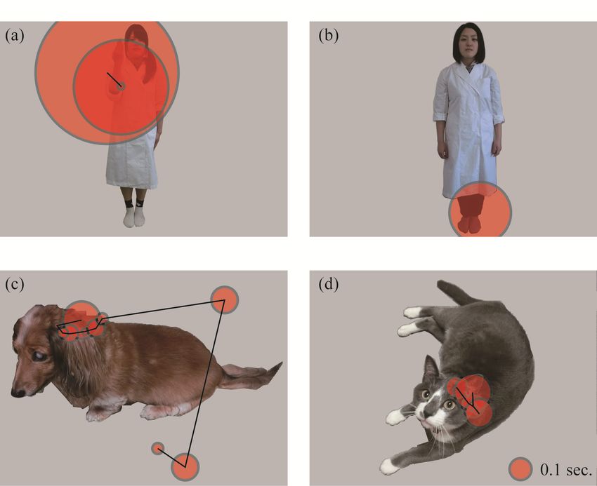
Animals 2020, 10, 755                                                                                 2 of 12

owner and strangers based on isolated internal features of a human face (i.e., eyes, nose, or mouth),
with configural information being highly important. During the process of domestication, dogs have
also been “worked” under various situation (for a review see [3]), so that they acquired the ability
to discern social information from human body signals. Dogs successfully follow human pointing
gestures at a much younger age than their closest wild-living relative (i.e., wolf, Canis lupus), even
when hand-reared [4]. Furthermore, dog-owners use a variety of hand signs as non-verbal signals
toward their dogs, which provide cues for following human intentions; however, little is known as to
dogs’ attentional patterns to these hand signs.
      Visual attention across various animal species has recently been investigated through eye-tracking
techniques. Eye-trackers are able to record subtle gaze movements, enabling researchers to detect small
changes in subjects’ interest and/or focal attention (for a review see [5]). Additionally, task-specific
pre-training is not required; therefore, naturalistic behavior toward stimuli can be investigated.
For example, one eye-tracking study revealed that among chimpanzees (Pan troglodytes), facial features
are prominently detected when viewing pictures of both chimpanzees and humans [6]. Specifically,
when exposed to photographs of conspecific and human faces, chimpanzees intensely focus on eye
and mouth regions [7]. Gorillas (Gorilla gorilla) and orangutans (Pongo abelii) show similar attentional
biases to facial images [8]. For non-primate animals, Schwarz et al. [9] developed a head-mounted
eye-tracking system for chickens (Gallus gallus domesticus), while Tyrrell et al. [10] developed a system for
starlings (Sturnus vulgaris) and was successfully able to detect valid eye movements. A head-mounted
eye-tracker was also used in a group of peahens (Pavo cristatus), revealing gaze preferences toward
the lower train relative to the head, crest, and upper train of peacocks [11]. Eye-trackers have also
been utilized with dogs, especially for purposes of investigating human face processing. For instance,
Somppi et al. [12] successfully employed an eye-tracker to investigate visual cognition in a sample of
privately owned dogs. The authors found that dogs preferred facial images of conspecifics relative to
human faces, children’s toys, and alphabetic characters, while also fixating longer on a familiar image
relative to novel stimuli. In a related study, kennel and privately owned dogs showed greater interest
in eye relative to adjacent face regions when viewing either conspecific or human faces [13]. Their
interest in eye region were not found in inverted faces, suggesting that dogs are able to recognize both
conspecific and human faces in photographs. More recently, Somppi et al. [14] observed that kennel and
pet dogs gaze toward conspecific and human faces differently depending on the emotional expression
displayed. Furthermore, oxytocin treatment can change pet dogs’ preferential looking patterns toward
human facial emotional images, particularly within the eye region [15]. Téglás et al. [16] also revealed
that pet dogs’ gaze follows a human’s referential signals (head-turning). Privately owned and kennel
dogs gaze longer at human actors within social settings as compared to when viewing other dogs,
which could indicate that processing social interactions within a non-conspecific context may be
more demanding for dogs [17]. Although these frontier studies have expanded knowledge as to
visual attention among dogs, research on gaze toward bodily signals outside a human face has not
been conducted.
      Human beings have been the most influential social partners for domestic dogs during their
domestication process. Comparison among visual attentional patterns of dogs towards human beings,
conspecifics, and other species will reveal dogs’ basic social competences and those specialized to
inter-species communication with humans. The present study investigated dogs’ visual selective
attention to human bodily signals, conspecifics, and cats (a species which lives in the same community
with dogs but seems to be less influential to dogs’ attentional patterns than humans). If the dogs’
human-social skills have been tuned to recognition of human bodily signals, their gaze to human
hands are more evident than the gaze to limbs of other species. We also examined the effect of stimulus
familiarity when dogs viewed pictures of hand signs expressed by humans. The goal of this study
was to determine how dogs’ fixation patterns could provide insight into cross-species communication
with humans.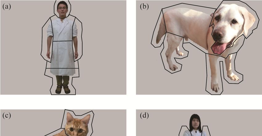
Animals 2020, 10, 755                                                                                                3 of 12

2. Materials and Methods

2.1. Subjects
     The subjects were 7 privately owned dogs, aged one to 18 years (Table 1). Before participating
in this study, all dogs were trained by their owner to lie on a floor until commanded to move. Prior
to the experimental protocol, the dogs visited the experimental rooms over at least two separate
days and were trained to settle calmly, without the aid of their owner, and to pass a calibration test
(see below) conducted by three experimenters (two handlers and one operator). During training,
the dogs were not encouraged to fix their eyes toward any monitor or images. All training procedures
were conducted based on shaping methods with positive reinforce training [18]. Four subjects did not
pass the calibration procedure; thus, only data from 3 dogs were analyzed.

                                                Table 1. Subject dog list.

                        ID            Breed             Age (Years)           Sex          Neutering
                        1a     Labrador retriever            13               Male          Neutered
                        2a       Norfolk terrier              5              Female         Neutered
                        3a           Mixed c                  2               Male          Neutered
                        4b       Unidentified d              18              Female         Neutered
                        5b     Shetland sheepdog              9               Male          Neutered
                        6b     German shepherd               2                Male          Neutered
                        7b          Mixed e                   1              Female          Intact
      a The dogs who passed the calibration procedure and participated in the experiment. b The dogs who did not

      pass the calibration procedure and were omitted from the experiment. c Cavalier King Charles Spaniel x Mixed
      d The breed was not identified because the dogs was a shelter dog and assumed as a golden retriever. e Chihuahua

      x Dachshund.

2.2. Apparatus and Experimental Set ups
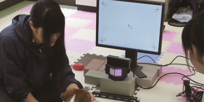
      The apparatus included an eye-tracking system (model ETL-300-HD, ISCAN, USA), which
consisted of two computers, two color displays, and an eye-tracking camera. One computer and
display was used to run the eye-tracking system and record eye movement data. The computer and
display (43.2 × 32.4 cm, 1600 × 1200 px; model S2133-HBK, Eizo Nanao, Ishikawa, Japan) were used
for stimulus presentation. The camera obtained images of each subject’s head and eyes. The camera
emitted infrared light in order to track subjects’ monocular pupil and corneal reflection at 60 Hz.
      All assessments were conducted in an experimental room at the School of Veterinary Medicine,
Kitasato University, by one operator and two handlers (Figure 1). During the experiment, the dogs
were released from a leash and could move around the room freely. The handlers guided the dogs to lie
down on the floor in front of the presentation display; once the dogs complied, the dogs were rewarded
with a treat. The distance between each dog’s head and the display was approximately 50 cm. During
the calibration and experimental presentation, each dog was positioned between the two handlers
staying closely with the dog. One of the handlers instructed each dog to put his/her jaw on a chinrest.
The other handler lightly held the dog’s body if needed. The operator in the same room controlled the
two computers. During the experimental procedure, the owners were absent from the room.

2.3. Stimuli
     Digital color photographs comprising three stimulus species—humans, dogs, and cats—were
used in the experiment (Figure 2). For each species, 12 images per subject were presented. All pictures
were 1600 × 1200 px and consisted of single, whole body images of an individual on a gray background.
The human pictures consisted of 3 novel individuals with a hand sign, 3 novel individuals without
a hand sign, 3 familiar individuals with a hand sign, and 3 familiar individuals without a hand
sign. A familiar person was defined as someone who spent time with the subject dog for longer than
one day, including the owners, operator, and handlers. Hand signs used commonly in the subjects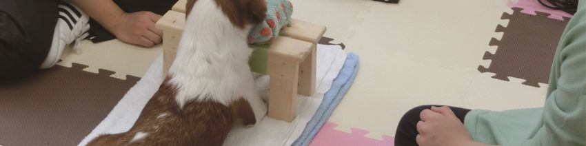
Animals 2020, 10, 755                                                                                                               4 of 12

(“hand”, “sit”, or “stay”) were depicted. All human pictures were front facing, upright, and laterally
symmetric except a hand showing a sign to avoid visuospatial attentional bias [19]. Each human
model wore a white, laboratory coat for consistency and to avoid color distraction. The human sex
was counterbalanced. All dog/cat pictures were novel to the subject dogs and were taken from a
front/lateral side with a natural posture. Dog and cat breeds varied across the images. We constructed
6 sets of stimuli,    comprising
            Animals 2020, 10, x  6 pictures each, with at least one picture from each stimulus
                                                                                            4 of species.
                                                                                                 13

      Figure 1. Subject
                 Figure 1. dogs
                           Subjectlied
                                    dogsdown     in front
                                         lied down           ofofan
                                                      in front    aneye-tracking    camera
                                                                     eye-tracking camera and and  display.
                                                                                             display. During During
                                                                                                               calibrationcalibration
      and theAnimals
              experimental
                 and2020,
                     the 10, x session,
                          experimental      the subject
                                        session,            put
                                                  the subject puthis/her   jawsonon
                                                                   his/her jaws     a purpose-designed
                                                                                  a purpose-designed         chin rest.5 of 13
                                                                                                      chin rest.

             2.3. Stimuli
                  Digital color photographs comprising three stimulus species—humans, dogs, and cats—were
             used in the experiment (Figure 2). For each species, 12 images per subject were presented. All pictures
             were 1600 × 1200 px and consisted of single, whole body images of an individual on a gray
             background. The human pictures consisted of 3 novel individuals with a hand sign, 3 novel
             individuals without a hand sign, 3 familiar individuals with a hand sign, and 3 familiar individuals
             without a hand sign. A familiar person was defined as someone who spent time with the subject dog
             for longer than one day, including the owners, operator, and handlers. Hand signs used commonly
             in the subjects (“hand”, “sit”, or “stay”) were depicted. All human pictures were front facing, upright,
             and laterally symmetric except a hand showing a sign to avoid visuospatial attentional bias [19]. Each
             human model wore a white, laboratory coat for consistency and to avoid color distraction. The human
             sex was counterbalanced. All dog/cat pictures were novel to the subject dogs and were taken from a
             front/lateral side with a natural posture. Dog and cat breeds varied across the images. We constructed
             6 sets of stimuli, comprising 6 pictures each, with at least one picture from each stimulus species.

                  Figure 2. Examples of stimuli used in the experiment and areas of interest (AOI) employed in the
      Figure 2. Examples        of stimuli used in the experiment and areas of interest (AOI) employed in the
                  analyses: (a) human without a hand-sign, (b) dog, (c) cat, and (d) human with a hand-sign. The frames
      analyses: (a) human without a hand-sign, (b) dog, (c) cat, and (d) human with a hand-sign. The frames
                  drawn on the pictures (a), (b), and (c) indicate the AOI utilized in the analysis comparing the three
      drawn on the     pictures
                  species and on the(a–c) indicate
                                      picture         the AOI
                                              (d) indicates       utilized
                                                            the AOI           inthe
                                                                    utilized in   the  analysis
                                                                                    analysis     comparing
                                                                                             comparing          theand
                                                                                                       stimuli with  three species
      and on the without
                  picturea (d)
                             hand-sign.
                                  indicates the AOI utilized in the analysis comparing stimuli with and without
      a hand-sign.
              2.4. Experimental Procedures
                   First, a 5-point calibration was conducted for each subject. Five black crosses were presented at
              the center, upper left, upper right, bottom left, and bottom right of the monitor on a gray background.
              A handler pointed to the crosses in that order using a treat. When the handler confirmed that the
              subject gazed on the cross, a vocal cue was provided to the operator in order to record the calibration
              point. This was repeated for the remaining 4 crosses. The calibration was then checked with a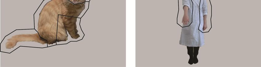
Animals 2020, 10, 755                                                                                5 of 12

2.4. Experimental Procedures
     First, a 5-point calibration was conducted for each subject. Five black crosses were presented at
the center, upper left, upper right, bottom left, and bottom right of the monitor on a gray background.
A handler pointed to the crosses in that order using a treat. When the handler confirmed that the subject
gazed on the cross, a vocal cue was provided to the operator in order to record the calibration point.
This was repeated for the remaining 4 crosses. The calibration was then checked with a validation
procedure: the handler pointed to the crosses again and the operator confirmed that the gaze points
were on the crosses through the eye-tracker system. The calibration was repeated until all gaze points
were validly recorded.
     After the calibration, the experimental session began. Each subject performed 6 sessions of the
task. Each session began with a blank gray screen. When the subject gazed anywhere on the screen
(not limited to any specific point to avoid the bias on the first fixation point described below), one
of the handlers provided a vocal cue for the operator to start the stimulus presentation. A picture
was then presented for 1.5 s, followed by a blank screen for 0.5 s. This routine was repeated for six
times and progressed automatically by a computer program assembled for this experiment. Image
presentation order was randomized. After each session was completed, the subject was rewarded
with a treat, regardless as to whether the dog gazed at the screen. Each subject was then allowed
to move freely around the room after each session to avoid to be stressed or habituated. Before the
next session started, the subject was guided to lie down at the same position and the calibration was
conducted again.
     This work complied with the laws of Japan, and the experiment was approved by the President
and Institutional Animal Care and Use Committee of Kitasato University (Approval no. 15-067).

2.5. Data Analyses
      We divided each picture into several features (areas of interest; AOI) to quantitatively analyze
subjects’ gaze patterns. For the analysis comparing the three species and that comparing human
familiarity, the human/dog/cat within each picture was divided into four AOIs: Head, body, buttocks,
and limbs (Figure 2a–c). For human pictures, the body indicated the upper third area from the neck
to the feet, because the dividing bounds between body parts were obscure due to a white lab coat.
The middle third area was included in the buttocks, and the bottom third and arms were included in the
“limbs” AOI. For dog and cat pictures, the buttocks included the tail, and the forelimbs and hindlimbs
were combined into the limbs AOI. For the analysis comparing stimuli with and without a hand-sign,
only the gaze at hands were incorporated (Figure 2d). To avoid errors in gaze estimation, the AOI
frame was drawn larger than the actual outline (approximately 50 pixels on the edge). From monocular
gaze data, the number of fixations and total fixation durations within each AOI per presentation were
calculated (Figure 3). For each picture, the AOI to which the first fixation was directed (hereafter, first
gazed AOI) were also analyzed. For each AOI, the number of pictures in which the first fixation was
directed was counted.
      The fixation number and total fixation duration for each AOI were analyzed using a Generalized
Linear Mixed Model (GLMM) (lmer, lme4 library, freeware package R, Version 2.14.2; R Development
Core Team, 2012). The models were constructed using a Poisson distribution for fixation number
data, as these were non-negative count data. A Gaussian distribution was used for analyzing total
fixation duration, as these were non-negative continuous data [20]. One data point indicated a fixation
number or total fixation duration for each picture for each subject. Stimulus species (human, dog,
or cat) and AOI (head, body, buttock, limbs) were set as fixed factors. For the stimulus familiarity
model, familiarity (familiar or novel) and AOI (head, body, buttock, limbs) were set as fixed factors.
In the hand sign analysis, the hand sign conditions (with or without hand signs) and AOI (head, body,
buttock, limbs) were set as fixed factors. Subjects were considered a random factor to control for
individual differences. The models with and without the target fixed factor were compared based on
the Akaike Information Criterion (AIC [20,21]). Model’s significance was tested using a likelihood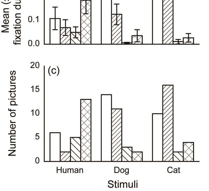
familiarity, the human/dog/cat within each picture was divided into four AOIs: Head, body, buttocks,
      and limbs (Figure 2(a), (b), and (c)). For human pictures, the body indicated the upper third area from
      the neck to the feet, because the dividing bounds between body parts were obscure due to a white
      lab coat. The middle third area was included in the buttocks, and the bottom third and arms were
      included in the “limbs” AOI. For dog and cat pictures, the buttocks included the tail, and the
Animals 2020, 10, 755
      forelimbs    and hindlimbs were combined into the limbs AOI. For the analysis comparing stimuli with6 of 12
      and without a hand-sign, only the gaze at hands were incorporated (Figure 2(d)). To avoid errors in
      gaze estimation, the AOI frame was drawn larger than the actual outline (approximately 50 pixels on
ration test. For multiple comparisons of the fixed factors, the models that contained every combination
      the edge). From monocular gaze data, the number of fixations and total fixation durations within
of theeach
        fixed
            AOIfactors   in the different
                   per presentation    were groups     were
                                              calculated     also 3).
                                                         (Figure  compared.     The number
                                                                      For each picture,  the AOIof to
                                                                                                    pictures categorized
                                                                                                      which the first
into each   firstwas
      fixation     gazed   AOI(hereafter,
                      directed   was analyzed       usingAOI)
                                             first gazed  a chi-square
                                                               were alsotest;  if significant,
                                                                           analyzed.  For each aAOI,
                                                                                                 standardized
                                                                                                      the numberresidual
                                                                                                                   of
post-hoc   analysis
      pictures         wasthe
                 in which   conducted      to investigate
                               first fixation  was directedany
                                                             wasdifference
                                                                 counted. from chance frequency.

          Figure
     Figure       3. Examples
            3. Examples         of subjects’
                            of subjects’      gazingpatterns:
                                           gazing     patterns: (a)
                                                                 (a) human
                                                                     humanwith
                                                                             witha ahand-sign, (b) (b)
                                                                                      hand-sign,   human
                                                                                                       humanwithout  a
                                                                                                                without a
          hand-sign,  (c) dog, and  (d) cat. Typical  fixations and  saccades are drawn  on the  pictures.
     hand-sign, (c) dog, and (d) cat. Typical fixations and saccades are drawn on the pictures. The circlesThe circles
          indicate fixations. The larger the circle, the longer the subject gazed in that area. The lines represent a
     indicate fixations. The larger the circle, the longer the subject gazed in that area. The lines represent a
          saccadic path.
     saccadic path.

3. ResultsThe fixation number and total fixation duration for each AOI were analyzed using a Generalized
      Linear Mixed Model (GLMM) (lmer, lme4 library, freeware package R, Version 2.14.2; R
3.1. Species
     The most gazed AOI differed between the stimulus species in the fixation number analysis.
The fixation number and total fixation duration within each AOI for all three stimulus species are
shown in Figure 4a,b, respectively. In the fixation number analysis, the models that included both
the stimulus species and AOI showed significantly lower AICs than the models that did not include
these factors (Table 2). In the fixation duration analysis, the models that included only the AOI
showed significantly lower AICs than the model that include both the stimulus species and AOI.
For multiple comparisons of both the fixation number and total fixation duration within the human
pictures, the models that included “limb” AOIs at a different group from the other AOIs had the lowest
AICs (164.4 and 16.08). For the dog pictures, the models that included a “head” AOI at a different
group from the other AOIs showed the lowest AICs (169.5 and 14.85). For cat pictures, the models that
included the “head” and “body” AOIs at a different group from the other AOIs showed the lowest
AICs (155.5 and 51.11). The number of pictures categorized into each first gazed AOI is shown in
Figure 4c. In all stimulus species, the numbers of pictures of each first gazed AOIs were significantly
different (human: χ2 = 10.00, p = 0.019, dog: χ2 = 14.00, p = 0.003, cat: χ2 = 15.00, p = 0.002). For the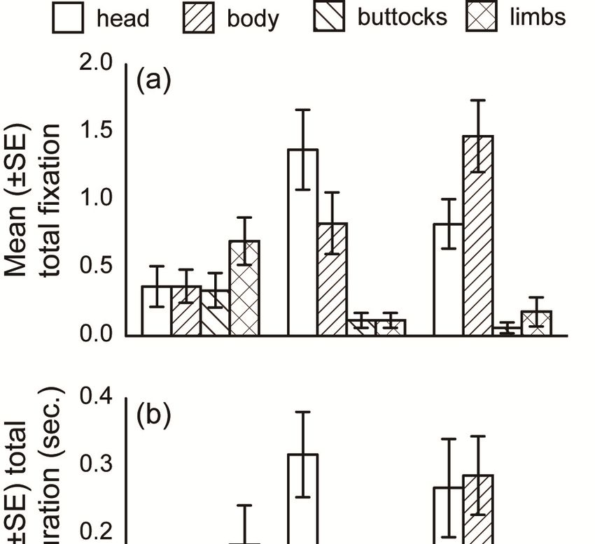
Animals 2020, 10, 755                                                                                                7 of 12

human pictures, the number of pictures where the first gazed AOI was on the “body” was significantly
less (p = 0.04), and the “limbs” was significantly greater (p = 0.003), than chance. For the dog pictures,
first gazed AOI for the “head” was significantly greater (p = 0.006), and the “limbs” was significantly
less (p = 0.02), than chance. For the cat pictures, the first gazed AOI for the “body” was significantly
greater (p = 0.001), and the “buttocks” was significantly less (p = 0.014) than chance. These results
suggest that the subjects predominantly oriented toward the “limbs” in human pictures, “head” in dog
pictures, and “head” and “body” in cat pictures.
       Animals 2020, 10, x                                                                                      8 of 13

             4. Mean
      FigureFigure     (± SE)
                   4. Mean  (± fixation    number,
                                SE) fixation        mean
                                              number,  mean(±(±SE)
                                                                SE)total
                                                                    total fixation duration,and
                                                                          fixation duration, and  number
                                                                                                number    of pictures
                                                                                                       of pictures
            categorized
      categorized       into each
                  into each   first first
                                     gazedgazed
                                             AOIAOI
                                                 forfor each
                                                     each    picturespecies.
                                                           picture   species.

3.2. Human Familiarity                Table 2. Generalized Linear Mixed Model results.

     For human pictures, gaze behavior did not differ         Responsible
                                                                    between Factor
                                                                                familiar and novel individuals.
                                             Fixation  number
The fixation number and total fixation duration in each AOI for familiar and  Total fixation duration
                                                                                         novel  human pictures are
                                                 Likelihood ratio test               Likelihood ratio test
shown in Figure 5a,b,    respectively.
                  Fixed factor
                                       Models
                                       AIC a
                                                 that
                                                    χ2
                                                       included   p
                                                                    both familiarity
                                                                          AIC a
                                                                                        and
                                                                                       χ2
                                                                                             AOIs   did
                                                                                                     p
                                                                                                           not show a
significantly lower AIC than the model     that comparing
                                       Analysis  only included     thespecies
                                                            the three  AOIs (Table 2). The number of pictures
categorized intoSpecies
                 each first  gazed AOI
                         and AOI         is shown in Figure 5c. For 106.9
                                       491.0                              both the familiar and novel pictures,
the number of pictures AOIdid not significantly
                                       547.8       72.80 between
                                                  differ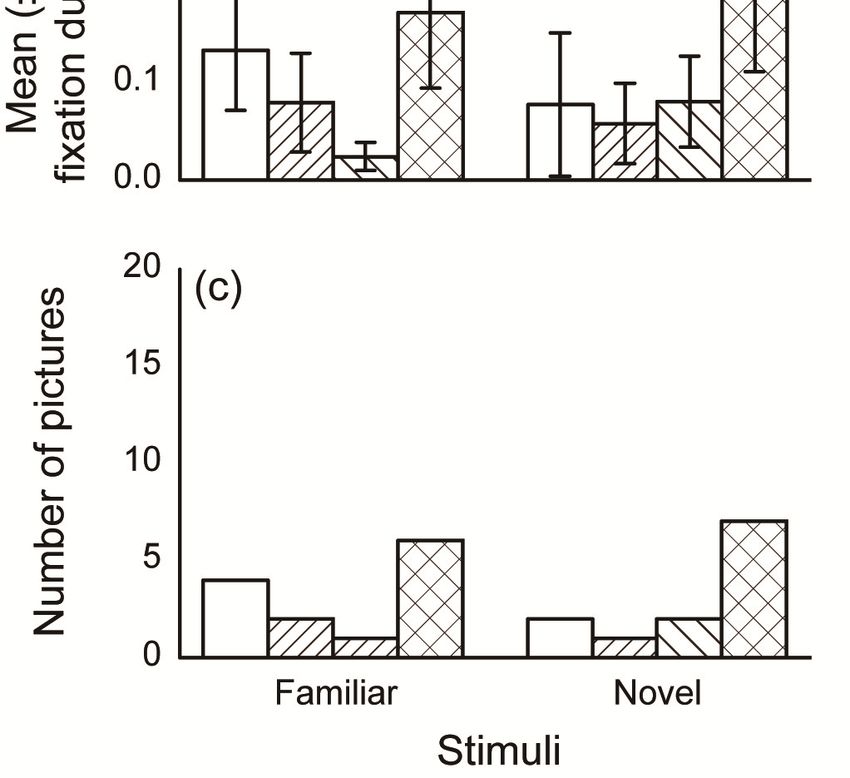
Animals 2020, 10, 755                                                                                                    8 of 12

                                    Table 2. Generalized Linear Mixed Model results.

                                                                  Responsible Factor
                                                 Fixation number                 Total fixation duration
                                                    Likelihood ratio test                Likelihood ratio test
                     Fixed factor        AIC a          χ2          p         AIC a         χ2           p
       Animals 2020, 10, x                                                                                          9 of 13
                                           Analysis comparing the three species
                  Species and AOI        491.0                             106.9
             a AIC = “Akaike Information Criterion,” an index used to compare the fitted models. The model with
                        AOI              547.8      72.80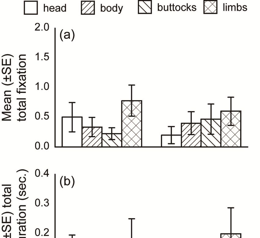
Animals 2020, 10, 755                                                                                                 9 of 12

3.3. Hand Sign
 Animals
     The2020, 10, x
          subject   dogs’ gaze behavior differed between the human pictures with and without a hand                   10 of 13
sign. The fixation number and total fixation duration to hands in the pictures with and without a
 hand-signare
hand-sign      are  shown
                 shown       in Figure
                         in Figure   6a,b,6(a)  and (b), respectively.
                                            respectively.                   For fixation
                                                            For fixation number,            number,
                                                                                     the models    thatthe    models
                                                                                                         included        that
                                                                                                                      AOIs
 included    AOIs    had  significantly    lower   AICs    than    models    that  did   not include
had significantly lower AICs than models that did not include AOIs (Table 2), although differences       AOIs    (Table    2),
 although    differences   between     the   AICs  did   not   differ significantly    in  the  total
between the AICs did not differ significantly in the total fixation duration analysis. The number of   fixation   duration
 analysis.
pictures  inThe
             whichnumber
                     “hands”of pictures
                                and other in body
                                              whichparts
                                                     “hands”
                                                           wereand    other
                                                                  gazed   firstbody  parts in
                                                                                is shown    were   gazed
                                                                                               Figure   6c. first is shown
                                                                                                             For pictures
 in Figure    6(c). For pictures    with   a  hand  sign,   the  number     of  pictures  did   not  differ
with a hand sign, the number of pictures did not differ significantly between pictures in which “hands”       significantly
 between
were  gazedpictures   in which
              and other           “hands”
                          body parts   werewere    gazed
                                               gazed        and
                                                      at first (χ2other body
                                                                   = 1.33,  p =parts   were
                                                                                 0.248). For gazed
                                                                                             pictures at without
                                                                                                         first (χ2 =a 1.33,
                                                                                                                      handp
 = 0.248).  For  pictures   without   a  hand   sign,  the  numbers     of pictures   of  each  first gazed
sign, the numbers of pictures of each first gazed AOIs were significantly different (χ = 9.31, p = 0.002).
                                                                                                    2           AOIs   were
 significantly   different  (χ2 = 9.31, p = 0.002). The number of pictures where “hands” were gazed at first
The number of pictures where “hands” were gazed at first was significantly less (p = 0.002), and gaze
 was significantly
toward  other parts less   (p body
                      of the  = 0.002),
                                     wasand    gaze toward
                                          significantly        other
                                                          greater  (pparts  of the
                                                                      = 0.002),     body
                                                                                  than     was significantly
                                                                                       chance.   These resultsgreater
                                                                                                                   suggest(p
 = 0.002),  than  chance.   These   results  suggest   that  the  dogs  gazed
that the dogs gazed at “hand” pictures more so when a hand sign was present.     at “hand”   pictures    more   so  when a
 hand sign was present.

       Figure6.6.Mean
      Figure     Mean(±(±SE)
                         SE)fixation
                              fixationnumber,
                                       number,mean
                                              mean(±
                                                   (±SE)
                                                      SE)total
                                                          totalfixation
                                                                fixationduration,
                                                                         duration,and
                                                                                   andthe
                                                                                       thenumber
                                                                                           numberofofpictures
                                                                                                      pictures
      categorized into each first gazed AOI for human pictures  with  and without  hand  signs.
       categorized into each first gazed AOI for human pictures with and without hand signs.

 4. Discussion
     Dogs’ gaze behavior differed depending on the species presented. The fixation number analyses
 revealed that the dogs gazed more toward human limbs while also gazing more toward dog heads
 and cat heads and bodies. The first gazed AOI analysis showed similar results for the human and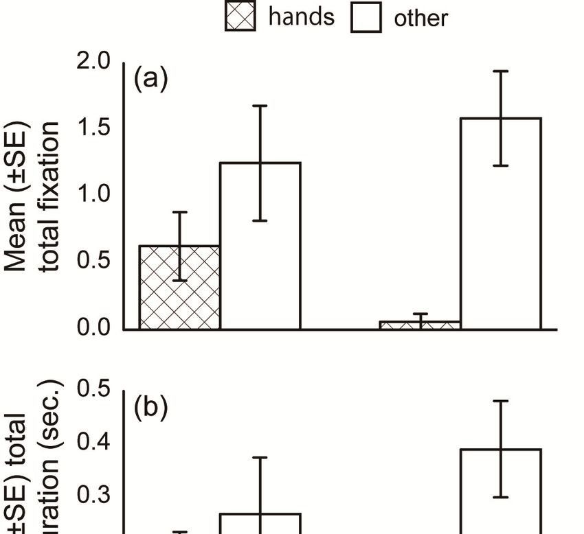
Animals 2020, 10, 755                                                                               10 of 12

4. Discussion
       Dogs’ gaze behavior differed depending on the species presented. The fixation number analyses
revealed that the dogs gazed more toward human limbs while also gazing more toward dog heads and
cat heads and bodies. The first gazed AOI analysis showed similar results for the human and dog
pictures. For the cat pictures, subjects first gazed toward the body. Notable gaze toward human limbs
relative to the dog and cat pictures suggests that dogs have acquired an attentional specialization for
non-verbal human limb gestures differed with attention pattern when facing with non-human species.
Contrary to these analysis, the total fixation duration did not differ among species, suggesting that the
subjects directed shorter gaze toward the AOIs with more fixation numbers repeatedly, but longer gaze
toward other AOIs. A fixed stare is interpreted as an expression of threat, while gaze alternation is
used in cooperative and affiliative interactions [22,23]. The AOIs with more fixation numbers such as
human limbs, dog heads, and cat heads might be affiliative signals for dogs. Conversely, when viewing
the faces of other dogs, the head and face provide more communicative information. Contrary to our
expectations, the subjects gazed toward other dogs’ tails less frequently than the other AOIs. A dog’s
tail movement could be modulated by the dog’s emotional state [24]. Although this modulation may be
unintentional, dogs are sensitive to other dogs’ tail expressions [25]. However, the present study used
still images that lacked information regarding tail movement, which may be why we failed to observe
significant attention toward tails in the present study. It should also be noted that our AOIs differed in
size, as it was our intent to present pictures depicting natural objects. Interestingly, the subjects gazed
more toward the smaller AOIs (e.g., dog head) than the larger AOIs (e.g., dog limbs), indicating that
gaze behavior was non-random and contingent on AOI saliency. In other words, the dogs changed
their gaze behavior depending on which features were most visually attractive within the particular
species presented.
       For pictures depicting familiar and novel humans, the fixation number, total fixation duration to
each AOI, and first gazed AOIs did not differ significantly. These results indicate that dogs did not
change their gaze behavior as a function of stimulus familiarity. There are two potential explanations
for this. One possibility is that dogs just do not discriminate between familiar and unfamiliar humans.
Alternatively, dogs might discriminate familiarity, but this does not impact their general gaze behavior.
Adachi et al. [26] demonstrated that pet dogs have a cross-modal (voice-face) representation of their
owners. Somppi et al. [13] also tested whether dogs gaze differently toward personally familiar
pictures and found that familiar faces attract attention more than unfamiliar ones. This aforementioned
evidence indicates that dogs are able to discriminate novel from familiar human faces. However, in
our study, the human stimulus features may have impacted performance. For instance, the images
were relatively small, and each human model was wearing the same white lab coat. This may have
made it difficult for the dogs to discriminate the novel from the familiar. Hence, differences in gaze
patterns on the entirety of a human image within novel and familiar stimuli should be tested in future
studies (particularly with a head-mounted eye-tracker; Williams et al. [27] examined gaze patterns
toward real people or life-sized images projected on a screen (see [28]).
       The fixation number varied significantly as a function of presenting a human hand sign. Here,
the dogs gazed more toward human hands when a hand sign was present compared to when it was
not. This was also the case for the first gazed AOI. However, total fixation duration did not differ
significantly between the hand sign conditions, but a trend level tendency in line with the other gaze
analyses was observed. These results suggest that a dog’s attention is enhanced when non-verbal
hand signals are present, indicating that hand signs serve as a reliable communication aid for pet
dogs. Humans and chimpanzees gaze more at human faces than other parts [6]. Hattori et al. [29]
revealed humans and chimpanzees gaze less at humans’ hands conveying human intentional signs.
The discrepancy between the results from these study on humans and chimpanzees and those from
our study on dogs could be due to a long phylogenetic distance from dogs to human and chimpanzees.
During the process of domestication, dogs might have acquired specialized attentional patterns to
discern social information from a human hand sign.
Animals 2020, 10, 755 11 of 12
This study aimed to clarify dogs’ attentional patterns to the signals produced by human hands
by comparing visual gaze toward still pictures showing human, dogs, and cats. The subjects in the
experiment were pet dogs, because any potential influence of pre-training to participate in any other
task than this experiment must be excluded. This limited the number of the subjects and the breeds
of the subject dogs, but the present study could successfully provide some useful insights on the
dogs’ domestication process. Following the previous eye-tracking studies in dogs, this study used still
pictures as experimental stimuli. This favorably highlighted dogs’ selective attention to human hand
signs. However, human bodily signals are usually expressed with movement which was lacking in the
still images. As well as information presented in the experimental stimuli, body movement might
be important for dogs to communicate with humans. Further experiments with movie stimuli may
enable deeper discussions on dogs’ visual cognition specialized to communication with humans.
The present experiment revealed that dogs gazed most notably toward human limbs, and this
differed depending on the presence of a hand sign. It appears that dogs have acquired an attentional
style that is differed when facing with human and non-human species and enables the collection of
information for adequately communicating with people living in their community. Thus, the present
study empirically demonstrates that a dog’s cognitive abilities can be adjusted to accommodate life
with humans.
Author Contributions: Conceptualization: T.O., M.M., S.N. (Saki Nagata) and S.N. (Sanae Nakamura); Data
curation, T.O., M.M., S.N. (Saki Nagata) and S.N. (Sanae Nakamura); Formal analysis, T.O.; Funding acquisition,
T.O.; Investigation, T.O., M.M., S.N. (Saki Nagata) and S.N. (Sanae Nakamura); Methodology, T.O., M.M.,
S.N. (Saki Nagata) and S.N. (Sanae Nakamura); Project administration, T.O.; Resources, T.O., M.M., S.N.
(Saki Nagata) and S.N. (Sanae Nakamura); Software, T.O.; Supervision, T.O.; Validation, T.O.; Visualization, T.O.;
Writing—original draft, T.O., M.M., S.N. (Saki Nagata) and S.N. (Sanae Nakamura); Writing—review & editing,
T.O. All authors have read and agreed to the published version of the manuscript.
Funding: This research was funded by JSPS KAKENHI Grant Number 16K16075 and 19K20649.
Acknowledgments: We wish to thank Shoko Izumi, Miku Imai, Sakurako Nagano, Azusa Oki, and Sayaka Murakami
for their contribution to the preliminary experiments. We are grateful to the students and veterinary technicians for
offering their dogs and to the members of the Laboratory of Animal Behavior, Department of Animal Science,
School of Veterinary Medicine, Kitasato University, for their valuable comments.
Conflicts of Interest: The authors declare no conflict of interest.
References
1. Miklósi, Á.; Topál, J. What does it take to become ‘best friends’? Evolutionary changes in canine social
competence. Trends Cogn. Sci. 2013, 17, 287–294. [CrossRef]
2. Pitteri, E.; Mongillo, P.; Carnier, P.; Marinelli, L.; Huber, L. Part-based and configural processing of owner’s
face in dogs. PLoS ONE 2014, 9, e108176. [CrossRef] [PubMed]
3. Miklósi, Á. Dog Behavior, Evolution, and Cognition; Oxford University Press: Oxford, UK, 2007.
4. Virány, Z.; Gácsi, M.; Kubinyi, E.; Topál, J.; Belényi, B.; Ujfalussy, D.; Miklósi, Á. Comprehension of human
pointing gestures in young human-reared wolves (Canis lupus) and dogs (Canis familiaris). Anim. Cogn. 2008,
11, 373. [CrossRef] [PubMed]
5. Winters, S.; Dubuc, C.; Higham, J.P. Perspectives: The looking time experimental paradigm in studies of
animal visual perception and cognition. Ethology 2015, 121, 625–640. [CrossRef]
6. Kano, F.; Tomonaga, M. How chimpanzees look at pictures: A comparative eye-tracking study. Proc. R. Soc.
B-Biol. Sci. 2009, 276, 1949–1955. [CrossRef]
7. Kano, F.; Tomonaga, M. Face scanning in chimpanzees and humans: Continuity and discontinuity. Anim. Behav.
2010, 79, 227–235. [CrossRef]
8. Kano, F.; Call, J.; Tomonaga, M. Face and eye scanning in gorillas (Gorilla gorilla), orangutans (Pongo abelii),
and humans (Homo sapiens): Unique eye-viewing patterns in humans among hominids. J. Comp. Psychol.
2012, 126, 388–398. [CrossRef]
9. Schwarz, J.S.; Sridharan, D.; Knudsen, E.I. Magnetic tracking of eye position in freely behaving chickens.
Front. Syst. Neurosci. 2013, 7, 91. [CrossRef]Animals 2020, 10, 755 12 of 12
10. Tyrrell, L.P.; Butler, S.R.; Yorzinski, J.L.; Fernández-Juricic, E. A novel system for bi-ocular eye-tracking in
vertebrates with laterally placed eyes. Methods Ecol. Evol. 2014, 5, 1070–1077. [CrossRef]
11. Yorzinski, J.L.; Patricelli, G.L.; Babcock, J.S.; Pearson, J.M.; Platt, M.L. Through their eyes: Selective attention
in peahens during courtship. J. Exp. Biol. 2013, 216, 3035–3046. [CrossRef]
12. Somppi, S.; Törnqvist, H.; Hänninen, L.; Krause, C.; Vainio, O. Dogs do look at images: Eye tracking in
canine cognition research. Anim. Cogn. 2012, 15, 163–174. [CrossRef] [PubMed]
13. Somppi, S.; Törnqvist, H.; Hänninen, L.; Krause, C.M.; Vainio, O. How dogs scan familiar and inverted faces:
An eye movement study. Anim. Cogn. 2014, 17, 793–803. [CrossRef] [PubMed]
14. Somppi, S.; Törnqvist, H.; Kujala, M.V.; Hänninen, L.; Krause, C.M.; Vainio, O. Dogs evaluate threatening
facial expressions by their biological validity—Evidence from gazing patterns. PLoS ONE 2016, 11, e0143047.
[CrossRef] [PubMed]
15. Kis, A.; Hernádi, A.; Miklósi, B.; Kanizsár, O.; Topál, J. The way dogs (Canis familiaris) look at human
emotional faces is modulated by oxytocin. An eye-tracking study. Front Behav. Neurosci. 2017, 11, 210.
[CrossRef]
16. Téglás, E.; Gergely, A.; Kupán, K.; Miklósi, Á.; Topál, J. Dogs’ gaze following is tuned to human communicative
signals. Curr. Biol. 2012, 22, 209–212. [CrossRef]
17. Törnqvist, H.; Somppi, S.; Koskela, A.; Krause, C.M.; Vainio, O.; Kujala, M.V. Comparison of dogs and
humans in visual scanning of social interaction. R. Soc. Open Sci. 2015, 2, 150341. [CrossRef]
18. Pryor, K. Don’t Shoot the Dog!: The New Art of Teaching and Training; Simon & Schuster: New York, NY,
USA, 1984.
19. Siniscalchi, M.; D’Ingeo, S.; Fornelli, S.; Quaranta, A. Relationship between visuospatial attention and paw
preference in dogs. Sci. Rep. 2016, 6, 31682. [CrossRef]
20. Dobson, A.J. An Introduction to Generalized Linear Models, 2nd ed.; Chapman & Hall: London, UK, 2002.
21. Akaike, H. A new look at the statistical model identification. IEEE Trans. Autom. Control 1974, 19, 716–723.
[CrossRef]
22. Topál, J.; Miklósi, Á.; Gácsi, M.; Dóka, A.; Pongrácz, P.; Kubinyi, E.; Virány, Z.; Csányi, V. The dog as a model
for understanding human social behavior. In Advances in the Study of Behavior; Brockmann, H.J., Roper, T.J.,
Naguib, M., Wynne-Edwards, K.E., Mitani, J.C., Simmons, L.W., Eds.; Academic Press: Burlington, NJ, USA,
2009; Volume 39, pp. 71–116.
23. Vas, J.; Topál, J.; Gácsi, M.; Miklósi, Á.; Csányi, V. A friend or an enemy? Dogs’ reaction to an unfamiliar
person showing behavioural cues of threat and friendliness at different times. Appl. Anim. Behav. Sci. 2005,
94, 99–115. [CrossRef]
24. Quaranta, A.; Siniscalchi, M.; Vallortigara, G. Asymmetric tail-wagging responses by dogs to different
emotive stimuli. Curr. Biol. 2007, 17, R199–R201. [CrossRef]
25. Siniscalchi, M.; Lusito, R.; Vallortigara, G.; Quaranta, A. Seeing left- or right-asymmetric tail wagging
produces different emotional responses in dogs. Curr. Biol. 2013, 23, 2279–2282. [CrossRef] [PubMed]
26. Adachi, I.; Kuwahata, H.; Fujita, K. Dogs recall their owner’s face upon hearing the owner’s voice. Anim. Cogn.
2007, 10, 17–21. [CrossRef]
27. Williams, F.J.; Mills, D.S.; Guo, K. Development of a head-mounted, eye-tracking system for dogs. J. Neurosci.
Methods 2011, 194, 259–265. [CrossRef] [PubMed]
28. Péter, A.; Miklósi, Á.; Pongrácz, P. Domestic dogs’ (Canis familiaris) understanding of projected video images
of a human demonstrator in an object-choice task. Ethology 2013, 119, 898–906. [CrossRef]
29. Hattori, Y.; Kano, F.; Tomonaga, M. Differential sensitivity to conspecific and allospecific cues in chimpanzees
and humans: A comparative eye-tracking study. Biol. Lett. 2010, 6, 610–613. [CrossRef] [PubMed]
© 2020 by the authors. Licensee MDPI, Basel, Switzerland. This article is an open access
article distributed under the terms and conditions of the Creative Commons Attribution
(CC BY) license (http://creativecommons.org/licenses/by/4.0/).You can also read


