Distribution of the CM-Dil-Labeled Human Umbilical Cord Vein Mesenchymal Stem Cells Migrated to the Cyclophosphamide-Injured Ovaries in C57BL/6 Mice
←
→
Page content transcription
If your browser does not render page correctly, please read the page content below
FULL LENGTH                                            Iranian Biomedical Journal 23 (3): 200-208 May 2019
[ DOI: 10.29252/.23.3.200 ]

                                                                                      Distribution of the CM-Dil-Labeled Human Umbilical Cord
                                                                                             Vein Mesenchymal Stem Cells Migrated to the
                                                                                         Cyclophosphamide-Injured Ovaries in C57BL/6 Mice

                                                                                      Ladan Jalalie1,2, Mohammad Jafar Rezaie2,3*, Ali Jalili4, Mohammad Ali Rezaee5,6,
                                                                                               Zakaria Vahabzadeh7,8, Mohammad Reza Rahmani4,5, Mojtaba
                                                                                                   Karimipoor9,10 and Mohammad Saeed Hakhamaneshi1,8
Downloaded from ibj.pasteur.ac.ir at 5:59 IRDT on Thursday July 25th 2019

                                                                                  1                                                                                                       2
                                                                                 Student Research Committee, Kurdistan University of Medical Sciences, Sanandaj, Iran; Cellular and Molecular
                                                                                     Research Center, Research Institute for Health Development, Kurdistan University of Medical Sciences,
                                                                                              3
                                                                              Sanandaj, Iran; Department of Anatomical Sciences, Faculty of Medicine, Kurdistan University of Medical Sciences,
                                                                                              4
                                                                              Sanandaj, Iran; Cancer and Immunology Research Center, Kurdistan University of Medical Sciences, Sanandaj, Iran;
                                                                               5
                                                                                Zoonoses Research center, Research Institute for Health Development, Kurdistan University of Medical Sciences,
                                                                                                 6
                                                                                  Sanandaj, Iran; Department of Medical Laboratory Sciences, Faculty of Paramedical, Kurdistan University of
                                                                                                                7
                                                                              Medical Sciences , Sanandaj, Iran; Liver and Digestive Research Center, Research Institute for Health Development,
                                                                                                                                                  8
                                                                                        Kurdistan University of Medical Sciences, Sanandaj, Iran; Department of Clinical Biochemistry,
                                                                                                                                                                   9
                                                                                    Faculty of Medicine, Kurdistan University of Medical Sciences, Sanandaj, Iran; Department of Anatomy,
                                                                                                                                                                10
                                                                                        Faculty of Medicine, Urmia University of Medical Sciences, Urmia, Iran; Cellular and Molecular
                                                                                            Research Center, Faculty of Medicine, Urmia University of Medical Sciences, Urmia, Iran

                                                                                                           Received 14 July 2018; revised 18 August 2018; accepted 20 August 2018

                                                                                                                                            ABSTRACT

                                                                            Background: Mesenchymal stem cells (MSCs) can be used to treat premature ovarian failure (POF). Different
                                                                            methods have already been applied to detect MSCs in tissues. This study aimed to investigate the quantitative
                                                                            distribution of CM-DiI-labeled human umbilical cord vein MSCs (hUCV-MSCs) in different regions of the ovarian
                                                                            tissue of the cyclophosphamide (CTX)-induced POF in mice. Methods: Adult female C57BL/6 mice (n = 40) were
                                                                            divided into four groups: (1) Mice receiving PBS as control (Ctrl) group; (2) mice receiving hUCV-MSCs
                                                                            intravenously as Ctrl + hUCV-MSCs group; (3) mice receiving CTX intraperitoneally (i.p.) as CTX group; (4) mice
                                                                            receiving CM-DiI-labeled hUCV-MSCs after CTX injection as CTX + hUCV-MSCs group. Histological changes and CM-
                                                                            DiI-labeled hUCV-MSCs distribution were analyzed in the ovarian tissues. Quantitative real-time PCR was
                                                                            performed to detect human mitochondrial cytochrome b (MTCYB) gene in the ovarian tissues of the mice.
                                                                            Results: The mean number of the fluorescent hUCV-MSCs was 20 ± 2.5 (57.1%) in the medulla, 11.3 ± 2.8 (32.2%)
                                                                            in the cortex, and 5.5 ± 1 (15%) in the germinal epithelium of the ovarian tissue (p < 0.05). Moreover, MTCYB gene
                                                                            was detected in the mice ovaries of the CTX + hUCV-MSCs group, but not in other groups. Conclusion: Our
                                                                            findings suggest that the distribution of the transplanted hUCV-MSCs in different regions of the ovarian tissue is
                                                                            not equal, and it is greater in the medulla than the cortex and germinal epithelium. This is the first report of
                                                                            quantitative distribution of MSCs in different regions of ovarian tissue in the POF model. DOI: 10.29252/.23.3.200

                                                                            Keywords: Cyclophosphamide, Mesenchymal stem cells, Premature ovarian failure, Transplantation

                                                                             Corresponding Author: Mohammad Jafar Rezaie
                                                                             Cellular and Molecular Research Center, Research Institute for Health Development, Kurdistan University of Medical Sciences, P.O. Box: 66177-
                                                                             13446, Sanandaj, Iran; Tel: (+98-873) 3664653, Fax: (+98-871) 6664663; E-mail: Rezaiemjafar@gmail.com

                                                                            200                                                                                                          Iran. Biomed. J. 23 (3): 200-208
Jalalie et al. MSCs Distribution in Cyclophosphamide-Injured Ovaries
INTRODUCTION study was obtained from the Animal Ethics Committee
[ DOI: 10.29252/.23.3.200 ]
of the Kurdistan University of Medical Sciences in
P
remature ovarian failure (POF) is a compliance with the guidelines published in the NIH
heterogeneous syndrome in which menopause (National Institutes of Health Publications) Guide for
occurs before the age of 40. This failure is the Care and Use of Laboratory Animals (Ethic code:
caused by a variety of genetic diseases such as IR.MUK.REC.1395.185).
fragile X syndrome and Turner syndrome, as well as
by some autosomal disorders or medical interventions, Isolation and expansion of hUCV-MSCs
including radiotherapy and chemotherapy[1,2]. POF is Following informed consents from the mothers,
characterized by the decreased number of follicles, hUCV-MSCs were obtained from an operating room.
increased FSH levels, decreased estrogen, and Briefly, the umbilical cord veins were collected
amenorrhea[3,4].
aseptically and transferred to the lab in sterile tubes on
Cyclophosphamide (CTX) is an alkylated
chemotherapy drug that can cause POF and ice. After washing with Hank's buffered salt solution
Downloaded from ibj.pasteur.ac.ir at 5:59 IRDT on Thursday July 25th 2019
degenerative changes in ovarian tissue[5]. The goal of (HBSS) containing 400 mg/L of KCl, 60 mg/L of
POF treatment is to increase the chance of fertility. KH2PO4 , 100 mg/L of MgSO4-7H2O, 8 g/L of NaCl, 60
Hormone replacement therapy has been used to treat mg/L of Na2HPO4-2H2O, 1 g/L of Glucose, 140 mg/L of
POF, but it has systemic and cardiovascular CaCl2, 100 mg /L of MgCl2-6H2O, 350 mg of NaHCO3,
complications[6]. In recent years, cell therapy has been the veins were filled with collagenase IV (Gibco,
proposed as a strategy for POF treatment with minimal USA) and incubated at 37 °C for 20 minutes. After
side effects and considered in clinical practice[7,8]. centrifugation at 600 g for 15 minutes, the cells were
Mesenchymal stem cells (MSCs) are highly removed, washed twice in sterile PBS and cultured in
important in regenerative medicine because of their tissue culture flasks containing Dulbecco's Modified
inherent regenerative properties[9,10]. MSCs could Eagles Medium (DMEM; Gibco, USA) supplemented
increase the reproductive capacity of sterilized female with 10% fetal bovine serum (Gibco, USA), 100 μg/ml
animals in practical researches. Restorative effects of of streptomycin, and 100U/ml of penicillin. The
MSCs on injured ovaries have been observed in animal cultures were incubated at 37 °C in a humidified
models of POF[7,11,12]. MSCs tracking and tissue environment containing 5% CO2. After 48 hours, the
distribution are interesting aspects of MSCs studies. non-adherent cells were discarded, and the media were
Some fluorochrome materials have been used for replaced every three days. When the hUCV-MSCs
labeling MSCs to be tracked in target tissues[13,14]. CM- reached 80-90% confluency, the adherent cells were
DiI is a very effective and available lipophilic dyes
trypsinized with 0.02% Trypsin (Sigma-Aldrich,
binding to the membrane phospholipids[15,16]. It has
USA), and then the cells were passaged up to passage
low cytotoxicity for long-term culturing of labeled
MSCs without significant effect on the cell 4[19].
properties[14,17].
Currently, MSCs have been known as a potential hUCV-MSCs morphology and immunophenotyping
choice for the treatment of many diseases and tissue hUCV-MSCs morphology was observed by an
injuries. It is important to determine the distribution of inverted microscope at passages 3-4. Moreover, the
MSCs in different parts of the target organ after expression of hUCV-MSCs-related surface markers
administration of these stem cells. Although MSCs such as CD105 and CD73 and the lack of the CD45
tracking has been conducted in previous studies[11,18], and CD34 markers were evaluated by flow cytometry.
there is no report on quantitative distribution of MSCs In brief, after reaching 70-80% confluency, the cells
in different regions of the ovarian tissue. This study were detached using Trypsin. The harvested cells were
aimed to investigate the quantitative distribution of washed and resuspended in PBS. Aliquots of 1 106
labeled human umbilical cord vein MSCs (hUCV- cells were incubated with FITC-labeled anti-CD45,
MSCs) in different parts of the ovarian tissues in a anti-CD105, anti-CD34, and PE-labeled anti-CD73 in
mice model of CTX-induced POF. the dark at 4 °C for 30 minutes. After staining, the cells
were fixed using paraformaldehyde, and the expression
MATERIALS AND METHODS of the cell surface markers were detected using flow
cytometry.
Animals and ethics
Female C57BL/6 mice (n = 40), at 7–8 weeks of age CM-DiI-labeled hUCV-MSCs preparation
and weighing 25-30 g, were obtained from Animal hUCV-MSCs were detached using Trypsin and
House Center at the Kurdistan University of Medical resuspended at a concentration of 1 106 cells/ml in
Sciences (Sanandaj, Iran). The ethical approval of the HBSS buffer. CM-DiI dye stock was prepared as
Iran. Biomed. J. 23 (3): 200-208 201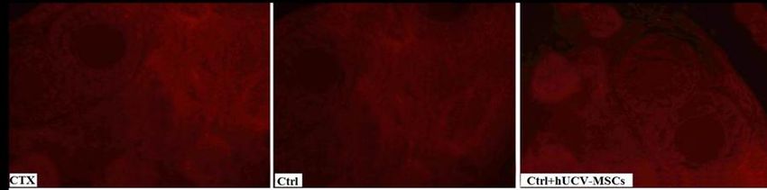
MSCs Distribution in Cyclophosphamide-Injured Ovaries Jalalie et al.
recommended by the manufacturer (Thermo Fisher calculated compared to the total counted fluorescent
[ DOI: 10.29252/.23.3.200 ]
Scientific Inc., Waltham, MA, USA). A confluent layer cells in the ovarian tissue.
of 1 × 10⁶ MSCs/ml was stained using CM-DiI
solution (5 µl) and was incubated at 37 °C for 15 DNA extraction and PCR
minutes, then at 4 °C for 15 minutes. Next, the cell Real-time PCR was performed to detect the human
suspension was centrifuged, the media were removed, mitochondrial cytochrome b (MTCYB) gene to
and the cells were washed twice in sterile PBS (pH confirm hUCV-MSCs migration to the target tissue.
7.4). Subsequently, the labeled cells were cultured, Mouse GAPDH gene was used as a reference for
maintained at sub-confluences and monitored for sample normalization and quantitative analysis in all
fluorescence using the Olympus BX51 microscope the study groups. Specific primers were used to
(Olympus, Tokyo, Japan). amplify the target genes. The forward and reverse
primer sequences used for MTCYB were 5΄-AGCCAC
POF model induction, grouping, and hUCV-MSCs TTTCCACACAGAC-3΄ and 5΄-AGTAGTATGGGAG
TGGGAG-3΄, and for GAPDH included 5´-AATGTG
Downloaded from ibj.pasteur.ac.ir at 5:59 IRDT on Thursday July 25th 2019
administration
To induce POF, mice were injected with 50 mg/kg TCCGTCGTGGATCTGA-3´ and 5´-GATGCCTGCT
CTX (Sigma-Aldrich, St. Louis, MO, USA) dissolved TCACCACCTTCT-3´, respectively. The amplicons
in PBS intraperitoneally (i.p.) for 15 consecutive days. predicted for MTCYB and GAPDH were 219 and 167
The animals were divided into the following four bp, respectively. DNA was extracted from 25 mg of the
groups, each group including 10 mice: (1) mice ovarian tissues in each group using QIAamp DNA
receiving PBS i.p. for 15 days and then 200 µl PBS via Mini Kit (QIAGEN, Hilden, Germany) according to
lateral tail vein as control (Ctrl) group; (2) mice the manufacturer’s instructions. The concentration and
receiving PBS i.p. for 15 days and then 1 106 CM- purity of the DNA extracted from each sample were
DiI-labeled hUCV-MSCs in 200 µl PBS via lateral tail determined micro-spectrophoto-metrically (BioTek
vein as Ctrl + hUCV-MSCs group; (3) mice receiving Instruments Inc., USA). The amplification and
50 mg/kg CTX i.p. for 15 consecutive days and then detection were performed using the Real Q Plus Master
200 µl PBS intravenously (i.v.) as CTX group; (4) Mix Green without ROX™ (Ampliqon, Denmark)
mice receiving CTX for 15 consecutive days and then according to the protocols provided by manufacturer
1 106 CM-DiI-labeled hUCV-MSCs as CTX + using a Rotor-Gene 6000 real-time PCR machine
hUCV-MSCs group. One week after the hUCV-MSCs (Corbett Life Science, Sydney, Australia). Briefly, the
injection, the mice in all the study groups were PCR reaction was performed in a 25-μl final volume.
euthanized by cervical dislocation, and their ovaries Each reaction was composed of 12.5 μl of real-time
were removed under sterile conditions to conduct PCR master mix, 7.5 μl of deionized water, 1 μl of
further experiments. each primer with a concentration of 10 μM, and 3 μl of
the template DNA (30 ng/μl). Thermocycler thermal
Histological examination conditions included primary denaturation at 90 °C for
Briefly, the ovaries of the mice were removed 15 minutes, followed by 40 repetitive cycles at 90 °C
aseptically, washed with sterile PBS, fixed with 4% for 60 seconds and then at 60 °C for 60 seconds.
paraformaldehyde (Sigma, St. Louis, MO, USA), Relative copy number of human MTCYB and mouse
embedded in paraffin, serially sectioned at 5 µm GAPDH genes were calculated using the open access
thickness and then dehydrated using graded ethanol. softwarbe LinRegPCR version 13[22].
Finally, the sections were stained with hematoxylin
(Merck KGaA, Darmstadt, Germany) and eosin (H & Statistical analysis
E; Sigma, St. Louis, MO, USA). The statistical analyses of the data were conducted
using SPSS 16 and one-way analysis of variance (one-
Quantification of labeled hUCV-MSCs way ANOVA). p value less than 0.05 was considered
CM-DiI-labeled hUCV-MSCs were counted in 10 statistically significant.
serial ovarian tissue sections (5 µm), in each mouse of
the study groups[20]. The fluorescent cells were counted
in different regions of the ovarian tissues with a RESULTS
fluorescent microscope (Olympus, Tokyo, Japan)[21].
The mean numbers of the CM-DiI-labeled hUCV- hUCV-MSCs characterization
MSCs were calculated in the total ovarian tissue as Morphology of the hUCV-MSCs was examined
well as in the medulla, cortex, and germinal epithelium under an inverted microscope. Colonies of the hUCV-
of the ovary, separately. Also, the percentage of the MSCs were observed two days after the initial
counted cells in each region of the ovarian tissue was isolation. After a week, the surfaces of the cell culture
202 Iran. Biomed. J. 23 (3): 200-208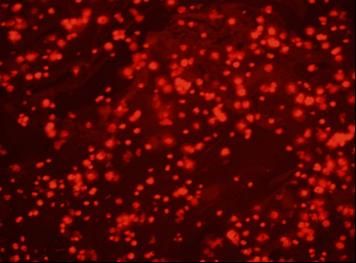
Jalalie et al. MSCs Distribution in Cyclophosphamide-Injured Ovaries
flasks were filled, and spindle-shaped cells were groups were used to evaluate the histological
[ DOI: 10.29252/.23.3.200 ]
observed in the passage 3 (Fig. 1A). Flow cytometry changes for the confirmation of POF induction. In mice
analysis indicated that hUCV-MSCs were positive for receiving CTX, ovarian tissue sections stained with H
CD73 and CD105 and negative for CD45 and CD34 & E showed a decrease in the number of the follicles,
(Fig. 1B). especially the primordial follicles. Also, many atretic
follicles were observed in the stromal region of the
Confirmation of POF after administration of CTX CTX group (quantitative data are not shown; Fig. 2).
The ovarian tissue sections of both CTX and Ctrl
(A)
Downloaded from ibj.pasteur.ac.ir at 5:59 IRDT on Thursday July 25th 2019
(B)
Fig. 1. Human umbilical cord vein mesenchymal stem cells (hUCV-MSCs) morphology and immunophenotyping at passage 3. (A)
The hUCV-MSCs showing an elongated and a fibroblast-like shape (magnification 100); (B) hUCV-MSCs indicating to be positive
for CD73 and CD105 and negative for the CD45 and CD34 surface markers.
Iran. Biomed. J. 23 (3): 200-208 203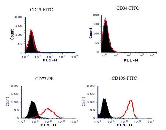
MSCs Distribution in Cyclophosphamide-Injured Ovaries Jalalie et al.
[ DOI: 10.29252/.23.3.200 ]
Downloaded from ibj.pasteur.ac.ir at 5:59 IRDT on Thursday July 25th 2019
Fig. 2. H & E staining of the mice ovarian tissue sections. (A) In the ovarian tissue section of the group receiving sterile PBS (Ctrl),
normal growing follicles were observed. (B) In the ovarian tissue section of the mice receiving cyclophosphamide (CTX), healthy
follicles decreased, and many atretic primordial and primary follicles were observed (magnification 200).
Counting CM-DiI-labeled hUCV-MSCs DISCUSSION
After staining with CM-DiI, hUCV-MSCs were
observed using a fluorescence microscope at the In the present study, the CTX-induced POF model
magnification of 100 (Fig. 3). Subsequently, the was used to assess the distribution of the hUCV-MSCs
ovarian tissue sections from the experimental groups in the mice ovarian tissues. POF model was confirmed
were examined to observe the CM-DiI-labeled hUCV- by observing degenerative changes in the ovarian
MSCs using a fluorescent microscope. CM-DiI-labeled tissues. This model was in accordance with the
hUCV-MSCs were detected in different parts of the previous studies[11,23]. MSCs-based cell therapy is an
ovarian tissue such as the medulla, especially near important research field in regenerative medicine for
the medullary veins, cortex, between the follicles, and the treatment of various diseases. MSCs can migrate to
germinal epithelium of the CTX + hUCV-MSCs group, damaged tissues and repair them by modulating the
but these cells were not found inside the follicles and immune system and secreting growth factors[24,25].
oocytes Fig. 4A) and in the tissue sections of other The healing effects of MSCs on injured ovarian
groups (Fig. 4B). The mean number of hUCV-MSCs tissues have been reported in the POF animal
was 35 ± 4.1 in the total ovarian region, 20 ± 2.5 in the models[26,27]. UC-MSCs are preferred for clinical
medulla, 11.3 ± 2.8 in the cortex, and 5.5 ± 1 in the
epithelium. The mean number of the fluorescent cells
in the medullary region was significantly higher than
the cortex and germinal epithelium (p ˂ 0.001). Also,
the mean number of CM-DiI-labeled hUCV-MSCs in
the cortex was significantly higher than the germinal
epithelium (p = 0.01), as shown in Figure 4C. The
percentage of the fluorescent cells in the medulla,
cortex, and epithelium were 57.1%, 32.2%, and 15%,
respectively.
Detection of human MTCYB gene in the ovarian
tissue
Real-time PCR was used for each tissue sample to
evaluate the presence of human MTCYB gene. Mouse
GAPDH was used as a reference gene to normalize the Fig. 3. Photomicrograph of human umbilical cord vein
results. CT, ΔCT, and the relative copy number of the mesenchymal stem cells (hUCV-MSCs) stained with CM-DiI
samples were also calculated. Human MTCYB gene fluorochrome. hUCV-MSCs were trypsinized at passage 3 and
was detected in the CTX + hUCV-MSCs group, but stained with CM-DiI in vitro, then the cells were observed under
not in other groups (Fig. 5). a fluorescence microscope (magnification 100).
204 Iran. Biomed. J. 23 (3): 200-208Jalalie et al. MSCs Distribution in Cyclophosphamide-Injured Ovaries
(A)
[ DOI: 10.29252/.23.3.200 ]
(B)
Downloaded from ibj.pasteur.ac.ir at 5:59 IRDT on Thursday July 25th 2019
(C)
Fig. 4. Red fluorescent CM-DiI-labeled human umbilical cord vein mesenchymal stem cells (hUCV-MSCs) detection in the mice
ovarian regions. (A) Labeled hUCV-MSCs were detected in different regions of the ovarian tissue sections from CTX + hUCV-MSCs
group; in the medulla (M) near the medullary veins, in the cortex (C), and in germinal epithelium (E); (B) fluorescent cells were not
observed in the tissue sections of the CTX, Ctrl, and Ctrl + hUCV-MSCs groups; (C) the number of florescent hUCV-MSCs in
different regions of the ovary in the CTX+ hUCV-MSC group was counted. The number of the labeled MSCs in the medulla is greater
than the cortex and germinal epithelium (p ˂ 0.001), and in the cortex, it is higher than germinal epithelium (p = 0.01). Data are shown
as mean ± SD. *p = 0.01, **p ˂ 0.001.
applications because of their accessibility and poor have demonstrated that hUC-MSCs could recover
immunogenic properties, which are attributed to their ovarian structure and improve ovarian function injured
low expression of major histocompatibility complex I by CTX in the mice. Ghadami et al.[29] have shown that
and the absence of major histocompatibility complex i.v.-injected BM-MSCs are able to increase the FSH
II. Moreover, UC-MSCs can be easily isolated and receptors, resume estrogen hormone production, and
expanded in vitro, and they have little ethical issues as restore folliculogenesis in POF mice. In addition,
compared to other types of MSCs[28]. Wang et al.[11] adipose-derived MSCs and amniotic fluid MSCs have
Iran. Biomed. J. 23 (3): 200-208 205MSCs Distribution in Cyclophosphamide-Injured Ovaries Jalalie et al.
compared with the cortex and the germinal epithelium.
[ DOI: 10.29252/.23.3.200 ]
Also, most of the cells migrated to the ovarian stromal
Relative copy number of
region. Fewer hUCV-MSCs reached the cortex, and
human MTCYB
smaller numbers of them were found in the germinal
epithelium tissue. Moreover, few hUCV-MSCs were
observed in the interface between primordial and
primary follicles. hUCV-MSCs were not detected
within primary follicles or even adult follicles. Wang
et al.[11] have detected the MSCs in the ovarian stromal
tissue, which is consistent with our findings. Sun et
al.[23] have found labeled adipose-derived MSCs in the
ovarian stromal tissue after i.v. injection, but not inside
the follicles or oocytes. On the other hand, Liu et al.[18]
Downloaded from ibj.pasteur.ac.ir at 5:59 IRDT on Thursday July 25th 2019
Groups
have detected green fluorescent protein-labeled BM-
MSCs in the ovarian tissue of the cisplatin-induced
Fig. 5. Detection of the human MTCYB gene in the mice POF mice model after MSCs transplantation. The
ovarian tissues after the administration of human umbilical cord majority of MSCs were replaced in the medulla, and
vein mesenchymal stem cells (hUCV-MSCs). Human MTCYB
fewer cells were observed in the cortical region of the
gene was detected only in the mice receiving cyclophosphamide
(CTX) and hUCV-MSCs (CTX + hUCV-MSCs group), but not ovarian tissue. The cells were absent in the follicle and
in the mice groups receiving CTX (CTX), PBS (Ctrl), and PBS corpus luteum. Additionally, MSCs were mainly
+ hUCV-MSCs (Ctrl + hUCV-MSCs). distributed along the blood vessels[18]. It has been
shown that some of the MSCs are found in the cortical
stromal region in the space between the follicles, and
been referred to as therapeutic agents for few numbers of cells are inside the follicles[32].
chemotherapy-induced ovarian damage[18,30]. The therapeutic effect of MSCs can be related to
In the current study, CM-DiI-labeled hUCV-MSCs more replacement of these cells in the stromal region
were injected into the POF mice. Red fluorescent cells of the injured ovaries. The effects of MSCs may be due
were observed in the ovarian sections of the CTX- to their active paracrine function by secreting growth
injured mice receiving hUCV-MSCs. Also, human factors to repair damaged follicles. Also, engrafted
MTCYB gene was detected in the ovarian tissues of MSCs around the blood vessels can induce
CTX + hUCV-MSCs group, but not in other groups. angiogenesis by secreting angiogenic growth
Therefore, our data confirmed the engraftment of factors[33,34]. The fact that MSCs were not seen in the
hUCV-MSCs in the ovaries of the CTX-injured mice. follicles or oocytes could indicate that MSCs do not
Wang et al.[11] have detected CM-DiI-labeled hUCV- differentiate into follicular cells, and they indirectly
MSCs in the POF mice ovaries one week after the i.v. affect follicular regeneration[33]. The most well-known
injection. It seems that the migration of MSCs growth factors secreted by MSCs are vascular
toward injured ovaries in the POF mice was due to the endothelial growth factor, hepatocyte growth factor,
degenerative changes after CTX administration. and insulin-like growth factor[33]. Vascular endothelial
Studies have confirmed these degenerative changes in growth factor is of paramount importance as it affects
POF models[18,29]. MSCs could migrate toward the the growth of blood vessels in the granulosa follicular
injured tissues due to increased chemokines in the cell layers, and it prevents apoptosis[34].
damaged and inflamed tissues. MSCs express Quantitative distribution of MSCs in different
molecules that mediate migration to the target tissues regions of the ovarian tissue in the POF mouse model
such as CXCR4 (C-X-C chemokine receptor type 4), has not been reported. We observed that the number of
CXCR7, and integrins. CXCR4 is one of the most the CM-DiI-labeled hUCV-MSCs in ovarian medulla
known chemokine receptors involved in the migration was greater than that of the ovarian cortex and
of MSCs. The stromal-derived factor 1 (SDF-1) is the germinal epithelium. Besides, the mean number of the
CXCR4 ligand increased in ischemic and injured labeled hUCV-MSCs in the cortex region was
tissues[24,31]. It appears that CXCR4/SDF-1 axis has an significantly higher than the germinal epithelium.
important role in the migration of the MSCs to the hUCV-MSCs migration towards the medulla may be
CTX-injured ovaries[18]. due to the fact that it is mostly composed of stromal
In our study, hUCV-MSCs were not distributed tissue rather than the cortex and germinal epithelium,
equally in different parts of the ovarian tissue. and also that medulla is rich in blood vessels. The
Fluorescent cells were mostly observed in the medulla stromal region of the ovarian tissue is the source of
206 Iran. Biomed. J. 23 (3): 200-208Jalalie et al. MSCs Distribution in Cyclophosphamide-Injured Ovaries
SDF-1 during injury and inflammation; as a result, it 4. Poggio F, Levaggi A, Lambertini M. Chemotherapy-
[ DOI: 10.29252/.23.3.200 ]
increases the migration of MSCs to this region[18]. induced premature ovarian failure and its prevention in
Meanwhile, physical barriers in ovarian tissue can also premenopausal breast cancer patients. Journal expert
be a limiting factor for the homing of MSCs in a review of quality of life in cancer care 2016; 1(1): 5-7.
5. Cox L, Liu JH. Primary ovarian insufficiency: an
particular region of the tissue. For instance, the reason
update. International journal of women's health 2014;
for the small numbers of MSCs engraftments in the 6(1): 235-243.
ovarian germinal epithelium could be related to the 6. Cartwright B, Robinson J, Seed PT, Fogelman I, Rymer
preventing effects of physical barriers such as basal J. Hormone replacement therapy versus the combined
membranes and cell-cell junction complexes[35]. oral contraceptive pill in premature ovarian failure: a
hUCV-MSCs were not observed in the follicles, randomized controlled trial of the effects on bone
cumulus cells, or oocytes. Tight junctions between the mineral density. The journal of clinical endocrinology
follicular cells may prevent the cells from entering the and metabolism 2016; 101(9): 3497-3505.
follicles. Furthermore, intracellular connections and 7. Fu X, He Y, Xie C, Liu W. Bone marrow mesenchymal
stem cell transplantation improves ovarian function and
Downloaded from ibj.pasteur.ac.ir at 5:59 IRDT on Thursday July 25th 2019
gap junctions in both inner and outer theca cell layers
structure in rats with chemotherapy-induced ovarian
in the mature follicles could be an obstacle to MSCs damage. Cytotherapy 2008; 10(4): 353-363.
homing. In addition, the presence of a barrier around 8. Edessy M, Hosni HN, Wafa Y, Bakry S, Shady Y,
the follicle like the basement membrane can prevent Kamel M. Stem cells transplantation in premature
MSCs from entering the follicles[36]. ovarian failure. World journal of medical sciences 2014;
The findings of the present study show that hUCV- 10(1): 12-16.
MSCs home in the CTX-injured mice ovaries with 9. Lee HJ, Selesniemi K, Niikura Y, Niikura T, Klein R,
more engraftment in the medulla and stromal region, Dombkowski DM, Tilly JL. Bone marrow
and a small number of hUCV-MSCs engraft in the transplantation generates immature oocytes and rescues
ovarian cortex and germinal epithelium. long-term fertility in a preclinical mouse model of
chemotherapy-induced premature ovarian failure.
Journal of clinical oncology 2007; 25(22): 3198-3204.
10. Mohammadi M, Jaafari M, Mirzaei H, Mirzaei H.
ACKNOWLEDGEMENTS Mesenchymal stem cell: a new horizon in cancer gene
therapy. Cancer gene therapy 2016; 23(9): 285-286.
This research was funded by Research Council of the 11. Wang S, Yu L, Sun M, Mu S, Wang C, Wang D, Yao Y.
Kurdistan University of Medical Sciences (grant The therapeutic potential of umbilical cord
number 13446-66177), Sanandaj, Iran. The authors mesenchymal stem cells in mice premature ovarian
thank the staff members of the Immunology Lab and failure. Biomed research international 2013; 2013:
690491.
Cellular and Molecular Research Center at Kurdistan
12. Li J, Yu Q, Huang H, Deng W, Cao X, Adu-Frimpong
University of Medical Sciences for their technical M, Yu J, Xu X. Human chorionic plate-derived
assistance. mesenchymal stem cells transplantation restores ovarian
function in a chemotherapy-induced mouse model of
CONFLICT OF INTEREST. None declared. premature ovarian failure. Stem cell research and
therapy 2018; 9(1): 81.
13. Guo Y, Su L, Wu J, Zhang D, Zhang X, Zhang G, Li T,
Wang J, Liu C. Assessment of the green florescence
REFERENCES protein labeling method for tracking implanted
mesenchymal stem cells. Cytotechnology 2012; 64(4):
1. Ayesha VJ, Goswami D. Premature ovarian failure: an 391-401.
association with autoimmune diseases. Journal of 14. Ji F, Duan HG, Zheng CQ, Li J. Comparison of
clinical and diagnostic research 2016; 10(10): QC10- chloromethyl-dialkylcarbocyanine and green fluorescent
QC12. protein for labeling human umbilical mesenchymal stem
2. Falcone T, Bedaiwy MA. Fertility preservation and cells. Biotechnology letters 2015; 37(2): 437-447.
pregnancy outcome after malignancy. Current opinion 15. Progatzky F, Dallman MJ, Lo Celso C. From seeing to
in obstetrics and gynecology 2005; 17(1): 21-26. believing: labelling strategies for in vivo cell-tracking
3. Del Mastro L, Ceppi M, Poggio F, Bighin C, Peccatori experiments. Interface focus 2013; 3(3): 20130001.
F, Demeestere I, Levaggi A, Giraudi S, Lambertini M, 16. Cao AH, Shi HJ, Zhang Y, Teng GJ. In vivo tracking of
D'Alonzo A, Canavese G, Pronzato P, Bruzzi P. dual‐labeled mesenchymal stem cells homing into the
Gonadotropin-releasing hormone analogues for the injured common carotid artery. The anatomical record
prevention of chemotherapy-induced premature ovarian (Hoboken) 2009; 292(10):1677-1683.
failure in cancer women: systematic review and meta- 17. Ferrari A, Hannouche D, Oudina K, Bourguignon M,
analysis of randomized trials. Cancer treatment reviews Meunier A, Sedel L, Petite H. In vivo tracking of bone
2014; 40(5): 675-683. marrow fibroblasts with fluorescent carbocyanine dye.
Iran. Biomed. J. 23 (3): 200-208 207MSCs Distribution in Cyclophosphamide-Injured Ovaries Jalalie et al.
Journal of biomedical materials research 2001; 56(3): cellular and molecular medicine 2015; 19(9): 2108-
[ DOI: 10.29252/.23.3.200 ]
361-367. 2117.
18. Liu J, Zhang H, Zhang Y, Li N, Wen Y, Cao F, Ai H, 28. Zhou C, Yang B, Tian Y, Jiao H, Zheng W, Wang J,
Xue X. Homing and restorative effects of bone marrow- Guan F. Immunomodulatory effect of human umbilical
derived mesenchymal stem cells on cisplatin injured cord Wharton’s jelly-derived mesenchymal stem cells
ovaries in rats. Molecules and cells 2014; 37(12): 865- on lymphocytes. Cellular immunology 2011; 272(1): 33-
872. 38.
19. Moradi M, Rezaee MA, Mohammadi M, Rezaie MJ, 29. Ghadami M, El-Demerdash E, Zhang D, Salama SA,
Jalili A, Rahmani MR. Attenuating effect of long-term Binhazim AA, Archibong AE, Chen X, Ballard BR,
culture of umbilical cord vein mesenchymal stromal Sairam MR, Al-Hendy A. Bone marrow transplantation
cells on pulmonary fibrosis in C57BL/6 mice. Iranian restores follicular maturation and steroid hormones
journal of allergy, asthma and immunology 2017; 16(6): production in a mouse model for primary ovarian
501-510. failure. PLoS one 2012; 7(3): e32462.
20. Teshima T, Matsumoto H, Michishita M, Matsuoka A, 30. Wang F, Wang L, Yao X, Lai D, Guo L. Human
Shiba M, Nagashima T, Koyama H. Allogenic adipose amniotic epithelial cells can differentiate into granulosa
Downloaded from ibj.pasteur.ac.ir at 5:59 IRDT on Thursday July 25th 2019
tissue-derived mesenchymal stem cells ameliorate acute cells and restore folliculogenesis in a mouse model of
hepatic injury in dogs. Stem cells international 2017; chemotherapy-induced premature ovarian failure. Stem
Article ID: 3892514. cell research and therapy 2013; 4(5): 124.
21. Campaner AB, Galvão MAL. Application of an easy 31. Kholodenko IV, Konieva AA, Kholodenko RV, Yarygin
and useful morphometric technique for immuno- KN. Molecular mechanisms of migration and homing of
histochemistry counting. Gynecologic oncology 2009; intravenously transplanted mesenchymal stem cells.
112(1): 282-283. Journal of regenerative medicine and tissue
22. Ruijter JM, Ramakers C, Hoogaars WM, Karlen Y, engineering. 2013; 2(1): 4.
Bakker O, Van den Hoff MJ, Moorman AF. 32. Noha A, Olfat NR. Role of mesenchymal stem cell
Amplification efficiency: linking baseline and bias in therapy in restoring ovarian function in a rat model of
the analysis of quantitative PCR data. Nucleic acids chemotherapy-induced ovarian failure: a histological
research 2009; 37(6): e45. and immunohistochemical study. The Egyptian journal
23. Sun M, Wang S, Li Y, Yu L, Gu F, Wang C, Yao Y. of histology 2013; 36(1): 114-126.
Adipose-derived stem cells improved mouse ovary 33. Lai D, Wang F, Dong Z, Zhang Q. Skin-derived
function after chemotherapy-induced ovary failure. Stem mesenchymal stem cells help restore function to ovaries
cell research and therapy 2013; 4(4): 80. in a premature ovarian failure mouse model. PLoS one
24. Patel DM, Shah J, Srivastava AS. Therapeutic potential 2014; 9(5): e98749.
of mesenchymal stem cells in regenerative medicine. 34. Abd-Allah SH, Shalaby SM, Pasha HF, El-Shal AS,
Stem cells international 2013; Article ID: 496218. Raafat N, Shabrawy SM, Awad HA, Amer MG, Gharib
25. Le Blanc K, Davies LC. Mesenchymal stromal cells and MA, El Gendy EA, Raslan AA, El-Kelawy HM.
the innate immune response. Immunology letters 2015; Mechanistic action of mesenchymal stem cell injection
168(2): 140-146. in the treatment of chemically induced ovarian failure in
26. Fu X, He Y, Wang X, Peng D, Chen X, Li X, Qing W. rabbits. Cytotherapy 2013; 15(1): 64-75.
Overexpression of miR-21 in stem cells improves 35. Auersperg N, Wong AS, Choi KC, Kang SK, Leung PC.
ovarian structure and function in rats with Ovarian surface epithelium: biology, endocrinology, and
chemotherapy-induced ovarian damage by targeting pathology. Endocrine reviews 2001; 22(2): 255-288.
PDCD4 and PTEN to inhibit granulosa cell apoptosis. 36. Tajima K, Orisaka M, Yata H, Goto K, Hosokawa K,
Stem cell research and therapy 2017; 8(1): 187. Kotsuji F. Role of granulosa and theca cell interactions
27. Zhu SF, Hu HB, Xu HY, Fu XF, Peng DX, Su WY, He in ovarian follicular maturation. Microscopy research
YL. Human umbilical cord mesenchymal stem cell and technique 2006; 69(6): 450-458.
transplantation restores damaged ovaries. Journal of
208 Iran. Biomed. J. 23 (3): 200-208Jalalie et al. MSCs Distribution in Cyclophosphamide-Injured Ovaries
[ DOI: 10.29252/.23.3.200 ]
Downloaded from ibj.pasteur.ac.ir at 5:59 IRDT on Thursday July 25th 2019
Iran. Biomed. J. 23 (3): 200-208 209You can also read