Contrast-Enhanced 18F-FDG PET/CT: 1-Stop- Shop Imaging for Assessing the Resectability of Pancreatic Cancer
←
→
Page content transcription
If your browser does not render page correctly, please read the page content below
Downloaded from jnm.snmjournals.org by on November 3, 2015. For personal use only.

Contrast-Enhanced 18F-FDG PET/CT: 1-Stop-
Shop Imaging for Assessing the Resectability of
Pancreatic Cancer
Klaus Strobel1, Stefan Heinrich2, Ujwal Bhure1, Jan Soyka1, Patrick Veit-Haibach1, Bernhard C. Pestalozzi3,
Pierre-Alain Clavien2, and Thomas F. Hany1
1Division of Nuclear Medicine, Department of Medical Radiology, University Hospital Zurich, Zurich, Switzerland; 2Department of

Surgery, University Hospital Zurich, Zurich, Switzerland; and 3Department of Oncology, University Hospital Zurich, Zurich, Switzerland

Patients with pancreatic cancer continue to have a poor progno-
sis, with a 5-y survival rate of less than 5%. Surgery is the only
                                                                                    P    atients with pancreatic cancer continue to have a poor
                                                                                    prognosis, with a 5-y survival rate of less than 5%. Surgery
treatment that offers a potential cure. Determining resectability
                                                                                    is the only treatment that offers a potential cure, but only
is the principal goal of staging in pancreatic cancer patients.
Our objective was to evaluate the value of combined contrast-                       15%220% of the patients are candidates for surgery (1,2).
enhanced 18F-FDG PET/CT in assessing the resectability of                           Determining resectability is the principal goal of staging in
pancreatic cancer and to compare enhanced PET/CT with the                           pancreatic cancer patients. The current preoperative imag-
performance of PET alone and unenhanced PET/CT. Methods:                            ing standard for pancreatic cancer staging is contrast-
Fifty patients (25 women and 25 men; mean age, 64.3 y; range,                       enhanced multidetector CT (3,4). Endoscopic ultrasound is
39–84 y) with biopsy-proven pancreatic adenocarcinoma under-                        also routinely used in many centers for local staging and
went enhanced 18F-FDG PET/CT for the evaluation of resectability.
                                                                                    biopsy guidance (5,6). 18F-FDG PET/CT is a powerful
Criteria for unresectability were distant metastases, peritoneal
carcinomatosis, arterial infiltration, or invasion of neighboring or-               imaging method for the staging of many cancers and also
gans other than the duodenum. The performance of enhanced                           has been shown to affect the oncologic management of
PET/CT regarding resectability was compared with that of PET                        pancreatic cancer patients (7). However, because of the
alone and unenhanced PET/CT. Histology, intraoperative findings,                    limited information available in the literature, the role of
and follow-up CT with clinical investigations were used as the ref-                 18F-FDG PET/CT in the management of pancreatic cancer
erence standard. Results: According to the reference standard,                      remains undefined. The 1-stop-shop imaging approach with
27 patients had disease that was not resectable because of distant
                                                                                    whole-body 18F-FDG PET combined with enhanced multi-
metastases (n 5 17), peritoneal carcinomatosis (n 5 5), or local in-
filtration (n 5 5). In the assessment of resectability, PET alone had a             detector CT in a single investigation is feasible with the
sensitivity of 100%, specificity of 44%, accuracy of 70%, positive                  current generation of scanners, is convenient for patients,
predictive value of 61%, and negative predictive value of 100%;                     and appears to be an attractive staging tool for pancreatic
unenhanced PET/CT had respective values of 100%, 56%, 76%,                          cancer. The aim of this study was to evaluate the value of
66%, and 100%; and enhanced PET/CT, 96%, 82%, 88%, 82%,                             combined enhanced 18F-FDG PET/CT in determining the
and 96%. In 5 patients, unresectability was missed by all imaging                   resectability of pancreatic cancer and to compare enhanced
methods and was diagnosed intraoperatively. Enhanced PET/CT
                                                                                    PET/CT with PET alone and with unenhanced PET/CT.
was significantly superior to PET alone (P 5 0.035), and there
was a trend for enhanced PET/CT to be superior to unenhanced
PET/CT (P 5 0.070). Conclusion: The use of enhanced PET/CT                          MATERIALS AND METHODS
as a 1-stop-shop imaging protocol for assessing the resectability                   Eligibility Criteria
of pancreatic cancer is feasible and accurate. Enhanced PET/CT                         Patients with biopsy-proven pancreatic cancer who underwent
is significantly superior to PET alone.                                             staging PET/CT at the University Hospital of Zurich were eligible
Key Words: oncology; PET/CT; pancreatic cancer; staging, con-                       for this retrospective analysis. The disease was considered resect-
trast enhancement                                                                   able in the absence of distant metastases, arterial infiltration, and
J Nucl Med 2008; 49:1408–1413                                                       infiltration of organs other than the duodenum or stomach. The
DOI: 10.2967/jnumed.108.051466                                                      study was conducted in accordance with the local guidelines estab-
                                                                                    lished by the ethics committee for retrospective evaluation, and
                                                                                    written informed consent was waived for all patients.

 Received Feb. 7, 2008; revision accepted May 16, 2008.                             PET/CT
 For correspondence or reprints contact: Klaus Strobel, Division of Nuclear            All data were acquired on a combined PET/CT in-line system
Medicine, University Hospital Zurich, Raemistrasse 100 8091, Zurich, Switzerland.
 E-mail: klaus.strobel@usz.ch                                                       (Discovery ST; GE Healthcare). This dedicated system integrates
 COPYRIGHT ª 2008 by the Society of Nuclear Medicine, Inc.                          a PET scanner (Advance Nxi; GE Healthcare) with a multislice

1408         THE JOURNAL        OF   NUCLEAR MEDICINE • Vol. 49 • No. 9 • September 2008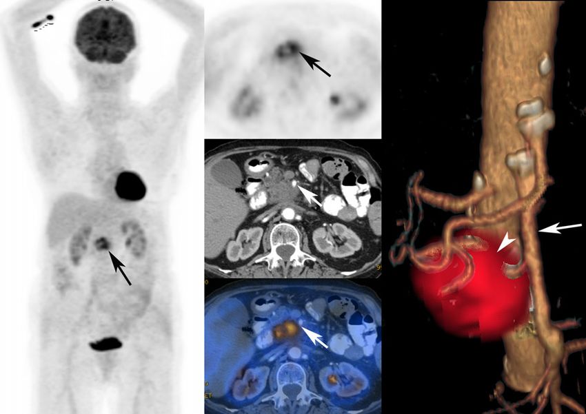
Downloaded from jnm.snmjournals.org by on November 3, 2015. For personal use only.

helical CT scanner (Lightspeed 16; GE Healthcare) and permits           for the presence and nature of lesions with focally increased
the acquisition of coregistered CT and PET images in a single           18F-FDG     uptake. For all patients, the attenuation-corrected PET
session.                                                                images were used for analysis. Lesions were interpreted as
   The patients fasted for at least 4 h before scanning, which          metastases if the uptake was higher than the uptake of the
started approximately 60 min after the injection of 370–400 MBq         surrounding background tissue so that a focal lesion was clearly
of 18F-FDG. All patients were tested for a normal glucose level         depicted. 18F-FDG uptake thought to be physiologic or due to
(range, 80–120 mg/dL [4.4–6.7 mmol/L]) before scanning. Pa-             benign variants such as uptake in muscles or brown fat or uptake
tients with elevated glucose levels were rescheduled, prepared          caused by pulmonary infiltration was considered nonmalignant.
with insulin, and scanned when they had normal glucose levels.          18F-FDG–negative pulmonary nodules without calcifications or

Patients were examined in the supine position. Initially, a low-        fatty content were diagnosed as pulmonary metastases (Fig. 1).
dose CT scan was acquired starting from the level of the head           The enhanced-CT part was analyzed using the established criteria
using the following parameters: 40 mAs, 140 kV, 0.5 s/tube              for the assessment of the primary tumor: vessel involvement
rotation, a slice thickness of 4.25 mm, a scan length of 867 mm,        (.180 of circumferential contiguity of tumor to vessel), organ
and a data acquisition time of 22.5 s. The CT scan was acquired         infiltration, and distant metastases (9,10).
during breath holding in the normal expiratory position. The low-
dose CT data were used for attenuation correction and lesion            Reference Standard
localization, and the images were reconstructed using a standard           The reference standard was intraoperative findings, histologic
iterative algorithm. Immediately after the CT acquisition, a PET        findings, or clinical and imaging follow-up of at least 6 mo if
emission scan was acquired with a time of 3 min per cradle              biopsies were judged too invasive to be performed.
position with a 1-slice overlap in 2-dimensional mode (matrix,
128 · 128). The 8–9 cradle positions starting from the head and         Statistical Analysis
continuing to the knees resulted in an acquisition time                    Data were analyzed on a patient basis using SPSS 15 for
of approximately 24–27 min. Afterward, enhanced CT of the               Windows (SPSS Inc.). Statistical significance was assessed with
abdomen was performed on the same scanner using a dual-phase            the sign test. A P value of less than 0.05 was considered to indi-
pancreatic protocol. The contrast agent (150 mL, Ultravist              cate a significant difference. Bonferroni correction was not pos-
300; Schering) was injected with a power injector at a rate of          sible because of the small number of patients.
3.0–4.0 mL/s through a 21-gauge catheter placed in the antecu-
bital vein. A bolus-tracking program (SmartPrep; GE Healthcare)         RESULTS
was used to monitor contrast enhancement after injection and
                                                                           Fifty consecutive patients (25 women and 25 men; mean
before initiation of the diagnostic scans. The region-of-interest
                                                                        age, 64.3 y; range, 39–84 y) with biopsy-proven adenocar-
cursor for bolus tracking was placed in the aorta at the level of the
diaphragmatic dome. Real-time low-dose serial monitoring scan-          cinoma of the pancreas underwent enhanced 18F-FDG PET/
ning was initiated 5 s after the start of the contrast injection.       CT for evaluation of resectability between April 2004 and
Sections 1.25 mm in nominal thickness were obtained from the            December 2006. All patients had biopsy-proven adenocar-
diaphragm to the inferior part of the duodenum after triggering of      cinoma of the pancreas. Eighteen patients had distant
60 Hounsfield units in the aortic region of interest. After a 70-s      metastases. Of these, metastases were histologically proven
delay, a portovenous phase from the diaphragm to the symphysis          in 10 patients. In 8 patients, metastases were confirmed by
pubis was obtained with a 2.5-mm thickness. In patients for whom        the radiologic appearance, with progression on follow-up
high-quality dual-phase abdominal CT had already been performed         PET/CT or CT scans and clinical investigations including
at an outside institution less than 2 wk before the PET/CT scan,        CA 19-9 measurements. Thirty-five patients underwent
enhanced CT was not repeated and these patients were excluded
                                                                        surgical exploration, of which 28 (56% of the 50 total)
from the study. The acquired images were reviewed with software
                                                                        underwent tumor resection (24 Whipple operations and 4
providing multiplanar reformatted images of PET alone, CT alone,
and fused unenhanced PET/CT and enhanced PET/CT imaged with             left resections) with a curative intent.
linked cursors using a Xeleris workstation (GE Healthcare). PET/           According to the reference standard, 27 patients had
CT was performed according to the recently published Procedure          disease that was not resectable because of distant metasta-
Guideline for Tumor Imaging with 18F-FDG PET/CT 1.0 (8).                ses (n 5 17), peritoneal carcinomatosis (n 5 2), or local
                                                                        infiltration (n 5 5); a combination of peritoneal carcino-
Image Interpretation                                                    matosis and metastasis (n 5 1); or a combination of
   The PET/CT images were analyzed by 2 dual–board-certified            peritoneal carcinomatosis and arterial infiltration (n 5 2).
nuclear radiology physicians with 7 and 3 y of experience in PET/       Twenty-three patients had resectable disease. The discrep-
CT reading, with specialization in tumor staging and abdominal          ancy that 5 patients underwent tumor resection despite
imaging. Images were evaluated by consensus. The only infor-            unresectability regarding the reference standard is ex-
mation the readers had was that the investigation was being done
                                                                        plained by an aggressive surgical approach in unclear
for pancreatic cancer staging. They were unaware of other clinical
                                                                        cases: tumor resection was performed in 2 patients with
information and the results of other imaging modalities (e.g.,
endoscopic ultrasound or MRI). First, the reader interpreted the        unclear lung nodules, which turned out to be lung metas-
PET images alone; in a second step, the reader interpreted the          tases during follow-up; in 1 patient with a liver metastasis
unenhanced PET/CT images; and in a third step, the reader               discovered during the Whipple procedure; and in 2 patients
interpreted the enhanced PET/CT images. There was at least a            with arterial infiltration, 1 of whom underwent arterial
2-wk delay between the readings. The PET images were analyzed           grafting. The sensitivity, specificity, accuracy, NPV, and

                                                         PANCREATIC CANCER STAGING         WITH   PET/CT • Strobel et al.            1409
Downloaded from jnm.snmjournals.org by on November 3, 2015. For personal use only.
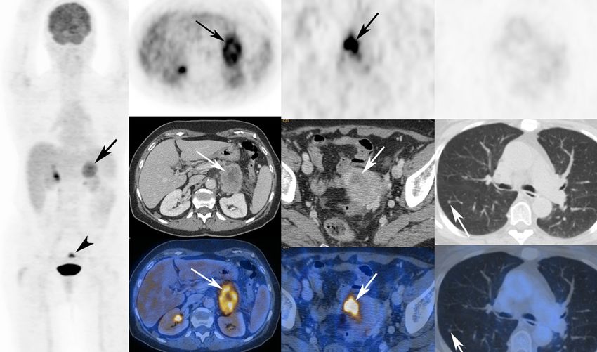
FIGURE 1. A 73-y-old woman with cancer of pancreatic tail. (First column) Primary tumor (arrow) and incidentally found small
uterine cancer (arrowhead) are seen on maximum-intensity projection. (Second column) Primary tumor is seen on axial PET,
enhanced CT, and fused enhanced PET/CT (arrows). (Third column) Incidentally found small uterine cancer is seen on axial PET,
enhanced CT, and fused enhanced PET/CT (arrows). (Fourth column) Small 18F-FDG–negative right lung metastasis is seen (arrows).
PPV of the various imaging methods were calculated on the ses in 9 (82%/97%). In 4 patients, all 3 imaging modalities
basis of the reference standard (Table 1). (PET, unenhanced PET/CT, and enhanced PET/CT) de-
tected the liver metastases. In 2 patients, all imaging
Distant Metastases
modalities failed to detect the liver metastases and they
Liver Metastases. Eleven patients had liver metastases.
were detected intraoperatively. In 1 patient, a liver metas-
In 6 patients, liver metastases were 18F-FDG–negative; in 5
tasis was diagnosed with enhanced PET/CT but was missed
patients, liver metastases were 18F-FDG–positive (mean
intraoperatively. Follow-up imaging clearly showed pro-
maximal standardized uptake value, 5.6; range, 3.8–6.9).
gressive liver metastases in this patient.
PET alone and unenhanced PET/CT detected liver metas-
Lung Metastases. Seven patients had lung metastases. In 6
tases in 5 of these 11 patients (sensitivity, 46%; specificity,
patients, lung metastases were 18F-FDG–negative (mean size,
100%), whereas enhanced PET/CT detected liver metasta-
4.6 mm; range, 3–7 mm). One patient had multiple 18F-FDG–
positive lung metastases (mean maximal standardized uptake
TABLE 1 value, 6.5; maximal size, 20 mm) With PET alone, lung
Value of PET, Unenhanced PET/CT, and Enhanced metastases were diagnosed in only 1 patient (14%/100%), but
PET/CT in Assessment of Overall Resectability all lung metastases were diagnosed with unenhanced PET/CT
in 50 Patients with Pancreatic Cancer and enhanced PET/CT (100%/100%). There was no case of
Unenhanced Enhanced false-positive diagnosis of lung metastases.
Index PET PET/CT PET/CT Bone Metastases. Three patients had bone metastases.
All bone metastases were 18F-FDG–positive (mean maxi-
Sensitivity 100% (23/23) 100% (23/23) 96% (22/23)
Specificity 44% (12/27) 56% (15/27) 82% (22/27)
mal standardized uptake value, 6.2; range, 4.5–6.9). PET,
Accuracy 70% (35/50) 76% (38/50) 88% (44/50) unenhanced PET/CT, and enhanced PET/CT detected the
PPV 61% (23/38) 66% (23/35) 82% (22/27) bone metastases in all 3 patients (100%/100%).
NPV 100% (12/12) 100% (15/15) 96% (22/23)
Peritoneal Carcinomatosis
PPV 5 positive predictive value, NPV 5 negative predictive Five patients had peritoneal carcinomatosis, which was
value.
detected by PET in 1 patient (20%/100%), by unenhanced
1410 THE JOURNAL OF NUCLEAR MEDICINE • Vol. 49 • No. 9 • September 2008Downloaded from jnm.snmjournals.org by on November 3, 2015. For personal use only.
PET/CT in 3 (60%/100%), and by enhanced PET/CT in 4 adjuvant chemotherapy can improve survival after a cura-
(80%/100%). In 1 patient, the peritoneal carcinomatosis tive resection (11). Furthermore, accurate staging is imper-
was diagnosed only intraoperatively. ative for optimal patient selection, which can be improved
by PET/CT, compared with standard staging (7).
Arterial Infiltration Our results show that the 1-stop-shop protocol with
Five patients had arterial infiltration of the celiac trunk or enhanced multislice 18F-FDG PET/CT is feasible and
the superior mesenteric artery. Enhanced PET/CT diag- accurate for preoperative pancreatic cancer staging.
nosed arterial infiltration in all 5 patients (100%/100%). In our patients, the liver was the organ most frequently
PET and unenhanced PET/CT failed to detect arterial affected by metastases, showing distant metastases in 11
infiltration in all 5 cases (0%/100%). patients. Other authors compared PET and CT in detecting
No patient had disease that was unresectable because of liver metastases from pancreatic cancer and described an
infiltration of organs other than the duodenum or stomach. accuracy of about 90% for PET alone, which was compa-
Table 2 summarizes the results for detection of metasta- rable to the accuracy of enhanced CT or ultrasound (12).
ses, peritoneal carcinomatosis, and arterial infiltration. For hepatic metastasis, Diederichs et al. described a PET
sensitivity of 70% and specificity of 95% caused by some
Overall Resectability Assessment metastases smaller than 1 cm being missed by PET (13).
Comparing Accuracy of Different Imaging Modalities. The additional use of contrast material increased the sen-
Although we did not find a significant difference between sitivity for liver metastases from 46% to 82% in our
unenhanced PET/CT and PET alone, a trend toward supe- patients. We also observed 1 patient with a false-positive
riority for enhanced PET/CT over unenhanced PET/CT was diagnosis of liver metastases on enhanced PET/CT. This
found (P 5 0.070). Furthermore, enhanced PET/CT was patient underlines the necessity of not only relying on
significantly superior to PET alone in our analysis (P 5 imaging results but also obtaining histologic confirmation
0.035) (Table 1): 12 patients (24%) with disease judged to of suspected liver metastases or at least performing ade-
be resectable on PET demonstrated local unresectability or quate imaging follow-up in selected cases. In 2 patients, all
distant metastases on enhanced PET/CT. imaging modalities failed to detect small liver metastases,
Despite the improved accuracy of enhanced PET/CT which were discovered during operative exploration to-
over PET, 5 patients (10%) were judged to have resectable gether with intraoperative ultrasound.
disease on enhanced PET/CT but had surgically unresect- It is known that PET alone is not sensitive enough to find
able disease at laparotomy: 2 patients had liver metastases, lung metastases smaller than 1 cm, especially if they are in
2 had infiltration of the mesenteric root, and 1 had perito- the lower parts of the lungs, where respiratory motion
neal carcinomatosis (Table 3). decreases their detectability. Therefore, some authors rec-
Incidental Detection of Simultaneous Other Cancers. ommend additional diagnostic lung CT for tumors that tend
PET/CT also detected simultaneous cancer in 2 patients to metastasize to the lungs, such as sarcomas (14). Our results
(4%). One patient had a non–small cell lung cancer (T1 emphasize that preoperative imaging should include a lung
stage), and the other patient had cancer of the uterus (T1 CT scan and that suggestive pulmonary nodules should be
stage), both of which were successfully resected. biopsied before a pancreatic resection is performed.
PET alone is clearly limited in the detection of peritoneal
DISCUSSION carcinomatosis, especially in cases of diffuse infiltration
Although the morbidity and mortality of pancreas sur- without formation of larger nodules. PET/CT can partially
gery has decreased during recent years, the long-term overcome the limitation of PET alone in diagnosing perito-
outcome of patients with pancreatic cancer remains poor. neal carcinomatosis by showing stranding, peritoneal nod-
This poor outcome is generally attributed to a relatively ules, or ascites. Exploration remains the gold standard for the
chemoresistant disease and undetected metastases at the diagnosis of peritoneal carcinomatosis, considering the lim-
time of surgery (2) A multimodality regimen including ited resolution of all established imaging methods (15,16).
TABLE 2
Value of PET, Unenhanced PET/CT, and Enhanced PET/CT in Detection of Metastases, Peritoneal Carcinomatosis,
and Arterial Infiltration in 50 Patients with Pancreatic Cancer
PET Unenhanced PET/CT Enhanced PET/CT
Site Sensitivity Specificity Sensitivity Specificity Sensitivity Specificity
Liver metastasis 46% (5/11) 100% (39/39) 46% (5/11) 100% (39/39) 82% (9/11) 97% (38/39)
Lung metastasis 14% (1/7) 100% (43/43) 100% (7/7) 100% (43/43) 100% (7/7) 100% (43/43)
Bone metastasis 100% (3/3) 100% (47/47) 100% (3/3) 100% (47/47) 100% (3/3) 100% (47/47)
Peritoneal carcinomatosis 20% (1/5) 100% (45/45) 60% (3/5) 100% (45/45) 80% (4/5) 100% (45/45)
Arterial infiltration 0% (0/5) 100% (45/45) 0% (0/5) 100% (45/45) 100% (5/5) 100% (45/45)
PANCREATIC CANCER STAGING WITH PET/CT • Strobel et al. 1411Downloaded from jnm.snmjournals.org by on November 3, 2015. For personal use only.
TABLE 3
Detection of Reasons for Unresectability with Different Imaging Methods in 27 Patients with Unresectable Disease
Patient Unenhanced Enhanced Operation or
no. Site PET PET/CT PET/CT Intraoperative* intervention
1 Liver met ND ND ND D Diagnostic LT
3 Celiac trunk infiltration ND ND D D Whipple with arterial graft
4 PC ND D D D Palliative GE
5 Lung met ND D D ND Left resection
7 PC, supraclavicular LN met ND D D — FNP supraclavicular LN
8 Liver met D D D — Transcutaneous liver biopsy
9 Bone and lung met Bone met D, D D — —
lung met ND
12 Liver met D D D — Transcutaneous liver biopsy
14 Lung met ND D D — Wedge resection lung
16 Lung met D D D —
17 Liver met D D D D Transcutaneous liver biopsy
18 Liver met D D D —
19 Liver, lung, and bone met Bone met D, Bone and D —
liver and lung met D,
lung met ND liver met ND
20 Liver met ND ND D ND Left resection
21 Celiac trunk infiltration, PC ND ND D — Transcutaneous ascites
puncture
22 PC, celiac trunk infiltration D D D —
23 Celiac trunk infiltration ND ND D —
24 Liver met D D D —
25 Liver met ND ND D D Palliative GE
26 AMS infiltration ND ND D —
27 Liver met ND ND D D Diagnostic LT
28 Lung and bone met Lung met ND, D D —
bone met D
40 Lung met ND D D ND Whipple, lung wedge
2 mo later
41 Liver met ND ND ND D Palliative GE
42 Dorsal infiltration ND ND ND D Whipple
43 Dorsal infiltration ND ND ND D Palliative GE
45 PC ND ND ND D Palliative GE
*Intraoperative during abdominal surgery.
met 5 metastasis or metastases; ND 5 not detected with this method; D 5 detected with this method; LT 5 laparotomy; PC 5
peritoneal carcinomatosis; GE 5 gastroenterotomy; LN 5 lymph node; FNP 5 fine-needle puncture; AMS 5 superior mesenteric artery.
Although infiltration of the portal vein is not generally vessels and the pancreatic tumor in a volume-rendered
considered a contraindication for surgery, infiltration of the 3-dimensional CT angiography/PET combination as dem-
superior mesenteric artery or celiac trunk precludes surgery onstrated in Figure 2.
in most centers. Therefore, the diagnosis of infiltration of In addition to improved staging of pancreatic cancer,
the superior mesenteric artery or celiac trunk preoperatively enhanced PET/CT also detects simultaneous cancers, as
is crucial for surgical planning. Arterial infiltration cannot reported previously (7). These incidental findings affect the
be detected by PET alone or unenhanced PET/CT because oncologic treatment of the patients, since undetected pri-
the vessels cannot be delineated. Thin-section helical CT is mary tumors may metastasize before their detection.
reliable in assessing local resectability, with a sensitivity Because the intravenous contrast protocol is used only
and specificity of 84% and 98%, respectively, if criteria for the abdominal part of the CT study, radiation exposure
such as the .180 circumferential contiguity of tumor to remains acceptable, at approximately 12 mSv. Also, en-
vessel are used (9). The ability to create high-resolution hanced CT is not repeated in our daily routine if high-
2-dimensional and 3-dimensional maximum-intensity pro- quality enhanced CT has been performed recently.
jections and volume-rendered images is an advantage of The higher resectability rate in our population (46%),
the thin-slice multidetector CT technique that has replaced compared with previous publications (10%220%), is pre-
conventional angiography (17,18). The 1-stop-shop en- sumably related to a referral bias and due to the aggres-
hanced-PET/CT approach offers the advantage of clear siveness of the pancreas surgeons at our institution (1): only
visualization of the relationship between the important patients whose disease was deemed resectable on standard
1412 THE JOURNAL OF NUCLEAR MEDICINE • Vol. 49 • No. 9 • September 2008Downloaded from jnm.snmjournals.org by on November 3, 2015. For personal use only.
FIGURE 2. A 72-y-old woman with
cancer of pancreatic head. (First column)
Maximum-intensity-projection image
shows 18F-FDG–active primary (arrow)
without 18F-FDG–active distant metasta-
ses. (Second column) Axial PET, enhanced
CT, and enhanced PET/CT in arterial phase
show that tumor is growing around supe-
rior mesenteric artery (arrows). (Third col-
umn) Software-fused volume-rendered CT
angiography and PET illustrate normal
variant, with additional branch (arrowhead)
from superior mesenteric artery (long ar-
row) supplying liver. Additional branch is
infiltrated by pancreatic-head cancer.
staging were referred for PET/CT; those with clear evi- endoscopic ultrasonography, helical computed tomography, magnetic resonance
imaging, and angiography. Am J Gastroenterol. 2004;99:492–501.
dence of metastatic disease on ultrasound or enhanced CT 7. Heinrich S, Goerres GW, Schafer M, et al. Positron emission tomography/
were not. This circumstance strengthens the value of computed tomography influences on the management of resectable pancreatic
enhanced PET/CT, which revealed mainly previously un- cancer and its cost-effectiveness. Ann Surg. 2005;242:235–243.
8. Delbeke D, Coleman RE, Guiberteau MJ, et al. Procedure guideline for tumor
detected metastases. imaging with 18F-FDG PET/CT 1.0. J Nucl Med. 2006;47:885–895.
Experience with other tracers, such as 18F-fluorothymidine 9. Lu DS, Reber HA, Krasny RM, Kadell BM, Sayre J. Local staging of pancreatic
for pancreatic adenocarcinomas, is limited (19), and com- cancer: criteria for unresectability of major vessels as revealed by pancreatic-
phase, thin-section helical CT. AJR. 1997;168:1439–1443.
parative studies with 18F-FDG are missing. It is known 10. Zamboni GA, Kruskal JB, Vollmer CM, Baptista J, Callery MP, Raptopoulos
that in neuroendocrine pancreatic tumors, alternative tracers VD. Pancreatic adenocarcinoma: value of multidetector CT angiography in
such as 68Ga-DOTA-NOC and 18F-DOPA PET work better preoperative evaluation. Radiology. 2007;245:770–778.
11. Neoptolemos JP, Stocken DD, Friess H, et al. A randomized trial of chemo-
than 18F-FDG (20). radiotherapy and chemotherapy after resection of pancreatic cancer. N Engl J Med.
2004;350:1200–1210.
12. Nakamoto Y, Higashi T, Sakahara H, et al. Contribution of PET in the
CONCLUSION detection of liver metastases from pancreatic tumours. Clin Radiol. 1999;54:
We have demonstrated for, what is to our knowledge, the 248–252.
13. Diederichs CG, Staib L, Vogel J, et al. Values and limitations of 18F-
first time the feasibility and accuracy of a 1-stop-shop fluorodeoxyglucose-positron-emission tomography with preoperative evaluation
imaging approach by combining enhanced CT and PET in a of patients with pancreatic masses. Pancreas. 2000;20:109–116.
single investigation in this analysis of 50 patients with 14. Franzius C, Daldrup-Link HE, Sciuk J, et al. FDG-PET for detection of
pulmonary metastases from malignant primary bone tumors: comparison with
histologically proven pancreatic cancer. Enhanced PET/CT spiral CT. Ann Oncol. 2001;12:479–486.
was significantly superior to PET alone for the preoperative 15. Drieskens O, Stroobants S, Gysen M, Vandenbosch G, Mortelmans L, Vergote I.
assessment of respectability. Positron emission tomography with FDG in the detection of peritoneal and
retroperitoneal metastases of ovarian cancer. Gynecol Obstet Invest. 2003;55:
130–134.
16. Dromain C, Leboulleux S, Auperin A, et al. Staging of peritoneal carcinoma-
REFERENCES
tosis: enhanced CT vs. PET/CT. Abdom Imaging. 2008;33:87–93.
1. Wray CJ, Ahmad SA, Matthews JB, Lowy AM. Surgery for pancreatic cancer: 17. Vargas R, Nino-Murcia M, Trueblood W, Jeffrey RB Jr. MDCT in pancreatic
recent controversies and current practice. Gastroenterology. 2005;128:1626–1641. adenocarcinoma: prediction of vascular invasion and resectability using a
2. Li D, Xie K, Wolff R, Abbruzzese JL. Pancreatic cancer. Lancet. 2004;363: multiphasic technique with curved planar reformations. AJR. 2004;182:419–
1049–1057. 425.
3. Smith SL, Rajan PS. Imaging of pancreatic adenocarcinoma with emphasis on 18. Prokesch RW, Chow LC, Beaulieu CF, et al. Local staging of pancreatic
multidetector CT. Clin Radiol. 2004;59:26–38. carcinoma with multi-detector row CT: use of curved planar reformations—
4. Long EE, Van Dam J, Weinstein S, Jeffrey B, Desser T, Norton JA. Computed initial experience. Radiology. 2002;225:759–765.
tomography, endoscopic, laparoscopic, and intra-operative sonography for 19. Quon A, Chang ST, Chin F, et al. Initial evaluation of 18F-fluorothymidine (FLT)
assessing resectability of pancreatic cancer. Surg Oncol. 2005;14:105–113. PET/CT scanning for primary pancreatic cancer. Eur J Nucl Med Mol Imaging.
5. Palazzo L, Roseau G, Gayet B, et al. Endoscopic ultrasonography in the diagnosis 2008;35:527–531.
and staging of pancreatic adenocarcinoma: results of a prospective study with 20. Ambrosini V, Tomassetti P, Castellucci P, et al. Comparison between 68Ga-
comparison to ultrasonography and CT scan. Endoscopy. 1993;25:143–150. DOTA-NOC and 18F-DOPA PET for the detection of gastro-entero-pancreatic
6. Soriano A, Castells A, Ayuso C, et al. Preoperative staging and tumor and lung neuro-endocrine tumours. Eur J Nucl Med Mol Imaging. April 17, 2008
resectability assessment of pancreatic cancer: prospective study comparing [Epub ahead of print].
PANCREATIC CANCER STAGING WITH PET/CT • Strobel et al. 1413Downloaded from jnm.snmjournals.org by on November 3, 2015. For personal use only. Contrast-Enhanced 18F-FDG PET/CT: 1-Stop-Shop Imaging for Assessing the Resectability of Pancreatic Cancer Klaus Strobel, Stefan Heinrich, Ujwal Bhure, Jan Soyka, Patrick Veit-Haibach, Bernhard C. Pestalozzi, Pierre-Alain Clavien and Thomas F. Hany J Nucl Med. 2008;49:1408-1413. Published online: August 14, 2008. Doi: 10.2967/jnumed.108.051466 This article and updated information are available at: http://jnm.snmjournals.org/content/49/9/1408 Information about reproducing figures, tables, or other portions of this article can be found online at: http://jnm.snmjournals.org/site/misc/permission.xhtml Information about subscriptions to JNM can be found at: http://jnm.snmjournals.org/site/subscriptions/online.xhtml The Journal of Nuclear Medicine is published monthly. SNMMI | Society of Nuclear Medicine and Molecular Imaging 1850 Samuel Morse Drive, Reston, VA 20190. (Print ISSN: 0161-5505, Online ISSN: 2159-662X) © Copyright 2008 SNMMI; all rights reserved.
You can also read