Incidence and Predictors of Sudden Cardiac Arrest in Adults With Congenital Heart Defects Repaired Before Adult Life
←
→
Page content transcription
If your browser does not render page correctly, please read the page content below
Incidence and Predictors of Sudden Cardiac Arrest in Adults With
       Congenital Heart Defects Repaired Before Adult Life
       Pastora Gallego, MD, PhDa,d, Ana Elvira Gonzalez, MDa,b, Angel Sanchez-Recalde, MDa,b,
               Rafael Peinado, MD, PhDb, Luz Polo, MDa,c, Carmen Gomez-Rubin, MDb,
               Jose Luis Lopez-Sendon, MD, PhDb, and Jose Maria Oliver, MD, PhDa,b,*
                  Many adult survivors of repaired congenital heart disease (CHD) are at premature risk of
                  death. Sudden cardiac arrest (SCA) is 1 of the leading causes of death but little is known
                  about determinants for SCA in adults with repaired lesions. We sought to determine
                  incidence and risk factors for SCA in a study population of 936 adults with previously
                  repaired CHD who had completed follow-up at a single tertiary center during a mean
                  period of 9 ⴞ 7 years. Mean age at first examination in our institution was 21 ⴞ 7 years.
                  Diagnostic categories included tetralogy of Fallot (216), coarctation of the aorta (157),
                  transposition complexes (99), single ventricle (55), and other CHD (409). During a total
                  follow-up of 8,387 person-years, 22 patients (2.6 per 1,000 person-years) presented with
                  SCA. Incidence of SCA varied widely between specific lesions; the highest incidence was
                  observed in transposition complexes (10 per 1,000 person-years). Independent predictors of
                  SCA were retrospectively identified using multivariate Cox proportional hazard modeling.
                  Age at initial examination and severely impaired subaortic ventricular systolic function
                  were independent risk factors for SCA (severe subaortic ventricular systolic dysfunction,
                  adjusted hazard ratio 29, 95% confidence interval 11 to 72, p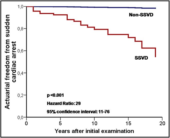
110
Table 1
Patient demographics, data on follow-up, and long-term outcomes in entire cohort and in each congenital heart disease category
Diagnostic Category       Cases (%)     Men               Age (years)               Follow-Up (years)                             SCA                                 Death or Transplantation

                                                      Initial        Last      Mean ⫾ SD      Person-Years      Cases     Incidence/1,000   Age at Sudden    Cases       Incidence/1,000   Age at End

                                                                                                                                                                                                        The American Journal of Cardiology (www.ajconline.org)
                                                   Examination    Follow-Up                                      (%)       Person-Years        Arrest         (%)         Person-Years       Point
                                                       (%)

Tetralogy of Fallot       216 (23%)   112 (52%)      22 ⫾ 9        32 ⫾ 8         10 ⫾ 7          2,127        3 (1%)            1.4          40 ⫾ 17       10 (5%)             4.7          31 ⫾ 11
Aortic coarctation        157 (17%)    87 (55%)      19 ⫾ 7        28 ⫾ 9          9⫾6            1,406        3 (2%)            2.1          35 ⫾ 29        7 (4%)             5            28 ⫾ 18
D-transposition            89 (10%)    56 (63%)      19 ⫾ 4        28 ⫾ 6          9⫾6              838        8 (9%)            9.5          26 ⫾ 10       10 (11%)           12            26 ⫾ 9
Aortic stenosis            85 (9%)     56 (66%)      19 ⫾ 6        30 ⫾ 8         11 ⫾ 8            971           0              0               —           3 (4%)             3.1          30 ⫾ 4
Pulmonic stenosis          85 (9%)     47 (55%)      22 ⫾ 8        30 ⫾ 9          8⫾7              719           0              0            20 ⫾ 0         2 (2%)             2.8          30 ⫾ 4
Atrioventricular septal    72 (8%)     27 (37%)      19 ⫾ 5        27 ⫾ 7          8⫾6              560        1 (1%)            1.8          20 ⫾ 0         2 (3%)             3.6          23 ⫾ 4
  defect
Ventricular septal         68 (7%)     30 (44%)      20 ⫾ 5        28 ⫾ 8          8⫾6             554         2 (3%)            3.6          34 ⫾ 1         2 (3%)             3.6          34 ⫾ 1
  defect
Atrial septal defect       57 (6%)     20 (35%)      21 ⫾ 7        26 ⫾ 8          5⫾5             287            0              0               —              0               0               —
Single ventricle           32 (3%)     14 (44%)      18 ⫾ 3        29 ⫾ 6         11 ⫾ 6           357         1 (3%)            2.8             36          5 (16%)           14            25 ⫾ 9
  (Fontan procedure)
Single ventricle           23 (2%)     10 (44%)      26 ⫾ 9        34 ⫾ 8          8⫾9             185         1 (4%)            5.4             22          3 (13%)           16            32 ⫾ 11
  (shunt/Glenn)
Patent ductus              21 (2%)      3 (14%)      28 ⫾ 14       36 ⫾ 13         8⫾7             159            0              0               —             0                0                —
  arteriosus
Congenitally corrected     10 (1%)      6 (60%)      24 ⫾ 9        32 ⫾ 9          8⫾7               79        2 (20%)           25           39 ⫾ 11        5 (50%)           63            31 ⫾ 9
  transposition of
  great arteries
Miscellaneous             21 (2%)      10 (48%)      21 ⫾ 10       28 ⫾ 11         7⫾5              145        1 (5%)            6.9          23 ⫾ 0         1 (5%)             6.9          23 ⫾ 0
Overall                   936         478 (51%)      21 ⫾ 7        30 ⫾ 8          9⫾7            8,387       22 (2.3%)          2.6          31 ⫾ 14       50 (5.3%)           5.9          29 ⫾ 10
Table 2
Variable distribution by diagnostic category in 936 patients with congenital heart disease repaired before 20 years of age
Congenital Heart Disease/Sudden Death in Adult Congenital Heart Disease
Diagnostic Category NYHA Endocarditis Reoperation SVSD Subaortic Clinical Severe SVR
Class ⬎II Right Sustained Pulmonary
Subaortic Subpulmonary Pulmonary Subpulmonary Subaortic Aorta
Ventricle Arrhythmia Hypertension
AV Valve AV Valve
Tetralogy of Fallot 32 (15%) 8 (4%) 52 (24%) 7 (3%) 34 (16%) 0 35 (16%) 6 (3%) 109 (50%) 25 (12%) 2 (1%) 11 (5%)
Aortic coarctation 11 (7%) 4 (3%) 18 (11%) 4 (3%) 2 (1%) 0 8 (5%) 10 (6%) 1 (1%) 1 (1%) 5 (3%) 9 (6%)
D-transposition 11 (12%) 2 (2%) 12 (13%) 28 (31%) 7 (8%) 74 (83%) 37 (42%) 2 (2%) 2 (2%) 4 (4%) 12 (13%) 4 (4%)
Aortic stenosis 5 (6%) 4 (5%) 13 (15%) 1 (1%) 0 0 4 (5%) 1 (1%) 0 1 (1%) 2 (2%) 23 (27%)
Pulmonic stenosis 9 (11%) 0 13 (15%) 0 8 (9%) 0 9 (11%) 2 (2%) 25 (29%) 8 (9%) 1 (1%) 1 (1%)
Atrioventricular septal 6 (8%) 1 (1%) 6 (8%) 3 (4%) 1 (1%) 0 10 (14%) 5 (7%) 1 (1%) 3 (4%) 18 (25%) 4 (6%)
defect
Ventricular septal 0 4 (6%) 3 (4%) 2 (3%) 0 0 5 (7%) 1 (1%) 0 1 (1%) 1 (1%) 5 (7%)
defect
Atrial septal defect 1 (2%) 0 1 (2%) 0 1 (2%) 0 3 (5%) 0 0 0 1 (2%) 0
Single ventricle 16 (50%) 0 11 (34%) 6 (19%) 0 3 (9%) 17 (53%) 0 0 0 4 (12%) 0
(Fontan procedure)
Single ventricle 14 (61%) 2 (9%) 8 (35%) 8 (35%) 8 (35%) 4 (17%) 15 (65%) 6 (26%) 2 (9%) 3 (13%) 2 (12%) 0
(shunt/Glenn)
Patent ductus 1 (5%) 0 2 (10%) 2 (10%) 0 0 1 (5%) 1 (5%) 0 0 0 1 (5%)
arteriosus
Congenitally corrected 4 (40%) 0 3 (30%) 7 (70%) 0 10 (100%) 7 (70%) 1 (10%) 0 2 (20%) 5 (50%) 1 (10%)
transposition of
great arteries
Miscellaneous 2 (10%) 0 3 (14%) 1 (5%) 1 (5%) 0 7 (33%) 1 (1%) 2 (10%) 3 (14%) 3 (14%) 0
Overall 111 (12%) 25 (2.7%) 144 (15%) 69 (7.4%) 62 (6.6%) 91 (10%) 158 (17%) 35 (3.7%) 142 (15%) 51 (5.4%) 55 (5.9%) 59 (6.3%)
AV ⫽ atrioventricular; NYHA ⫽ New York Heart Association; SVR ⫽ severe valve regurgitation; SVSD ⫽ severe ventricular systolic dysfunction.
111112 The American Journal of Cardiology (www.ajconline.org)
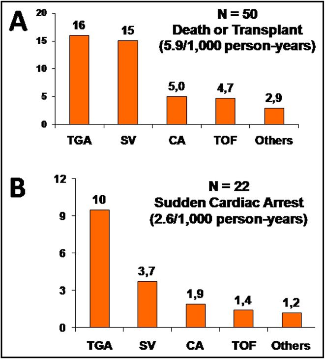
Figure 2. Incidence of all-cause of death or transplantation (A) and sudden
cardiac arrest (B) for each diagnostic category. Data are presented as
number of events per 1,000 person-years. CA ⫽ coarctation of aorta;
SV ⫽ single-ventricle physiology.
agnosis was the lesion considered the indication for the
Figure 1. Cause of death in 50 deceased patients by lesion (percentages): initial surgical approach.
perioperative death or death within 30 days after cardiac surgery; heart Details of demographic characteristics, clinical status,
failure or death from progressive symptoms of fluid congestion or de- Doppler echocardiography, cardiac magnetic resonance im-
creased cardiac output; other cardiovascular (CV) deaths included aortic
aging, and cardiac catheterization were abstracted from pa-
rupture, endocarditis, acute myocardial infarction, pulmonary embolism, or
stroke.
tients’ clinical records. The most recent data preceding the
end points were requested. Evaluation of systolic ventricu-
lar function was based on M-mode and 2-dimensional echo-
Senning, Mustard, Rastelli, and/or Jatene procedures; (4) cardiographic measurements and expert visual assessment
surgical or percutaneous relief of valvular, discrete subval- supplemented by objective measurements including ventric-
var, and supravalvar aortic stenosis; (5) repaired obstructive ular diameters, fractional shortening, tricuspid or mitral
right ventricular outflow tract lesions after surgical or bal- annular plane systolic excursion, and tissue Doppler param-
loon valvulotomy or patch reconstruction; (6) surgically eters. Because ventricular function in the morphologic right
repaired atrioventricular septal defect (including primum ventricle and for single-ventricle physiology may be as-
atrial septal defect); (7) surgically repaired ventricular sep- sessed inaccurately by echocardiography, for the previous
tal defect; (8) repaired ostium secundum or sinus venosus 10 years those patients with significantly decreased subaor-
defect; (9) anatomic or functional single-ventricle physiol- tic or subpulmonary right ventricular systolic function un-
ogy after Fontan palliation; (10) single-ventricle physiology derwent cardiac magnetic resonance whenever possible to
after aortopulmonary shunts or Glenn shunt palliation; (11) confirm the severity of ventricular dysfunction. Subaortic
patent ductus arteriosus repaired by surgery or catheteriza- and subpulmonary systolic ventricular functions were visu-
tion; and (12) congenitally corrected TGA after surgical ally graded by echocardiography in 4 degrees: (1) normal
repair of a ventricular septal defect, left ventricular outflow (ejection fraction ⱖ55%), (2) borderline to mild (ejection
tract obstructive lesions, or left atrioventricular valve dys- fraction 45% to 54%), (3) moderate (ejection fraction 35%
function. Diagnoses that did not belong in 1 of these 12 to 44%), and (4) severe (ejection fraction ⬍35%) dysfunc-
diagnostic categories such as total pulmonary venous anom- tion. Consistent with recently modified recommendations
alous connection or coronary anomalies were categorized as for implantable cardioverter– defibrillator (ICD) implanta-
miscellaneous. If ⬎1 diagnostic category applied, the he- tion in heart failure,15 an ejection fraction ⬍35% was used
modynamically most important lesion for each patient re- in this study to define severe impairment.
garding long-term outcome was classified as the principal For univariate and multivariate models, the following
diagnosis. If ⬎1 lesion was considered to have a major variables were assessed: (1) gender; (2) age at first exami-
impact on morbidity, the driver to assign the principal di- nation in our adult CHD clinic; (3) age at last examinationCongenital Heart Disease/Sudden Death in Adult Congenital Heart Disease 113
Table 3
Proportional distribution of variables and diagnostic categories according to outcomes (sudden cardiac arrest, death or heart transplantation, or survivors
free of transplantation)
Variables SCA All-Cause Death or Transplantation Survival Free of Transplantation
(n ⫽ 22) (n ⫽ 50) (n ⫽ 886)
Age at first examination (years) 24 ⫾ 14 22 ⫾ 9 21 ⫾ 7
Men 15 (68%) 28 (56%) 450 (51%)
New York Heart Association class ⬎II (%) 7 (32%)† 27 (54%)* 84 (9%)
Left ventricular outflow tract obstruction 3 (14%) 10 (20%) 232 (26%)
Tetralogy of Fallot 3 (14%) 10 (20%) 206 (23%)
Systemic-to-pulmonary shunt 3 (14%) 4 (8%)† 214 (24%)
Transposition of great arteries 10 (45%)* 15 (30%)* 84 (9%)
Single-ventricle physiology 2 (9%) 8 (16%)† 47 (5%)
Severe subaortic ventricular systolic dysfunction 16 (73%)* 28 (56%)* 41 (5%)
Severe subpulmonary ventricular systolic dysfunction 5 (23%)† 12 (24%)* 50 (6%)
Morphologic subaortic right ventricle 9 (41%)* 14 (28%)* 77 (9%)
Clinical sustained arrhythmias 14 (64%)* 23 (46%)* 135 (15%)
Severe pulmonary hypertension 3 (14%)† 10 (20%)* 25 (3%)
Severe pulmonary valve regurgitation 3 (14%) 8 (16%) 134 (15%)
Severe subpulmonary atrioventricular valve regurgitation 2 (9%) 3 (6%) 48 (5%)
Severe subaortic atrioventricular valve regurgitation 3 (14%) 9 (18%)* 47 (5%)
Severe aortic valve regurgitation 0 4 (8%) 55 (6%)
Infective endocarditis 0 2 (4%) 23 (3%)
Need for reintervention 5 (23%) 21 (42%)* 123 (14%)
* p ⬍0.001; † p ⬍0.01 compared to survival free of transplantation.
or at end-point event; (4) time from first visit to any end or believed to be arrhythmic in nature. Aborted cardiac
point or to last visit; (5) New York Heart Association arrest included successful cardiopulmonary resuscitation of
functional classes I to II or ⬎II at the last examination; (6) a cardiovascular collapse resulting from ventricular fibril-
clinically sustained arrhythmias such as supraventricular or lation and documented appropriate ICD discharge delivered
ventricular tachycardia, atrioventricular block, or sinus node in response to polymorphic ventricular tachycardia or ven-
disease that required pacemaker implantation (nonsustained tricular fibrillation. A secondary composite end point in-
arrhythmias captured from Holter monitoring or data on cluded all-cause of death and heart or heart–lung transplan-
ambient ectopy were not considered in analysis); (7) infec- tation.
tive endocarditis during adult follow-up; (8) need for sur- SPSS 15.0 for Windows (SPSS, Inc., Chicago, Illinois)
gical or percutaneous reintervention during follow-up; (9) was used for statistical analyses. Quantitative values are
severely impaired systolic function of the subaortic ventri- summarized as mean ⫾ SD. Categorical variables are pre-
cle at last cardiac imaging study; (10) severely impaired sented as percentage. For purposes of the analyses, age and
subpulmonary ventricular function at last cardiac imaging time computed since the first visit at our clinic were con-
study; (11) subaortic right ventricle (single or biventricular sidered continuous variables and all other variables were
physiology); (12) severe pulmonary hypertension defined as considered categorical. Patients fulfilling inclusion criteria
a pulmonary-to-systemic systolic pressure ratio ⬎50% on were retrospectively included in the analysis at the time of
cardiac catheterization or Doppler evaluation; (13) severe their first visit at the adult CHD clinic during the study
pulmonary regurgitation assessed by echocardiography or period. Person-years were accrued from time of entry until
cardiac magnetic resonance; (14) severe aortic regurgitation an end point or until the last follow-up visit before study
assessed by echocardiography or cardiac magnetic reso- termination. Prevalence was presented as percentage and
nance; (15) severe subaortic atrioventricular valve regurgi- defined as total number of events that occurred during total
tation assessed qualitatively by Doppler echocardiography; follow-up period divided by total number of cases or by
and (16) severe subpulmonary atrioventricular valve regur- number of patients in each diagnostic category. Incidence
gitation assessed qualitatively by Doppler echocardiogra- was measured by relating the number of new events to
phy. Diagnostic categories were also entered into the mod- person-years at risk and calculated by summing the periods
els. during which patients in the total population or in each
According to previous publications,2,4 cause of death diagnostic category were at risk during the observation
was classified as sudden death, death secondary to heart period. It was represented by events per 1,000 person-years.
failure, perioperative death, other cardiovascular death, or Comparison of variables between groups was performed
noncardiovascular death. Other causes of cardiovascular with Mann–Whitney U test. To assess predictors for SCA in
death included aortic rupture, endocarditis, acute myocar- longitudinal analysis, univariate analyses of individual vari-
dial infarction, pulmonary embolism, and stroke. The pri- ables were performed using Cox proportional hazards mod-
mary end point was SCA defined as the combined end point els from which hazard ratios and 95% confidence intervals
of sudden death or aborted cardiac arrest. Sudden death was were generated. To determine independent risk factors, vari-
defined as death within 1 hour of symptom onset and proved ables with a p value ⬍0.2 in univariate analysis were en-114 The American Journal of Cardiology (www.ajconline.org)
Table 4
Univariate and multivariate Cox proportional models for survival free from sudden cardiac arrest
Analysis Variable HR 95% CI p Value
Univariate age at first examination 1.07 1.0–1.1 ⬍0.001
gender 0.5 0.2–1.2 0.14
New York Heart Association class ⬎II 2.9 1.2–7.2 0.02
left ventricular outflow tract obstruction 0.4 0.1–1.3 0.14
tetralogy of Fallot 0.5 0.1–1.6 0.22
systemic-to-pulmonary shunt 0.7 0.2–2.3 0.55
transposition 6.8 2.9–16 ⬍0.001
single-ventricle physiology 1.4 0.3–6.2 0.62
severe subaortic ventricular systolic dysfunction 33 13–84 ⬍0.001
severe subpulmonary ventricular systolic dysfunction 3.9 1.4–10 0.008
morphologic subaortic right ventricle 5.9 2.5–14 ⬍0.001
clinical sustained arrhythmias 7.6 3.2–18 ⬍0.001
severe pulmonary hypertension 3.9 1.2–13 0.03
severe pulmonary valve regurgitation 0.8 0.2–2.7 0.70
severe subpulmonary atrioventricular valve regurgitation 1.7 0.4–7.3 0.48
severe subaortic atrioventricular valve regurgitation 2.5 0.8–8.6 0.13
severe aortic valve regurgitation 0.4 0–32 0.35
infective endocarditis 0.0 0.0 0.57
need for reintervention 1,3 0.5–3.5 0.60
Multivariate severe subaortic ventricular systolic dysfunction 29 11–76 ⬍0.001
age at first examination 1.05 1.0–1.1 0.019
CI ⫽ confidence interval; HR ⫽ hazard ratio.
tered into the multivariate Cox proportional model (forward
stepwise, p ⫽ 0.05 for entry and p ⫽ 0.1 for removal).
Two-tailed p values ⬍0.05 were considered statistically
significant.
Results
In total 936 patients (73%) were eligible for inclusion
and comprised the study population. Distribution of diag-
nostic categories and duration of follow-up for the entire
sample and separately by CHD category are presented in
Table 1. Although the mean follow-up period ranged from
5 ⫾ 5 years in patients with atrial septal defects to 11 ⫾ 8
years in patients with repaired aortic stenosis, differences in
follow-up were not statistically significant between diagnos-
tic categories. Further details on patient characteristics are
presented in Table 2. It should be noted that 83% of patients
with TGA repaired by a Mustard/Senning procedure plus 10
patients with congenitally corrected TGA and 7 of 55 pa- Figure 3. Multivariate Cox proportional model comparing actuarial free-
dom from sudden cardiac arrest in patients with severe systemic ventricular
tients with a single ventricle had morphologic right ventri-
dysfunction (SSVD) versus patients with nonsevere subaortic ventricular
cles supporting their systemic circulation. dysfunction (non-SSVD).
During a total follow-up period of 20 years (8,387 per-
son-years) 50 patients died or underwent transplantation.
SCA occurred in 22 patients: 15 patients developed sudden
death and 5 patients were successfully resuscitated after In another 22 patients, nonsudden cardiac death occurred
SCA and subsequently received an ICD for secondary pre- from perioperative death (9 patients), heart failure (8 pa-
vention. In addition, 11 patients underwent ICD implanta- tients), and other cardiovascular causes (5 patients). Nnon-
tion for primary prevention (7 patients) or secondary pre- cardiovascular death occurred in 2 patients and 4 patients
vention (4 patients). Of these 11 patients, appropriate ICD underwent a heart or heart–lung transplantation at follow-
shocks for ventricular fibrillation were recorded in 2 pa- up. After excluding aborted sudden death and transplanta-
tients; consistent with our definition of the primary end tion, overall mortality was 4.2%, incidence of death was 4.7
point, these patients were included in the SCA subgroup. per 1,000 person-years, and primary cause of death was
Thus, prevalence and incidence of SCA in the entire sample SCA (38%) followed by reoperation (23%), heart failure
of repaired CHD were 2.4% and 2.6 per 1,000 person-years, (21%), and other cardiovascular causes (13%). Causes of
respectively. death by lesions are shown in Figure 1.Congenital Heart Disease/Sudden Death in Adult Congenital Heart Disease 115
Table 5 aortic systolic ventricular dysfunction as a risk factor for
Comparative data on mortality between patients with and those without SCA in patients with CHD. Although left ventricular sys-
severe subaortic ventricular systolic dysfunction tolic dysfunction is considered a powerful prognostic factor
Cause of Death SSVSD p and an indication for ICD therapy in adults with ischemic or
Value dilated cardiomyopathy,21,22 this statement requires eluci-
Yes No
(n ⫽ 69) (n ⫽ 867)
dation in patients with CHD. Although the retrospective
study design prevented the inclusion of other potential risk
Sudden cardiac arrest 16 (23%) 6 (0.7%) ⬍0.001 factors, in our series severely impaired subaortic (left, right,
Incidence/1,000 person-years 26 0.8 ⬍0.001 or single) systolic ventricular function was a multivariate
Total death or transplantation 28 (41%) 22 (2.5%) ⬍0.001 determinant for SCA among the proposed predictors.
Incidence/1,000 person-years 45 2.8 ⬍0.001
In our study, risk for late SCA varied widely between
Sudden cardiac arrest/total death 57% 27% 0.047
ratio
lesions. Almost 80% of cases occurred in patients with
TGA, single-ventricle physiology, coarctation of aorta, or
SSVSD ⫽ severe subaortic ventricular systolic dysfunction. TOF. Interestingly, there appeared to be a relation between
the incidence of SCA and the development of subaortic
Figure 2 illustrates the incidence of the composite end ventricular systolic dysfunction between different lesions.
point of all-cause death or transplantation and of SCA by The highest incidence was found in patients with transpo-
diagnostic category. Prevalence and incidence varied sition complexes, most of which had been repaired using
widely by the specific lesion (Table 1). The highest Mustard/Senning procedures that result in a subaortic mor-
prevalence of SCA was observed in TGA complexes: 10 phologic right ventricle. The long-term prognosis of pa-
of 99 patients (10%) presented with SCA at follow-up, tients after atrial switch procedures is a topic of concern and
resulting in an incidence of SCA in combined TGA and a late decrease of systemic right ventricular function is a
congenitally corrected TGA of 10 per 1,000 person-years well-recognized outcome in TGA after Mustard/Senning
(Figure 2). procedures and congenitally corrected TGA.10 –12,16
Table 3 presents the proportional distribution of pro- The total mortality of our population with single-ventri-
posed risk factors according to outcomes in the entire cle physiology including patients after Fontan surgery and
cohort. Univariate and multivariate predictors of SCA are cyanotic patients with or without pulmonary hypertension
listed in Table 4. Independent factors associated with palliated with surgical shunts was also high. However, there
SCA among the proposed variables were age at first was a relatively smaller proportion of SCA deaths in pa-
examination and severe subaortic ventricular systolic tients with a single ventricle. Despite the high incidence of
dysfunction (adjusted hazard ratio 29, 95% confidence arrhythmias and advanced functional capacity impairment,
interval 11 to 72; Figure 3). Comparative data on mor- the primary causes of death were heart failure and periop-
tality in patients with severe versus nonsevere subaortic erative mortality. Although our patients with a single ven-
ventricular systolic dysfunction are presented in Table 5. tricle had developed some degree of ventricular dysfunc-
Interestingly, annual risks of SCA in patients with se- tion, severely impaired ventricular systolic function was
verely impaired subaortic ventricular systolic function relatively uncommon, which is consistent with previously
were 2.6% per year in the total CHD population, 3.0% reported data on the prevalence of ventricular dysfunction in
per year for patients with subaortic right ventricular sys- this population.23,24
tolic dysfunction, and 3.1% per year for patients with In accordance with previously published studies,1,2 we
TGA and severely decreased subaortic ventricular sys- observed the third highest incidence of SCA in patients with
tolic function. repaired coarctation of the aorta. Left ventricular systolic
function impairment without clinical evidence of coronary
Discussion artery disease was also the primary risk factor for SCA in
those studies. Finally, a strong association between left
To our knowledge, this is the first longitudinal cohort ventricular dysfunction and sudden death late after repair of
study to analyze predictors of SCA in a large population of the TOF has recently been reported.19,20,25 Because left
adult survivors after operation for CHD at ⬍20 years of age. ventricular dysfunction is rather less common than right
Results indicated that severe systemic ventricular dysfunc- ventricular function impairment in repaired TOF,26 this
tion has an outstanding role as a dominant multivariate might explain the relatively low incidence of SCA of our
predictor of SCA for all combined diagnoses in this heter- study.
ogenous population. There is controversy as to whether patients with CHD
Until recently, limited data existed on risk factors for and severe subaortic ventricular systolic dysfunction should
SCA during long-term follow-up after surgery in the total undergo prophylactic ICD implantation.27,28 To date, the
adult CHD population. Despite a multitude of predictors of clinical decision-making process for risk stratification in
ventricular arrhythmia and/or SCA in studies that focused primary prevention of SCA integrates a multitude of pre-
on patients with TOF8,9 and those who had undergone atrial dictors, but no risk factor had been individually demon-
switch procedures for TGA,10 –12 their predictive values are strated to possess high discriminative power to prompt ICD
relatively low. Observational and registry studies in TGA consideration. The present study provides evidence that
with Mustard/Senning repair,10 –13 congenitally corrected advanced subaortic ventricular systolic dysfunction has an
TGA,16 left heart obstructive lesions,17,18 and TOF14,19,20 outstanding role as an independent risk factor for SCA in
have provided some support for considering advanced sub- the postoperative adult CHD population seeking care at an116 The American Journal of Cardiology (www.ajconline.org)
adult clinic. Based on results of multicenter and major and mortality after the Mustard procedure: a 30-year single-center
randomized clinical trials, Khairy29 provided a probabilistic experience. J Am Coll Cardiol 1997;29:194 –201.
11. Kammeraad JA, van Deurzen CH, Sreeram N, Bink-Boelkens MT,
approach to risk stratification in patients with TOF. Of Ottenkamp J, Helbing WA, Lam J, Sobotka-Plojhar MA, Daniels O,
studies that reported a mortality decrease, the benefit of ICD Balaji S. Predictors of sudden cardiac death after Mustard or Senning
has been demonstrated if the baseline population risk for repair for transposition of the great arteries. J Am Coll Cardiol 2004;
SCA is ⬎3.5% per year. The findings in our study on the 44:1095–1102.
12. Schwerzmann M, Salehian O, Harris L, Siu SC, Williams WG,
annual risk of SCA for the total population and for patients Webb GD, Colman JM, Redington A, Silversides CK. Ventricular
with TGA or for patients with subaortic morphologic right arrhythmias and sudden death in adults after a Mustard operation
ventricle indicate that subaortic ventricular systolic dys- for transposition of the great arteries. Eur Heart J 2009;30:1873–
function may be a more important predictor for SCA than 1879.
ventricular structure or type of lesion. Therefore, severe 13. Gatzoulis MA, Walters J, McLaughlin PR, Merchant N, Webb GD,
Liu P. Late arrhythmia in adults with the Mustard procedure for
subaortic ventricular dysfunction could be ideally suited for transposition of great arteries: a surrogate marker for right ventricular
initial risk stratification in our CHD population. dysfunction? Heart 2000;84:409 – 415.
This study has limitations inherent to any retrospective 14. Ghai A, Silversides C, Harris L, Webb GD, Siu SC, Therrien J. Left
cohort study including the fact that only 73% patients had ventricular dysfunction is a risk factor for sudden cardiac death in
adults with repaired tetralogy of Fallot. J Am Coll Cardiol 2002;40:
complete follow-up. Because most excluded patients had 675– 680.
been examined at the adult CHD unit ⱖ1 time and presented 15. Jessup M, Abraham WT, Casey DE, Feldman AM, Francis GS, Gani-
with simple CHD, this bias likely does not have a major ats TG, Konstam MA, Mancini DM, Rahko PS, Silver MA, Stevenson
impact on risk factor analysis. Evaluation of ventricular LW, Yancy CW. 2009 Focused update: ACCF/AHA guidelines for the
function by echocardiography also poses a challenge for diagnosis and management of heart failure in adults: a report of the
American College of Cardiology Foundation/American Heart Associ-
risk stratification in our retrospective study. Unfortunately, ation Task Force on Practice Guidelines: developed in collaboration
most episodes of SCA occurred during the first decade of with the International Society for Heart and Lung Transplantation.
follow-up when cardiac magnetic resonance was not fully Circulation 2009;119:1977–2016.
available at our institution. However, when possible, refer- 16. Connelly MS, Liu PP, Williams WG, Webb GD, Robertson P,
McLaughlin PR. Congenitally corrected transposition of the great
ence was made to cardiac magnetic resonance estimates of arteries in the adult: functional status and complications. J Am Coll
ventricular function, which provides a more accurate assess- Cardiol 1996;27:1238 –1243.
ment of morphologically right or single ventricular func- 17. Toro-Salazar OH, Steinberger J, Thomas W, Rocchini AP, Carpenter
tion. The definition of SCA might be controversial,30 but B, Moller JH. Long-term follow-up of patients after coarctation of the
aborted sudden death was also considered a primary end aorta repair. Am J Cardiol 2002;89:541–547.
18. Keane JF, Driscoll DJ, Gersony WM, Hayes CJ, Kidd L, O’Fallon
point for providing data on the actual incidence of SCA. WM, Pieroni DR, Wolfe RR, Weidman WH. Second natural history
study of congenital heart defects. Results of treatment of patients with
1. Silka MJ, Hardy BG, Menashe VD, Morris CD. A population-based aortic valvar stenosis. Circulation 1993;87(suppl):I16 –I27.
prospective evaluation of risk of sudden cardiac death after operation 19. Khairy P, Aboulhosn J, Gurvitz MZ, Opotowsky AR, Mongeon FP,
for common congenital heart defects. J Am Coll Cardiol 1998;32:245– Kay J, Valente AM, Earing MG, Lui G, Gersony DR, Cook S, Ting G,
251. Nickolaus MJ, Webb G, Landzberg MJ, Broberg CS; Alliance for
2. Oechslin EN, Harrison DA, Connelly MS, Webb GD, Siu SC. Mode of Adult Research in Congenital Cardiology (AARCC). Arrhythmia bur-
death in adults with congenital heart disease. Am J Cardiol 2000;86: den in adults with surgically repaired tetralogy of Fallot: a multi-
1111–1116. institutional study. Circulation 2010;122:868 – 875.
3. Engelfriet P, Boersma E, Oechslin E, Tijssen J, Gatzoulis MA, Thilén 20. Yap SC, Roos-Hesselink JW, Hoendermis ES, Budts W, Vliegen HW,
U, Kaemmerer H, Moons P, Meijboom F, Popelová J, Laforest V, Mulder BJ, van Dijk AP, Schalij MJ, Drenthen W. Outcome of im-
Hirsch R, Daliento L, Thaulow E, Mulder B. The spectrum of adult plantable cardioverter defibrillators in adults with congenital heart
congenital heart disease in Europe: morbidity and mortality in a 5 year disease: a multi-centre study. Eur Heart J 2007;28:1854 –1861.
follow-up period. The Euro Heart Survey on adult congenital heart 21. Moss AJ, Zareba W, Hall WJ, Klein H, Wilber DJ, Cannom DS,
disease. Eur Heart J 2005;26:2325–2333. Daubert JP, Higgins SL, Brown MW, Andrews ML; Multicenter
4. Nieminen HP, Jokinen EV, Sairanen HI. Causes of late deaths after Automatic Defibrillator Implantation Trial II Investigators. Prophy-
pediatric cardiac surgery: a population-based study. J Am Coll Cardiol lactic implantation of a defibrillator in patients with myocardial
2007;50:1263–1271. infarction and reduced ejection fraction. N Engl J Med 2002;346:
5. Verheugt CL, Uiterwaal CS, van der Velde ET, Meijboom FJ, Pieper 877– 883.
PG, van Dijk AP, Vliegen HW, Grobbee DE, Mulder BJ. Mortality in 22. Bardy GH, Lee KL, Mark DB, Poole JE, Packer DL, Boineau R,
adult congenital heart disease. Eur Heart J 2010;31:1220 –1229. Domanski M, Troutman C, Anderson J, Johnson G, McNulty SE,
6. Harrison DA, Connelly M, Harris L, Luk C, Webb GD, McLaughlin Clapp-Channing N, Davidson-Ray LD, Fraulo ES, Fishbein DP,
PR. Sudden cardiac death in the adult with congenital heart disease. Luceri RM, Ip JH; Sudden Cardiac Death in Heart Failure Trial
Can J Cardiol 1996;12:1161–1163. (SCD-HeFT) Investigators. Amiodarone or an implantable cardio-
7. Gatzoulis MA, Till JA, Somerville J, Redington AN. Mechanoelectri- verter-defibrillator for congestive heart failure. N Engl J Med 2005;
cal interaction in tetralogy of Fallot. QRS prolongation relates to right 352:225–237.
ventricular size and predicts malignant ventricular arrhythmias and 23. Khairy P, Fernandes SM, Mayer JE Jr, Triedman JK, Walsh EP, Lock
sudden death. Circulation 1995;92:231–237. JE, Landzberg MJ. Long-term survival, modes of death, and predictors
8. Gatzoulis MA, Balaji S, Webber SA, Siu SC, Hokanson JS, Poile C, of mortality in patients with Fontan surgery. Circulation 2008;117:
Rosenthal M, Nakazawa M, Moller JH, Gillette PC, Webb GD, Red- 85–92.
ington AN. Risk factors for arrhythmia and sudden cardiac death late 24. Norozi K, Wessel A, Alpers V, Arnhold JO, Geyer S, Zoege M,
after repair of tetralogy of Fallot: a multicentre study. Lancet 2000; Buchhorn R. Incidence and risk distribution of heart failure in adoles-
356:975–981. cents and adults with congenital heart disease after cardiac surgery.
9. Nollert GD, Däbritz SH, Schmoeckel M, Vicol C, Reichart B. Risk Am J Cardiol 2006;97:1238 –1243.
factors for sudden death after repair of tetralogy of Fallot. Ann Thorac 25. Khairy P, Harris L, Landzberg MJ, Viswanathan S, Barlow A,
Surg 2003;76:1901–1905. Gatzoulis MA, Fernandes SM, Beauchesne L, Therrien J, Chetaille
10. Gelatt M, Hamilton RM, McCrindle BW, Connelly M, Davis A, Harris P, Gordon E, Vonder Muhll I, Cecchin F. Implantable cardioverter-
L, Gow RM, Williams WG, Trusler GA, Freedom RM. Arrhythmia defibrillators in tetralogy of Fallot. Circulation 2008;117:363–370.Congenital Heart Disease/Sudden Death in Adult Congenital Heart Disease 117
26. Broberg CS, Aboulhosn J, Mongeon FP, Kay J, Valente AM, Khairy lactic implantation of an ICD? Implantable cardioverter defibrillator
P, Earing MG, Opotowsky AR, Lui G, Gersony DR, Cook S, Ting JG, implantation guidelines based solely on left ventricular ejection
Webb G, Gurvitz MZ; Alliance for Adult Research in Congenital fraction do not apply to adults with congenital heart disease. Circ
Cardiology (AARCC). Prevalence of left ventricular systolic dysfunc- Arrhythm Electrophysiol 2008;1:307–316.
tion in adults with repaired tetralogy of Fallot. Am J Cardiol 2011; 29. Khairy P. Programmed ventricular stimulation for risk stratification in
107:1215–1220. patients with tetralogy of Fallot. A bayesian perspective. Nat Clin
27. Silka MJ, Bar-Cohen Y. Should patients with congenital heart disease Practice Cardiovasc Med 2007;4:292–293.
and a systemic ventricular ejection fraction less than 30% undergo 30. Ellenbogen KA, Levine JH, Berger RD, Daubert JP, Winters SL,
prophylactic implantation of an ICD? Patients with congenital heart Greenstein E, Shalaby A, Schaechter A, Subacius H, Kadish A;
disease and a systemic ventricular ejection fraction less than 30% Defibrillators in Non-Ischemic Cardiomyopathy Treatment Evalu-
should undergo prophylactic implantation of an implantable cardio- ation (DEFINITE) Investigators. Are implantable cardioverter de-
verter defibrillator. Circ Arrhythm Electrophysiol 2008;1:298 –306. fibrillator shocks a surrogate for sudden cardiac death in patients
28. Triedman JK. Should patients with congenital heart disease and a with nonischemic cardiomyopathy? Circulation 2006;113:776 –
systemic ventricular ejection fraction less than 30% undergo prophy- 782.You can also read