Lysophosphatidylcholine aggravates contact hypersensitivity by promoting neutrophil infiltration and IL17 expression - Korea Science
←
→
Page content transcription
If your browser does not render page correctly, please read the page content below
BMB Rep. 2021; 54(4): 203-208
BMB www.bmbreports.org
Reports
Lysophosphatidylcholine aggravates contact hypersensitivity by
promoting neutrophil infiltration and IL17 expression
Mi Hye Song , Anupriya Gupta , Hye One Kim & Kwonik Oh *
1 1 2 1,3,
1
Department of Pathology, Hallym University College of Medicine, Chuncheon 24252, 2Department of Dermatology, Kangnam Sacred
Heart Hospital, Hallym University College of Medicine, Seoul 07441, 3Institute of Medical Science, Hallym University College of Medicine,
Chuncheon 24252, Korea
Lysophosphatidylcholine (LPC) is a bioactive lysolipid known in the pathology of CHS, other immune cells, such as neutro-
to contribute to the development of lung allergic diseases. How- phils (3), innate lymphoid cells (4), T cells (5, 6), NKT cells
ever, it remains unknown whether LPC possesses proinflamma- (7) and even B cells (8), can be involved.
tory properties in the skin as well. Here, we investigated this LPC is a bioactive lysolipid produced by the cell-membrane
issue by injection of LPC into the murine contact hypersensiti- metabolism by hydrolyzing phosphatidylcholine by phospholipase
vity (CHS) model induced by 2,4-dinitrofluorobenzene (DNFB). A2 (PLA2) in the Lands cycle (9). It can be divided into short-
LPC increased the expression of IL17, recruited more neutrophils, chain LPC or saturated/unsaturated LPC according to the acyl-chain
and eventually aggravated the CHS in the skins. Moreover, the length and degree of saturation (10). The levels of LPC can also
effects of LPC diminished after neutralizing IL17 or depleting be increased by lipid metabolism by means of the secretory
neutrophils. Mechanistically, LPC upregulated not only IL17 but PLA2 or the oxidative modification of lipoprotein phospholipids
also CXCL1 and CXCL2 in a G2A-dependent manner. Taken under inflammatory conditions (11, 12). In asthmatic patients (13)
together, our study demonstrated that the upregulation of LPC and animal models with lung injury (14), the concentrations of
could contribute to allergic skin inflammation by increasing LPC were elevated in the lung fluids, accompanied by the
IL17 expression and neutrophil recruitment via G2A receptor. increased activity of PLA2. Further study showed that the cell
[BMB Reports 2021; 54(4): 203-208] population responsible for the increase of LPC was the bron-
chial epithelial cells (15) and that LPC played a critical role in
airway inflammation (16). LPC has various biological functions,
INTRODUCTION including inflammatory responses, oxidative stress, and apoptosis
(10), and the effects of LPC on immune cells are diverse, including
Contact hypersensitivity (CHS) is a representative type of T-cell- increased bactericidal activity of neutrophils (17, 18), phagosome
mediated skin inflammation and also a murine model of human maturation of macrophages (19), integrin–mediated adhesion
allergic contact dermatitis (1, 2). As in other T-cell-mediated of eosinophils (20), activation of NKT cells (21, 22), and immune
inflammatory models, CHS is divided into two distinct stages: regulation (23). Altogether, these reports suggest that LPC can
sensitization and elicitation. During sensitization, haptens applied be upregulated under inflammatory conditions and play diverse
to abdominal skin are taken up by antigen-presenting cells, roles, depending on environments.
such as Langerhans cells and dermal dendritic cells, and bind Recently, it was reported that the proportion of short-chain
to and modify self-antigens; then the modified self-antigens sensi- LPC (C16 or C18) was increased significantly in atopic dermatitis,
+ +
tize CD4 and CD8 T cells. After 5 to 7 days, we induced an which is accompanied by downregulation of fatty-acid elongation
elicitation response by application of the same haptens to the enzymes, ELOVL3/ELOVL6 (24). Although these findings provide
ear, to induce skin swelling; the extent of swelling was correlated insight about how the expression of short-chain LPC is regulated,
with the severity (1). Although T-cell responses are predominant they did not show whether the ‘increase of short-chain LPC’
contributes to the development of skin inflammation.
Here, we examined the role of LPC in skin allergic inflamma-
*Corresponding author. Tel: +82-33-238-2564; Fax: +82-33-251-8250;
tion in the DNFB-induced CHS model. We directly injected
E-mail: kwonik@hallym.ac.kr
the short-chain LPC (C18:0) into the CHS mice and monitored
https://doi.org/10.5483/BMBRep.2021.54.4.193 the severity of the skin inflammation. Interestingly, LPC aggra-
vated CHS by upregulating IL17 and CXCL1/2 and recruiting
Received 8 September 2020, Revised 29 September 2020, more neutrophils in a G2A-dependent manner.
Accepted 4 November 2020
Keywords: Cytokine, Inflammation, Lysophosphatidylcholine, Neutro-
phil, Skin
ISSN: 1976-670X (electronic edition)
Copyright ⓒ 2021 by the The Korean Society for Biochemistry and Molecular Biology
This is an open-access article distributed under the terms of the Creative Commons Attribution Non-Commercial License (http://creativecommons.org/li-
censes/by-nc/4.0) which permits unrestricted non-commercial use, distribution, and reproduction in any medium, provided the original work is properly cited.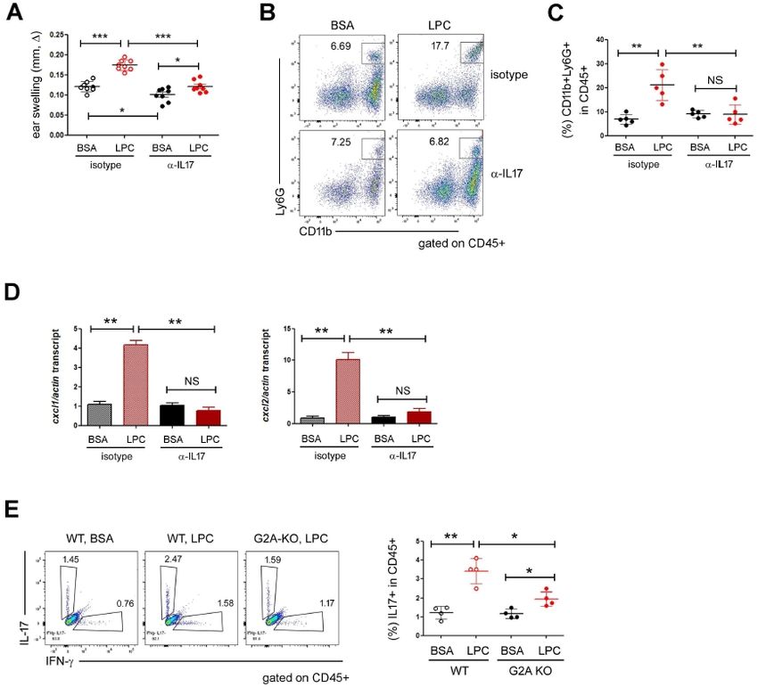
The inflammatory roles of LPC in CHS
Mi Hye Song, et al.
RESULTS AND DISCUSSION BSA (vehicle of LPC). Ears treated with DNFB swelled signifi-
cantly at d7-8 and the swelling was aggravated by LPC (Fig. 1B
LPC aggravates DNFB-induced skin inflammation and 1C).
To investigate the roles of LPC in skin inflammation, we decided
to use the DNFB-induced CHS model mimicking human allergic More neutrophils were recruited into the ear skin by LPC
dermatitis. We applied a high dose of DNFB to the abdominal Since the DNFB-induced skin inflammation is carried out by
skin two times at d0 and d1 for sensitization and challenged immune cells, we analyzed the phenotypes of immune cells in
+
the ear with a low dose of DNFB at d5 (Fig. 1A). On the next the skins by using flow cytometry. The percentage of total CD45
day, we measured the ear thickness every day as a readout of cells in the ear skins was increased significantly by DNFB, but
the skin inflammation. During the period of the experiment, we was not changed further by LPC treatment (Fig. 1D, left). Next,
injected LPC into the test-group mice every day. We also pre- we analyzed the adaptive immune-cell populations, such as
+ + +
pared three different control groups: the first one we treated CD4 helper, CD8 cytotoxic, and Foxp3 regulatory T cells,
with acetone (vehicle of DNFB) only; the second we sensitized but did not find any significant difference between BSA and
with acetone and challenged it with DNFB to assess the ear LPC (data not shown). Instead, the percentage of an innate cell
+ +
swelling induced by nonspecific irritation. Last, we sensitized population, like neutrophils (CD11b Ly6G ; Fig. 1D, right),
+ +
the third group, challenged it with DNFB, but treated it with but not macrophages (CD11b F4/80 ; Fig. 1D, middle), were
increased by LPC. Since LPC had been reported to promote
the activity of neutrophils, as in H2O2 production (17), we
speculated that LPC could aggravate skin inflammations by
increasing neutrophil infiltration. However, it was reported that
LPC was not chemotactic for neutrophils in vitro (25), prompting
us to analyze the expression of chemokines in the skin. Consis-
tent with the assumption, the expressions of neutrophil chemo-
kines, such as CXCL1 and CXCL2, were increased significantly
by DNFB and even further after DNFB plus LPC treatment (Fig.
1E). Next, we depleted neutrophils by using anti-Ly6G monoclonal
antibody (mAb) and repeated the DNFB-induced CHS experi-
ments (Supplementary Fig. 1). Interestingly, LPC failed to increase
skin inflammation when neutrophilswere depleted (Fig. 1F),
suggesting that LPC could control the recruitment of neutrophils
in vivo by regulating chemokine expression, which aggravates
skin inflammation.
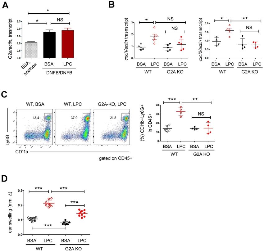
G2A is essential for the effects of LPC on CHS
Although G2A (G2 Accumulation, an orphan G protein coupled
Fig. 1. LPC aggravated DNFB-induced skin inflammation. (A) Experi-
receptor) is not a bona fide receptor of LPC, many functions of
mental design. We sensitized WT mice with 0.5% DNFB on d0 and LPC depend on G2A, which led us to investigate the roles of
d1, and then challenged them with 0.2% DNFB on d5. During the G2A in CHS. The expression of G2A increased significantly in
whole period, we injected LPC s.c. into the mice. The extent of CHS DNFB-treated ears but was not changed further by LPC (Fig.
is shown as the increase of the ear thickness (ear swelling, ), which
we calculated by subtracting the ear thickness of the treated mice
2A). Next, we sought to investigate whether G2A contributes
(DNFB sensitization and DNFB challenge) from that of the control to LPC-induced chemokine expressions in the skin. The expres-
mice (acetone sensitization and DNFB challenge: representing the non- sion of CXCL2 of skin tissues was similar in both WT and G2A
specific irritation). (B) Ear thickness results over time. We pooled data KO before LPC treatment. However, CXCL2 was significantly
from five independent experiments. The difference of ear swelling
between BSA and LPC was statistically significant on d7 and d8. (C)
upregulated in WT but not in KO after LPC treatment (Fig. 2B,
Ear thickness results on d7. We pooled data; each circle represents right). In the case of CXCL1, we also observed similar trends,
a single mouse. (D) FACS analysis of the ear skin. The percentages although the difference between WT and G2A KOwas not
of hematopoietic cells (CD45+, left), macrophages (CD11b+F4/80+, statistically significant (Fig. 2B, left). Consistent with the chemo-
middle) and neutrophils (CD11b+Ly6G+, right) are shown. We pooled
data; each circle represents a single mouse. (E) RT-qPCR analysis
kine expression results, in contrast to WT mice, the neutrophil
on the expressions of CXCL1 and CXCL2 in ear skin. We pooled data; infiltration into the ear skin was not increased by LPC
each circle represents a single mouse. (F) We treated naïve mice treatment in G2A KO mice (Fig. 2C). We also monitored the
with anti-Ly6G mAb to deplete neutrophils and then sensitized and extent of ear swelling and found that although the ears of both
challenged them with DNFB. The ear thickness results are shown. Data
are representative of two independent experiments, and each circle
WT and G2A KO mice swelled after LPC treatment, the ears
represents a single mouse. Data are presented as the mean ± SD. were swollen less in G2A KO mice (Fig. 2D). Interestingly, the
NS, not significant; *P < 0.05; **P < 0.01; ***P < 0.001. ears of G2A KO mice swelled less than did those of WT even
204 BMB Reports http://bmbreports.org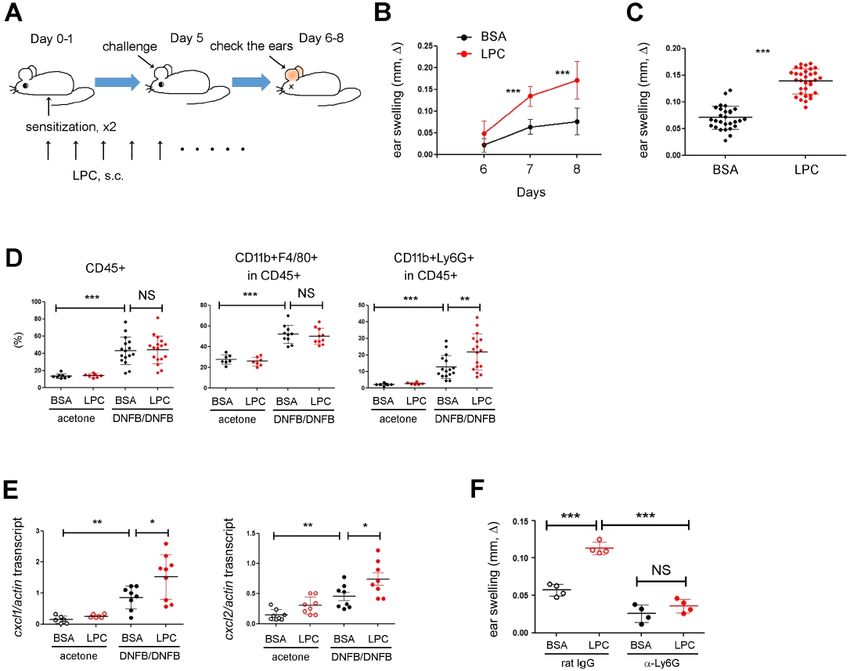
The inflammatory roles of LPC in CHS
Mi Hye Song, et al.
Fig. 3. LPC upregulated IL17. (A) The FACS analysis of IL17 and IFN-
in CD45+ cells of the ears. Statistical analysis is shown in the right.
We pooled data; each circle represents a single mouse. (B) RT-qPCR
analysis on the expressions of IL17 and IFN-. We subjected the
whole skin tissues to RNA extraction and used them for RT-qPCR
study. Data are presented as the mean ± SD. NS, not significant;
*P < 0.05; **P < 0.01; ***P < 0.001.
CHS experiments in lymphocyte-deficient RAG-1 KO mice to
Fig. 2. G2A mediated the effects of LPC on neutrophils. (A) RT-qPCR confirm the role of T cells. RAG-1 KO mice developed the much-
analysis on the expressions of G2A. We subjected the whole ear skin
tissues to RNA extraction and used it for RT-qPCR study. Data are milder CHS, as shown previously (27), and the ear swelling of
representative of two independent experiments. (B) RT-qPCR analysis RAG-1 KO mice was not increased significantly by LPC (Supple-
on the expressions of CXCL1 and CXCL2 in ear skin. We pooled mentary Fig. 2B). Moreover, the percentage of IL17-expressing
data; each circle represents a single mouse. (C) FACS analysis of cells was pretty low in RAG-1 KO mice treated with DNFB
the ear skins of WT and G2A KO mice in DNFB-induced CHS.
The percentages of neutrophils (CD11b+Ly6G+) in CD45+ popula- plus BSA andwas not increased by LPC (Supplementary Fig.
tion are shown. Statistical analysis is shown in the right panel. We 2C), indicating that LPC upregulated IL17 indirectly in T cells.
pooled data; each circle represents a single mouse. (D) Ear thickness
results of WT and G2A KO mice. Statistical analysis is shown. We The effects of LPC on CHS is mediated by IL17
pooled data; each circle represents a single mouse. Data are presented
as the mean ± SD. NS, not significant; *P < 0.05; **P < 0.01; IL17 is a signature cytokine in type 3 inflammation where neutro-
***P < 0.001. phils play an important role. We realized in this study that LPC
upregulated IL17 (Fig. 3A), recruited neutrophils more efficiently
(Fig. 1D), and exacerbated DNFB-induced skin inflammation
when stimulated with DNFB plus BSA (Fig. 2D), implying that (Fig. 1C). These findings led us to assume that LPC could recruit
G2A might play non-redundant roles in the DNFB-induced skin neutrophils by using IL17, which aggravated skin inflammation.
inflammation quite apart from exogenous LPC stimulation (26). To confirm the above hypothesis, we neutralized IL17 cytokine
Altogether, our study demonstrated that LPC aggravated the by using anti-IL17 mAb and did the CHS experiments. In mice
DNFB-induced skin inflammation in a G2A-dependent manner. treated with DNFB plus BSA, IL17 neutralization reduced the
ear swelling slightly (Fig. 4A) but did not prevent the neutro-
IL17 was upregulated by LPC treatment phil accumulation (Fig. 4B and 4C). However, the effects of
Next, we sought to investigate the expressions of IFN- and LPC on skin inflammation were dramatically reduced by IL17
IL17 as signature cytokines of T-cell-mediated inflammations. neutralization. The ears treated with anti-IL17 mAb did not
Interestingly, the percentage of IL17-expressing cells increased swell as much as did those treated with isotype control mAb in
in LPC-treated ear skins (Fig. 3A). We also observed similar the presence of LPC (Fig. 4A). Furthermore, IL17 neutralization
results in RT-qPCR analysis (Fig. 3B). The subsequent FACS abolished the neutrophil recruitment (Fig 4B and 4C) and the
analysis revealed that the majority of IL17-expressing cells upregulation of CXCL1/2 (Fig. 4D) induced by LPC, implying
+
were TCR cells. In contrast, TCR cellswere the major that the inflammatory effects of LPC depend on IL17.
cellular sources of IFN- (Supplementary Fig. 2A). To check Last, we investigated whether the upregulation of IL17 induced
whether LPC is directly involved in the regulation of IL17 by LPC also depended on G2A and found that the expression
+
expression in T cells, we sought to culture naïve CD4 T and of IL17 did not increase in G2A KO mice as much as in WT in
+
TCR cells in the presence of LPC under T helper type 17 the presence of LPC stimulation (Fig. 4E).
(TH17) conditions and checked the expression of IL17. Unex- In this study, we investigated the effect of LPC in a gain-of-
pectedly, IL17 was not upregulated by LPC (data not shown), function approach and found that LPC upregulated CXCL1 and
which caused concern that there might be another type of CXCL2 (Fig. 1E), recruited neutrophils (Fig. 1D), and aggravated
immune cell, such as innate lymphoid cells, not T cells, that DNFB-induced CHS (Fig. 1C). Moreover, once neutrophils were
produce IL17 in respond to LPC. Therefore, we repeated the depleted, LPC did not cause the ear swelling anymore (Fig.
http://bmbreports.org BMB Reports 205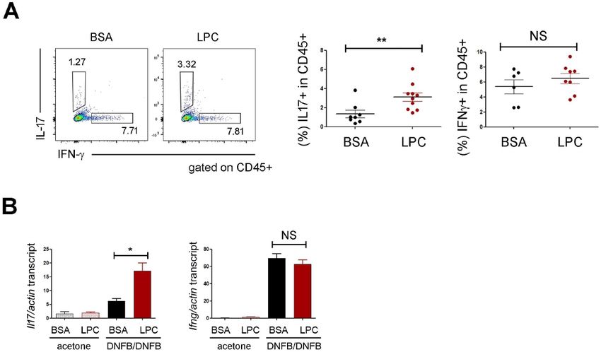
The inflammatory roles of LPC in CHS
Mi Hye Song, et al.
intriguing that G2A KO mice developed less-severe CHS even
in the absence of LPC treatment. Given that diverse G2A ligands
including LPC and oxidized fatty acids (35) are available in
skin, these findings imply the importance of G2A and its lipid
ligands in skin homeostasis.
In conclusion, we demonstrated that the upregulation of LPC
could exacerbate allergic skin inflammation by increasing IL17
expression and neutrophil recruitment via G2A receptor. Further
study on LPC and G2A would help our understanding of the
roles of lipid metabolites in skin immunology.
MATERIALS AND METHODS
Mice
We purchased WT C57BL/6 mice from Koatech (Pyeongtaek,
Korea). The G2A knockout (KO) mice (36) on the C57BL/6 back-
ground, as we described previously, we received from Dr. DK
Song (Hallym University). We did all animal experiments in
accordance with guidelines and approval of the International
Animal Care and Use Committees of Hallym University (Hallym
Fig. 4. LPC exacerbated CHS in an IL17-dependent manner. (A) Ear 2018-9, 2019-18).
thickness results after anti-IL17 mAb treatment. We pooled data; each
circle represents a single mouse. (B, C) FACS analysis of the ear skin
after anti-IL17 mAb treatment. The percentages of neutrophils (CD11b+ Sensitization and elicitation of CHS
Ly6G+) in CD45+ population are shown in (B). (D) RT-qPCR analysis We did the induction of CHS in mice as described previously
on the expressions of CXCL1 (left) and CXCL2 (right). We subjected (37, 38). The extent of CHS was shown as the increase of the
the whole skin tissues to RNA extraction and used them for RT-qPCR
study. Data are representative of two independent experiments. (E) ear thickness (ear swelling), which we calculated by subtrac-
FACS analysis on the expressions of IL17 and IFN-in the WT and ting the ear thickness of the treated mice from that of the
G2A KO skins. The frequency of the cytokine-expressing cells in control mice (mice challenged with DNFB (Sigma Korea, Seoul,
the CD45+ population is shown. Statistical analysis is shown in the Korea) without sensitization), which we measured every 24 h
right panel. Data are representative of two independent experiments
and each circle represents one mouse. Data are presented as the after challenging them using a micrometer (Mitutoyo, Kanagawa,
mean ± SD. NS, not significant; *P < 0.05; **P < 0.01; ***P < Japan). We injected LPC (18:0 Lyso PC, 1-stearoyl-2-hydroxy-sn-
0.001. glycero-3-phosphocholine, Avanti Polar Lipids, Alabaster, AL)
or 2% BSA (vehicle of LPC, Sigma) subcutaneously for the whole
period of the experiments.
1F). These findings clearly indicated that neutrophils recruited
by LPC and CXCL1/2 exacerbated CHS. Then, how was CXCL1/2 Tissue preparation and flow cytometry
upregulated by LPC? Based on our finding that IL17was upre- We removed ears, split them in half, and cut them into small
gulated by LPC (Fig. 3) and the previous reports that IL17 drives pieces. We treated skin tissues in RPMI media containing 0.1
neutrophil infiltration by inducing the expression of neutrophil- mg/ml DNase I and 0.1 mg/ml Liberase TL (Sigma) for 2 h at
o
attracting chemokines such as CXCL1/2 (28-31), we hypothesized 37 C. We filtered digested tissues with a 70-m cell strainer
that LPC upregulated IL17, which subsequently increased the (SPL, Seoul, Korea). For cytokine analysis, we cultured cells for
CXCL1/2 expressions and neutrophil infiltration. This hypothe- 4 h in the presence of PMA/ionomycin plus monensin (BD
siswas supported by the findings of our IL17 neutralization Biosciences, San Jose, CA) before intracellular cytokine staining
experiments (Fig. 4). However, we failed to identify the detailed unless otherwise specified. We acquired data by means of FACS
mechanisms by which LPC upregulated IL17, which need further Canto-II (BD Biosciences) and analyzed the data with FlowJo
study. software (BD Biosciences).
The putative LPC receptor is the G protein coupled receptors,
G2A. Although G2A KO mice developed a late-onset autoimmune Quantitative RT-PCR (RT-qPCR)
disease that looked like Systemic Lupus Erythematosus (32), We isolated RNA using the RNeasy Mini kit (Qiagen, Germantown,
recent studies have shown evidence that G2A can work as MD) or Trizol (Thermo Fisher Scientific Korea, Seoul, Korea),
both pro- and anti-inflammatory mediators (26, 33, 34). Here, and reverse-transcribed it into cDNA using QuantiTect Reverse
we demonstrated that LPC exacerbated CHS in a G2A-depen- Transcription kit (Qiagen). We normalized all data to actin. We
dent manner (Fig. 2D), suggesting the proinflammatory roles of checked non-specific amplification by the use of melting curves
LPC and G2A in skin inflammations. Particularly, it was and agarose gel electrophoresis (39). The sequences of primers
206 BMB Reports http://bmbreports.orgThe inflammatory roles of LPC in CHS
Mi Hye Song, et al.
(Genotech, Daejon, Korea) are as follows. Il17a forward, 5’-AC hypersensitivity in BALB/c mice. J Invest Dermatol 134,
TACCTCAACCGTTCCACGTC-3’; Il17a reverse, 5’-ATGTGGTGG 2709-2718
TCCAGCTTTCC-3’; Ifng forward, 5’-GATGCATTCATGAGTATT 8. Kim HS, Lee MB, Lee D et al (2019) The regulatory B
GCCAAGT-3’; Ifng reverse, 5’-GTGGACCACTCGGATGAGCTC- cell-mediated peripheral tolerance maintained by mast
3’; Cxcl1 forward, 5’-TGAGCTGCGCTGTCAGTGCCT-3’; Cxcl1 cell IL-5 suppresses oxazolone-induced contact hypersensi-
tivity. Sci Adv 5, eaav8152
reverse, 5’-AGAAGCCAGCGTTCACCAGA-3’; Cxcl2 forward, 5’-
9. Law SH, Chan ML, Marathe GK, Parveen F, Chen CH and
GAGCTTGAGTGTGACGCCCCCAGG-3’; Cxcl2 reverse, 5’-GTT Ke LY (2019) An updated review of lysophosphatidylcho-
AGCCTTGCCTTTGTTCAGTATC-3’; G2a forward, 5’-AAGTGT line metabolism in human diseases. Int J Mol Sci 20, 1149
CCAGAATCCACACAGGGT-3’; G2a reverse, 5’-AGTAAACCTA 10. Liu P, Zhu W, Chen C et al (2020) The mechanisms of
GCTTCGCTGGCTGT-3’; actin forward, 5’-CATCCGTAAAGACC lysophosphatidylcholine in the development of diseases.
TCTATGCCAAC-3’; actin reverse, 5’-ATGGAGCCACCGATCCA Life Sci 247, 117443
CA-3’. 11. Fuchs B, Schiller J, Wagner U, Hantzschel H and Arnold
K (2005) The phosphatidylcholine/lysophosphatidylcholine
Statistical analyses ratio in human plasma is an indicator of the severity of
rheumatoid arthritis: investigations by 31P NMR and MALDI-
We used a two-tailed, unpaired, student’s t-test to calculate the
TOF MS. Clin Biochem 38, 925-933
statistical significance of differences between groups unless 12. Kabarowski JH (2009) G2A and LPC: regulatory functions
specified. We represented P values as follows: ***P < 0.001; in immunity. Prostaglandins Other Lipid Mediat 89, 73-81
**P < 0.01; *P < 0.05, whereas we used NS, not significant, 13. Yoder M, Zhuge Y, Yuan Y et al (2014) Bioactive lysophos-
to denote P > 0.05. Error bars indicate s.d. phatidylcholine 16:0 and 18:0 are elevated in lungs of
asthmatic subjects. Allergy Asthma Immunol Res 6, 61-65
ACKNOWLEDGEMENTS 14. Arbibe L, Koumanov K, Vial D et al (1998) Generation of
lyso-phospholipids from surfactant in acute lung injury is
This work was supported by NRF of Korea (2018R1D1A1B070 mediated by type-II phospholipase A2 and inhibited by a
direct surfactant protein A-phospholipase A2 protein interac-
4642813), Hallym University (H20160646) and Korea Healthcare
tion. J Clin Invest 102, 1152-1160
R&D project (HI17C0597). 15. Zhuge Y, Yuan Y, van Breemen R et al (2014) Stimulated
bronchial epithelial cells release bioactive lysophosphatidyl-
CONFLICTS OF INTEREST choline 16:0, 18:0, and 18:1. Allergy Asthma Immunol Res
6, 66-74
The authors have no conflicting interests. 16. Bansal P, Gaur SN and Arora N (2016) Lysophosphatidyl-
choline plays critical role in allergic airway disease mani-
festation. Sci Rep 6, 27430
REFERENCES 17. Yan JJ, Jung JS, Lee JE et al (2004) Therapeutic effects of
lysophosphatidylcholine in experimental sepsis. Nat Med
1. Kaplan DH, Igyarto BZ and Gaspari AA (2012) Early immune 10, 161-167
events in the induction of allergic contact dermatitis. Nat 18. Hong CW, Kim TK, Ham HY et al (2010) Lysophos-
Rev Immunol 12, 114-124 phatidylcholine increases neutrophil bactericidal activity
2. Honda T, Egawa G, Grabbe S and Kabashima K (2013) by increasement of azurophil granule-phagosome fusion
Update of immune events in the murine contact hyper- via glycine. GlyR alpha 2/TRPM2/p38 MAPK signaling. J
sensitivity model: toward the understanding of allergic Immunol 184, 4401-4413
contact dermatitis. J Invest Dermatol 133, 303-315 19. Lee HJ, Ko HJ, Song DK and Jung YJ (2018) Lysophos-
3. Weber FC, Nemeth T, Csepregi JZ et al (2015) Neutrophils phatidylcholine promotes phagosome maturation and regu-
are required for both the sensitization and elicitation lates inflammatory mediator production by means of the
phase of contact hypersensitivity. J Exp Med 212, 15-22 protein kinase A-phosphatidylinositol 3 kinase-p38 mitogen-
4. Rafei-Shamsabadi DA, van de Poel S, Dorn B et al (2018) activated protein kinase signaling pathway during myco-
Lack of type 2 innate lymphoid cells promotes a type bacterium tuberculosis infection in mouse macrophages.
I-driven increased immune response in contact hyper- Front Immunol 9, 920
sensitivity. J Invest Dermatol 138, 1962-1972 20. Zhu X, Learoyd J, Butt S et al (2007) Regulation of eosino-
5. Jiang X, Park CO, Geddes Sweeney J, Yoo MJ, Gaide O phil adhesion by lysophosphatidylcholine via a non-store-
and Kupper TS (2017) Dermal gammadelta T cells do not operated Ca2+ channel. Am J Respir Cell Mol Biol 36,
freely re-circulate out of skin and produce IL-17 to pro- 585-593
mote neutrophil infiltration during primary contact hyper- 21. Fox LM, Cox DG, Lockridge JL et al (2009) Recognition of
sensitivity. PLoS One 12, e0169397 lyso-phospholipids by human natural killer T lymphocytes.
6. Nielsen MM, Lovato P, MacLeod AS et al (2014) IL-1beta- PLoS Biol 7, e1000228
dependent activation of dendritic epidermal T cells in 22. Maricic I, Girardi E, Zajonc DM and Kumar V (2014)
contact hypersensitivity. J Immunol 192, 2975-2983 Recognition of lysophosphatidylcholine by type II NKT
7. Shimizuhira C, Otsuka A, Honda T et al (2014) Natural cells and protection from an inflammatory liver disease. J
killer T cells are essential for the development of contact Immunol 193, 4580-4589
http://bmbreports.org BMB Reports 207The inflammatory roles of LPC in CHS
Mi Hye Song, et al.
23. Hasegawa H, Lei J, Matsumoto T, Onishi S, Suemori K duced by innate lymphoid cells is essential for intestinal
and Yasukawa M (2011) Lysophosphatidylcholine increases ischemia-reperfusion injury. J Immunol 199, 2921-2929
the suppressive function of human naturally occurring 32. Le LQ, Kabarowski JH, Weng Z et al (2001) Mice lacking
regulatory T cells by means of TGF-beta production. Biochem the orphan G protein-coupled receptor G2A develop a
Biophys Res Commun 415, 526-531 late-onset autoimmune syndrome. Immunity 14, 561-571
24. Berdyshev E, Goleva E, Bronova I et al (2018) Lipid abnor- 33. Kern K, Schafer SMG, Cohnen J et al (2018) The G2A
malities in atopic skin are driven by type 2 cytokines. JCI receptor controls polarization of macrophage by deter-
Insight 3, e98006 mining their localization within the inflamed tissue. Front
25. Quinn MT, Parthasarathy S and Steinberg D (1988) Lyso- Immunol 9, 2261
phosphatidylcholine: a chemotactic factor for human mono- 34. Frasch SC, McNamee EN, Kominsky D et al (2016) G2A
cytes and its potential role in atherogenesis. Proc Natl Acad signaling dampens colitic inflammation via production of
Sci U S A 85, 2805-2809 IFN-gamma. J Immunol 197, 1425-1434
26. Hattori T, Obinata H, Ogawa A et al (2008) G2A plays pro- 35. Obinata H and Izumi T (2009) G2A as a receptor for oxidized
inflammatory roles in human keratinocytes under oxidative free fatty acids. Prostaglandins Other Lipid Mediat 89, 66-
stress as a receptor for 9-hydroxyoctadecadienoic acid. J 72
Invest Dermatol 128, 1123-1133 36. Li HM, Jang JH, Jung JS et al (2019) G2A protects mice
27. Paust S, Gill HS, Wang BZ et al (2010) Critical role for the against sepsis by modulating kupffer cell activation: coo-
chemokine receptor CXCR6 in NK cell-mediated antigen- perativity with adenosine receptor 2b. J Immunol 202,
specific memory of haptens and viruses. Nat Immunol 11, 527-538
1127-1135 37. Engeman T, Gorbachev AV, Kish DD and Fairchild RL
28. Ye P, Rodriguez FH, Kanaly S et al (2001) Requirement of (2004) The intensity of neutrophil infiltration controls the
interleukin 17 receptor signaling for lung CXC chemokine number of antigen-primed CD8 T cells recruited into cuta-
and granulocyte colony-stimulating factor expression, neutro- neous antigen challenge sites. J Leukoc Biol 76, 941-949
phil recruitment, and host defense. J Exp Med 194, 519-527 38. He D, Wu L, Kim HK, Li H, Elmets CA and Xu H (2009)
29. Miyamoto M, Prause O, Sjostrand M, Laan M, Lotvall J IL-17 and IFN-gamma mediate the elicitation of contact
and Linden A (2003) Endogenous IL-17 as a mediator of hypersensitivity responses by different mechanisms and
neutrophil recruitment caused by endotoxin exposure in both are required for optimal responses. J Immunol 183,
mouse airways. J Immunol 170, 4665-4672 1463-1470
30. Sun D, Novotny M, Bulek K, Liu C, Li X and Hamilton T 39. Kim G, Jeong H, Youn H et al (2020) Anti-inflammatory
(2011) Treatment with IL-17 prolongs the half-life of chemo- mechanisms of suppressors of cytokine signaling target
kine CXCL1 mRNA via the adaptor TRAF5 and the splicing- ROS via NRF-2/thioredoxin induction and inflammasome
regulatory factor SF2 (ASF). Nat Immunol 12, 853-860 activation in macrophages. BMB Rep 53, 640-645
31. Geha M, Tsokos MG, Bosse RE et al (2017) IL-17A pro-
208 BMB Reports http://bmbreports.orgYou can also read