Expression of ADAMTS13 and PCNA in the Placentas of Gestational Diabetic Mothers
←
→
Page content transcription
If your browser does not render page correctly, please read the page content below
Int. J. Morphol.,
39(1):38-44, 2021.

                       Expression of ADAMTS13 and PCNA in the
                         Placentas of Gestational Diabetic Mothers

           Expresión de ADAMTS13 y PCNA en las Placentas de Madres Diabéticas Gestacionales

                                                   Süleyman Cemil Oglak1 & Mehmet Obut1

OGLAK, S. C. & OBUT, M. Expression of ADAMTS13 and PCNA in the placentas of gestational diabetic mothers. Int. J. Morphol.,
39(1):38-44, 2021.

        SUMMARY: GDM is linked with overexpression of inflammatory cytokines and increased oxidative stress, leading to endothelial
dysfunction and vascular disorder. Weaimed to examine the expression of ADAMTS13 and PCNA in the placentas of gestational diabe-
tes mellitus (GDM) patients to investigate the effects of hypoxia, induced by GDM, on proliferation and extracellular matrix formation
in the maternal and fetal placenta cells. A total of 60 placentas were collected from pregnant women admitted to the obstetrics clinic.
Thirty of them were diagnosed with GDM, and 30 of them were diagnosed with non-GDM patients. Samples were fixed in 10 %
formaldehyde, after routine follow-up, embedded in paraffin wax. Sections of 5 µm were cut stained with Mayer Hematoxylin-Eosin,
examined under a light microscope. Sections for immunohistochemical analysis were cut and processed for antigen retrievalin citrate
solution. Sections were incubated with ADAMTS13 and PCNA primary antibodies, counterstained with hematoxylin, and evaluate
under a light microscope. In histopathological examination, the non-diabetic placentas showed that decidua cells in the maternal region
were polygonal with oval nuclei and organized in groups. In the GDM group, there were pyknosis and apoptotic changes in decidua cell
nuclei. Vacuolar areas were observed in large cavities in maternal connective tissue. Inflammation and dilatation with congestion were
observed in the blood vessels of the villus. In the GDM group, positive ADAMTS13 expression was observed in the decidua cells
vascular endothelial cells, and surrounding connective tissue fibroblast cells. In the GDM group, a significant increase in PCNA expression
was observed in decidua cells, connective tissue cells and endothelial cells. Functional changes in ADAMTS13 proteases and PCNA
were thought to induce maternal and fetal complications by stimulating extracellular matrix development.

           KEY WORDS: Gestational diabetes mellitus, ADAMTS13, PCNA

INTRODUCTION

        Gestational diabetes mellitus (GDM) is one of the                     traumatic vaginal delivery, insufficient nursing, maternal/
most critical gestation complications affecting 7 % of all                    fetal hyperglycemia (Crowther et al., 2005). Morphological
pregnancies. GDM is defined as any glucose intolerance with                   examinations of GDM placentas have shown structural
the onset or first recognition during gestation can be                        alterations in the syncytiotrophoblasts, cytotrophoblasts,
diagnosed with a glucose tolerance test during gestation.                     trophoblastic basement membrane, and fetal vessels (Hon-
Hyperglycemia is characterized by several degrees of ma-                      da et al., 1992).
ternal (cesarean delivery, future diabetes, maternal death)
and fetal (premature birth, macrosomia, neonatal morbidity,                          A Disintegrin and Metalloproteinase with
perinatal mortality) complications (Shaat & Groop, 2007).                     Thrombospondin motifs (ADAMTS) proteases are expressed
                                                                              in many tissues beginning from the development of the
       GDM is linked with overexpression of inflammatory                      embryo. They are involved in many processes, including
cytokines and increased oxidative stress, leading to                          restructuring the extracellular matrix, remodeling tissue,
endothelial dysfunction and vascular disorder (Di Fulvio et                   angiogenesis, organogenesis, and invasion of tumor cells
al., 2014). GDM pregnancies carry high risk and require                       (Kelwick et al., 2015). Disintegrin or cysteine-rich domain
close maternal and fetal follow-up. GDM can cause                             in ADAMTS family members have been shown to regulate
morbidity and mortality such as polyhydramnios, intrauterine                  migration and cell adhesion. The active metalloproteinase
growth restriction, fetal hypoxia, fetal macrosomia with                      domain has been reported to disrupt extracellular matrix

1
    Department of Obstetrics and Gynecology, Health Sciences University, Diyarbakır Gazi Yasargil Training and Research Hospital, Diyarbakır, Turkey .

38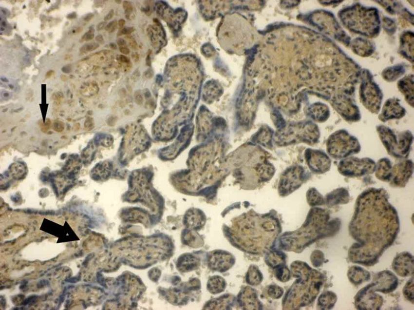
OGLAK, S. C. & OBUT, M. Expression of ADAMTS13 and PCNA in the placentas of gestational diabetic mothers. Int. J. Morphol., 39(1):38-44, 2021.

components, thereby promoting cell proliferation, cell                                  test was performed after a fasting period of 8 to 14 hours in
migration, and angiogenesis by inducing the secretion of                                the morning. Patients experienced the OGTT after three days
cytokines and growth factors (Martin et al., 2015; Ota et al.,                          of unlimited physical activity and free diet (>150 grams
2016). ADAMTS13 performs an essential role in regulating                                carbohydrates per day). One or more of the test’s values must
thrombosis and hemostasis by cleaving the von Willebrand                                be equaled or exceeded for the diagnosis of GDM (Committee
factor (VWF) (Dong et al., 2002). ADAMTS13 mRNA was                                     on Practice Bulletins—Obstetrics, 2018). Patients with health
detected in human placentas (Plaimauer et al., 2002). Once                              conditions related to intrauterine hypoxia (smoking,
a cell undergoes apoptosis, its chromatin begins to conden-                             preeclampsia, maternal obesity, anemia, advanced maternal
se, and its size begins to shrink. Later the cell divides into                          age, inflammatory diseases during pregnancy, infections, and
smaller apoptotic bodies. Fragmented nuclei and all                                     asthma) were excluded.
structures of the cell are eliminated.
                                                                                        Histopathological examination. Placental parts were fixed
        Proliferative cell nuclear antigen (PCNA) is a marker                           in 10 % formaldehyde, followed by washing, dehydration,
for the cell cycle. Although PCNA is an accessory protein                               clearing in xylene and incubated at 58 °C in paraffin wax.
required for DNA synthesis, its deficiency prevents cell                                Five mm sections were taken from paraffin blocks, and
division (Jónsson & Hübscher, 1997). Besides DNA                                        sections were transported to distilled water through
replication, PCNA is also related tochromatin modification,                             deparaffinization and descending alcohol series. We stained
DNA repair, sister-chromatid adhesion, and cell cycle con-                              the sections with Mayer Hematoxylin-Eosin and mounted
trol (Maga & Hubscher, 2003). In their study, Acar et al.                               with Entellan™. Zeiss Imager A-2 Axio (Germany) light
(2008) reported that PCNA immunopositive cell frequency                                 microscope was used for the evaluation of the tissue sections.
decreased in diabetic rat placentas and the control group. In
another study, it was observed that the number of PCNA                                  Immunohistochemical examination. Five µm sections
immunopositive cells in the placenta decreased in parallel                              were taken from paraffin-embedded placenta blocks, then
with the gestational age. These findings support that the                               deparaffinized passed through the alcohol series and brought
placenta loses its proliferative character as it approaches term                        to distilled water. Sections were held in the microwave oven
(Maruo et al., 2001).                                                                   for 3 x 5 min in citrate solution for antigen retrieval. After
                                                                                        allowing to cool for 10 min at room temperature, we washed
       In this study, we aimed to examine the expression of                             the sections with a phosphate buffer solution (PBS). They
ADAMTS13 and PCNA in the placentas of gestational dia-                                  were then treated with 3 % hydrogen peroxide (H2O2) for
betes mellitus (GDM) patients to investigate the effects of                             10 minutes. Samples were rinsed in distilled water and
hypoxia, induced by GDM, on proliferation and extracellular                             washed with PBS (pH 7.6). Then, we incubated the sections
matrix formation in the maternal and fetal placenta cells.                              with mouse monoclonal anti-CD44 antibody (Santa Cruz,
                                                                                        1:100) and mouse monoclonal anti-PCNA antibody (Santa
                                                                                        Cruz, 1:100). Washing three times in PBS, secondary
MATERIAL AND METHOD                                                                     antibody solution (Biotinylated Goat Anti-Mouse,
                                                                                        LabVision) was dropped on slides for 10 minutes. The
                                                                                        streptavidin peroxidase solution (Streptavidin Peroxidase,
       The study protocol was approved by the Health                                    LabVision) was used in the sections for 15 minutes. After
Sciences University Diyarbakır Gazi Yas¸argil Training and                              washing with PBS, a 3-amino-9-ethylcarbazole (AEC)
Research Hospital Ethical Committee, and all patients signed                            chromogen solution was applied to slides for 8 minutes. The
an informed consent form. A total of 60 placentas were                                  sections were washed with distilled water and counterstained
collected from pregnant women who were admitted to the                                  with hematoxylin for 2 minutes to evaluated under a light
obstetrics clinic. Only placentas between 28-38 weeks of                                microscope (Nikon).
gestation were included. These placental tissues were
obtained from non-diabetic and diabetic pregnancies                                     Statistical analysis. Statistical analyses of the data were
immediately after cesarean section.                                                     conducted using IBM SPSS 15.0 for Windows (SPSS Inc.,
                                                                                        Chicago, IL, USA). Measured variables were presented as
       The groups were assigned 30placentas of GDM                                      mean±standard deviation. Shapiro-Wilk tests were used to
patients and 30placentas of non-diabetic patients. A fasting                            determine whether the numerical data matched the normality
plasma glucose >126mg/dl is diagnostic of overt diabetes.                               distribution. The independent t-test was used to compare the
GDM was diagnosed with 75 g oral glucose tolerance test                                 normally distributed data. We used the Mann-Whitney U test
(OGTT) venous plasma/serum threshold values as follows:                                 to compare the non-normally distributed data. A P-value of
Fasting 92mg/dl, 1 hour 180mg/dl, 2 hours 153mg/dl. The                                 less than 0.05 was accepted as statistically significant.
                                                                                                                                                                39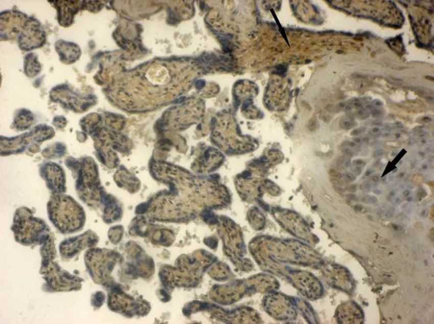
OGLAK, S. C. & OBUT, M. Expression of ADAMTS13 and PCNA in the placentas of gestational diabetic mothers. Int. J. Morphol., 39(1):38-44, 2021.

RESULTS

         The demographic characteristics and laboratory                                 (Fig. 1). In the GDM group, there were pyknosis and
values of the patients were shown in Table I. There were no                             apoptotic changes in decidua cell nuclei. Vacuolar areas were
statistically significant differences between the two groups                            observed in large cavities in maternal connective tissue.
in terms of age, gravidity, parity, blood pressure, hemoglobin,                         There was an increase in the number of syncytial nodes and
platelets, urea, creatinine, ALT, and AST values. Plasma                                bridges in the periphery of the root villi. Inflammation and
glucose and HbA1c levels were significantly higher in the                               dilatation with congestion were observed in the blood vessels
GDM group than in the control group (p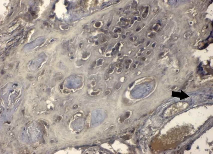
OGLAK, S. C. & OBUT, M. Expression of ADAMTS13 and PCNA in the placentas of gestational diabetic mothers. Int. J. Morphol., 39(1):38-44, 2021.
Fig. 1. Control group: Normal appearance in decidua cells, syncytial Fig. 2. GDM group: Picnosis and apoptotic changes in decidua
cells and blood vessels. Hematoxylin an Eosin staining x40. cell (thin arrow), an increase in the number of syncytial nodes and
bridges of root villi(thick arrow).Hematoxylin-Eosin staining x40.
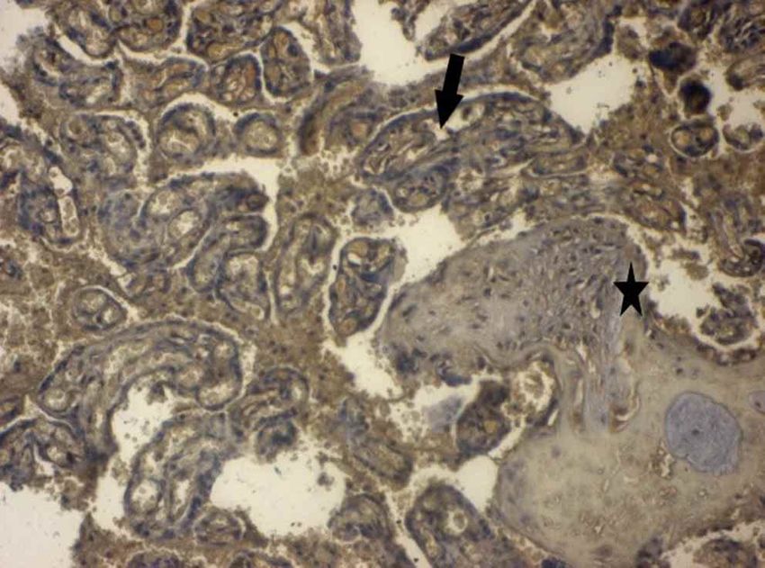
Fig. 3. Control group: positive in connective tissue fibers and
fibroblast cells (thin arrow) ,negative ADAMTS-13 expression in Fig. 4. GDM group: Positive ADAMTS-13 expression in the
decidua cells (thick arrow) and trophoblast cells of both root villi decidua cells (thin arrow), vascular endothelial cells, and Hofbauer
and free villi of maternal region. ADAMTS13 immun-staining x40. cells (thick arrow). ADAMTS13 immun-staining x40.
Fig. 5. Control group: Negative PCNA expression in decidua cells
(thin arrow), connective tissue cells, trophoblast, and vascular Fig. 6. GDM group: A significant increase of PCNA expression in
endothelial cells in the maternal region cells (thick arrow). decidua cells (star), positive PCNA expression connective tissue cells,
PCNAimmun-staining x40. and endothelial cells (thick arrow). PCNAimmun-staining x40.
41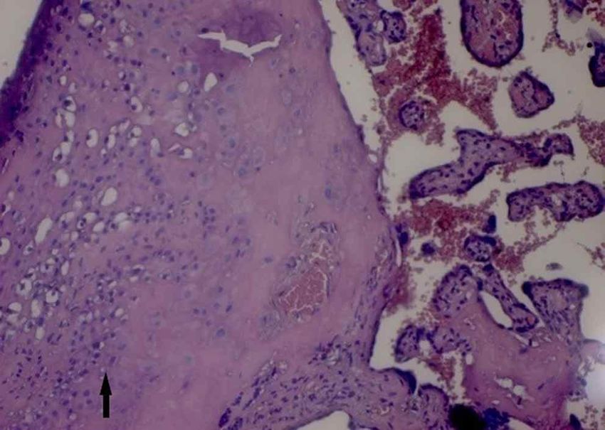
OGLAK, S. C. & OBUT, M. Expression of ADAMTS13 and PCNA in the placentas of gestational diabetic mothers. Int. J. Morphol., 39(1):38-44, 2021.

DISCUSSION

        Maternal diabetes induces the development of                                    placental weight in GDM may be associated with markedly
abnormal fetal organogenesis. Hyperglycemia of the mother                               reduced trophoblast apoptosis and apoptosis of villous
directly increases perinatal problems in diabetic pregnant                              cytotrophoblast and syncytiotrophoblast nuclei (Magee et
women. Increased glucose in the mother leads to fetal                                   al., 2014). A significant increase in PCNA activity was
hyperglycemia since it passes to the fetus by facilitated                               observed in our study, especially in decidua cells in the GDM
diffusion. Glycogen storage of the placenta in diabetic                                 group. PCNA expression showed positive reactions in
pregnant women causes decreased glycogen levels in the                                  syncytial cells, villous connective tissue cells, and endothelial
mother's liver. Insulin resistance in pregnant women causes                             cells (Fig. 6). This may affect glucose metabolism in decidua
hyperinsulinemia, hyperglycemia, and hypertriglyceridemia,                              cells due to oxygen deficiency and trophoblastic
especially in the last three months (Committee on Practice                              proliferation, so we thought endothelial dysfunction might
Bulletins—Obstetrics). In diabetic placentas, there is an                               continue in the late periods of placenta. Although all
increase in the incidence of pathological vascular changes,                             ADAMTS family member’s biological functions are not
and these changes are most frequently seen in the blood                                 fully understood, ADAMTS1, 4, and 5 proteins are stated to
vessels of the root villi. These lesions affect fetoplacental                           have the ability to activate the proteolytic modification of
circulation, blood flow in the degenerated umbilical cord                               extracellular matrix proteins via cell surface proteins (Tang,
with ischemic histiocytic villitis, and placental infarction.                           2001; Lee et al., 2006). In the last trimester of pregnancy in
They also trigger syncytial necrosis and perivillous fibrin                             preeclampsia, ADAMTS proteases play an essential role in
accumulation with chronic fetal hypoxia. An increased                                   rearranging these structures with defective pituitary
number of chorionic villus branching and syncytial nodes                                trophoblast infestation, hypoxic spiral artery formation, and
in diabetic pregnancies was reported (Farrar, 2016). It has                             impaired extracellular matrix formation. ADAMTS13
been suggested that these changes develop as a counter                                  promotes trophoblast cell proliferation, migration, invasion,
mechanism to fetal hypoxemia to increase the surface where                              and tube formation. Placental ADAMTS13 mRNA levels
oxygen exchange between the fetus and the mother occurs.                                were highest in the first trimester and decreased in the second
In our study, the increased syncytial nodesin chorionic villi,                          and third trimesters. These authors have shown significantly
accumulation of necrotic fibrin, congestion in the vessels,                             lower ADAMTS13 protein levels in placental villi tissues
and elevated inflammation are significant histopathological                             of patients with severe preeclampsia than those in the con-
findings (Fig. 2).                                                                      trol group by the western blot method. They thought
                                                                                        decreased ADAMTS13 synthesis and secretion in the
        Brownlee (2001) reported that early intracellular                               placentas of patients with severe preeclampsia might be
hyperglycemia during diabetes causes abnormalities in blood                             associated with tissue hypoxia and placental ischemia
flow and decreased vasodilator activity, thus leading to                                associated with hypertension (Xiao et al., 2017).
increased vasoconstrictor activity. Reduced production of                               ADAMTS13 is expressed in the trophoblast and fetal blood
endothelial and neurotrophic factors and excessive                                      vessel endothelium. During pregnancy, the human placenta
accumulation of extracellular matrix contributed to                                     synthesizes and secretes the active proteolytic ADAMTS13.
microvascular damage and consequently occluded                                          It is also produced at the highest levels in the first trimester
capillaries.                                                                            and progressively decreases in the plasma during the second
                                                                                        and third trimesters of pregnancy (Feys et al., 2007). Chang
       Trophoblast proliferation induced by free radicals and                           & Yang (2013) showed that the levels of Perlecan increased
low oxygen pressure may lead to early preeclampsia. This                                in the third trimesterin the placentas of pregnant women with
may be a significant cause of maternal morbidity and                                    GDM. Perlecan is a crucial component of the basement
mortality (Gosseye & Fox, 1984). The PCNA protein is one                                membrane and plays a role in regulating growth factors,
of the central molecules responsible for the cell’s life and                            extracellular matrix, and angiogenesis.It has been suggested
death of the cell and is involved in DNA repair. Unek et al.                            that ADAMTS13 plays a potential role in normal pregnancy
(2014) reported that PCNA increased in villous                                          and the pathogenesis of pregnancy-related complications.
cytotrophoblasts in the preeclampsia group.                                             Therefore, it is stated that ADAMTS13 promotes the
                                                                                        invasion, migration, proliferation, and network formation
       Sgarbosa et al. (2006) reported that hyperglycemia                               of trophoblasts (Xiao et al.). The effect of ADAMTS13 on
might be an essential factor inducing apoptosis in placental                            angiogenesis is essential. In our study’s GDM group,
trophoblasts and therefore associated with diabetic placenta                            ADAMTS13 expression was positive in the maternal decidua
function. Some authors have also reported that increased                                cells, vascular endothelial cells, and connective tissue
42
OGLAK, S. C. & OBUT, M. Expression of ADAMTS13 and PCNA in the placentas of gestational diabetic mothers. Int. J. Morphol., 39(1):38-44, 2021.
fibroblast cells. Also, the expression of ADAMTS-13 was REFERENCES
positive in the vascular endothelial cells of free villi,
Hoffbauer cells, and some inflammatory cells. With the
influence of GDM,it was observed that proteases and Acar, N.; Korgun, E. T.; Cayli, S.; Sahin, Z.; Demir, R. & Ustunel, I. Is
there a relationship between PCNA expression and diabetic placental
proteoglycans like ADAMTS13 cause changes in cells and
development during pregnancy? Acta Histochem., 110(5):408-17,
extracellular matrix structure. 2008.
Brownlee, M. Biochemistry and molecular cell biology of diabetic
In conclusion, hyperglycemia has been shown to complications. Nature, 414(6865):813-20, 2001.
Chang, S. C. & Yang, W. C. V. Hyperglycemia induces altered expressions
modify the expression of apoptosis-inducing proteins and
of angiogenesis associated molecules in the trophoblast. Evid. Based
genes encoding cell proliferation and alter vascular Complement. Alternat. Med., 2013:457971, 2013.
endothelial growth. Functional changes in ADAMTS13 Committee on Practice Bulletins—Obstetrics. ACOG Practice Bulletin
proteases and PCNA were thought to induce maternal and No. 190: Gestational Diabetes Mellitus. Obstet. Gynecol., 131(2):e49-
e64, 2018.
fetal complications by stimulating extracellular matrix
Crowther, C. A.; Hiller, J. E.; Moss, J. R.; McPhee, A. J.; Jeffries, W. S.;
development. Further studies are needed at the genetic level Robinson, J. S. & Australian Carbohydrate Intolerance Study in
to prevent the development of GDM or reduce early and Pregnant Women (ACHOIS) Trial Group. Effect of treatment of
severe maternal and fetal complications associated with gestational diabetes mellitus on pregnancy outcomes. N. Engl. J. Med.,
352(24):2477-86, 2005.
hyperglycemia.
Di Fulvio, P.; Pandolfi, A.; Formoso, G.; Di Silvestre, S.; Di Tomo, P.;
Giardinelli, A.; De Marco, A.; Di Pietro, N.; Taraborrelli, M.; Sancilio,
S.; et al. Features of endothelial dysfunction in umbilical cord vessels
OGLAK, S. C. & OBUT, M. Expresión de ADAMTS13 y PCNA en of women with gestational diabetes. Nutr. Metab. Cardiovasc. Dis.,
las placentas de madres diabéticas gestacionales. Int. J. Morphol., 24(12):1337-45, 2014.
39(1):38-44, 2021. Dong, J. F.; Moake, J. L.; Nolasco, L.; Bernardo, A.; Arceneaux, W.;
Shrimpton, C. N.; Schade, A. J.; McIntire, L. V.; Fujikawa, K. &
RESUMEN: La diabetes gestacional está relacionada con la López, J. A. ADAMTS-13 rapidly cleaves newly secreted ultralarge
sobreexpresión de citocinas inflamatorias y aumento del estrés oxidativo, von Willebrand factor multimers on the endothelial surface under
flowing conditions. Blood, 100(12):4033-9, 2002.
lo que lleva a una disfunción endotelial y un trastorno vascular. Nuestro
Farrar, D. Hyperglycemia in pregnancy: prevalence, impact, and
objetivo fue examinar la expresión de ADAMTS13 y PCNA en las management challenges. Int. J. Womens Health, 8:519-27, 2016.
placentas con diabetes mellitus gestacional (DMG) para investigar los Feys, H. B.; Canciani, M. T.; Peyvandi, F.; Deckmyn, H.; Vanhoorelbeke,
efectos de la hipoxia inducida por DMG sobre la proliferación y forma- K. & Mannucci, P. M. ADAMTS13 activity to antigen ratio in
ción de matriz extracelular en células placentarias maternas y fetales. physiological and pathological conditions associated with an increased
Se recolectaron un total de 60 placentas de mujeres embarazadas ingre- risk of thrombosis. Br. J. Haematol., 138(4):534-40, 2007.
sadas a la consulta de obstetricia. Treinta de ellas fueron diagnosticadas Gosseye, S. & Fox, H. An immunohistological comparison of the secretory
con DMG y 30 diagnosticadas sin DMG. Las muestras se fijaron en capacity of villous and extravillous trophoblast in the human placenta.
formaldehído al 10 %, y luego de un seguimiento de rutina, fueron em- Placenta, 5(4):329-47, 1984.
bebidas en parafina. Se cortaron secciones de 5 µm teñidas con Honda, M.; Toyoda, C.; Nakabayashi, M. & Omori, Y. Quantitative
hematoxilina-eosina de Mayer, las que fueron examinadas bajo un mi- investigations of placental terminal villi in maternal diabetes mellitus
croscopio óptico. Se cortaron y procesaron las secciones para el análisis by scanning and transmission electron microscopy. Tohoku J. Exp.
inmunohistoquímico para la recuperación de antígeno en solución de Med., 167(4):247-57, 1992.
Jónsson, Z. O. & Hübscher, U. Proliferating cell nuclear antigen: more
citrato. Las secciones se incubaron con anticuerpos primarios
than a clamp for DNA polymerases. Bioessays, 19(11):967-75, 1997.
ADAMTS13 y PCNA, se contratiñeron con hematoxilina y se evalua-
Kelwick, R.; Desanlis, I.; Wheeler, G. N. & Edwards, D. R. The ADAMTS
ron con un microscopio óptico. En el examen histopatológico, las (A Disintegrin and Metalloproteinase with Thrombospondin motifs)
placentas no diabéticas mostraron que las células de la decidua en la family. Genome Biol., 16(1):113, 2015.
región materna eran poligonales con núcleos ovalados y organizadas en Lee, N. V.; Sato, M.; Annis, D. S.; Loo, J. A.; Wu, L.; Mosher, D. F. &
grupos. En el grupo de DMG, se observó picnosis y cambios apoptóticos Iruela-Arispe, M. L. ADAMTS1 mediates the release of
en los núcleos de las células de la decidua. Se observaron áreas vacuolares antiangiogenic polypeptides from TSP1 and 2. EMBO J.,
en el tejido conectivo materno. En los vasos sanguíneos de las 25(22):5270-83, 2006.
vellosidades se observó inflamación y dilatación con congestión. En el Maga, G. & Hubscher, U. Proliferating cell nuclear antigen (PCNA): a
grupo de DMG, se observó expresión positiva de ADAMTS13 en las dancer with many partners. J. Cell Sci., 116(Pt. 15):3051-60, 2003.
células de la decidua, en las células endoteliales vasculares y en los Magee, T. R.; Ross, M. G.; Wedekind, L.; Desai, M.; Kjos, S. &
fibroblastos del tejido conectivo circundante. En el grupo de DMG se Belkacemi, L. Gestational diabetes mellitus alters apoptotic and
observó un aumento significativo de la expresión de PCNA en células inflammatory gene expression of trophobasts from human term
de la decidua, células de tejido conectivo y en las células endoteliales. placenta. J. Diabetes Complications, 28(4):448-59, 2014.
Martin, A. C. B. M.; Cardoso, A. C. F.; Selistre-de-Araujo, H. S. &
Se considera que los cambios funcionales en las proteasas ADAMTS13
Cominetti, M. R. Recombinant disintegrin domain of human ADAM9
y PCNA inducen a complicaciones maternas y fetales al estimular el
inhibits migration and invasion of DU145 prostate tumor cells. Cell
desarrollo de la matriz extracelular. Adh. Migr., 9(4):293-9, 2015.
Maruo, T.; Ishihara, N.; Samoto, T.; Murakoshi, H.; Laoag-Fernandez, J.
PALABRAS CLAVE: Diabetes mellitus gestacional; B. & Matsuo, H. Regulation of human trophoblast proliferation and
ADAMTS13; PCNA. apoptosis during pregnancy. Early Pregnancy, 5(1):28-9, 2001.
43OGLAK, S. C. & OBUT, M. Expression of ADAMTS13 and PCNA in the placentas of gestational diabetic mothers. Int. J. Morphol., 39(1):38-44, 2021.
Ota, M.; Mochizuki, S.; Shimoda, M.; Abe, H.; Miyamae, Y.; Ishii, K.; Corresponding Author:
Kimura, H. & Okada, Y. ADAM23 is downregulated in side Süleyman Cemil Oglak
population and suppresses lung metastasis of lung carcinoma cells. Department of Obstetrics and Gynecology
Cancer Sci., 107(4):433-43, 2016.
Health Sciences
Plaimauer, B.; Zimmermann, K.; Völkel, D.; Antoine, G.; Kerschbaumer,
R.; Jenab, P.; Furlan, M.; Gerritsen, H.; Lämmle, B.; Schwarz, H. P.; et
University Diyarbakır
al. Cloning, expression, and functional characterization of the von Gazi Yasargil Training and Research Hospital
Willebrand factor-cleaving protease (ADAMTS13). Blood, Diyarbakır
100(10):3626-32, 2002. TURKEY
Sgarbosa, F.; Barbisan, L. F.; Brasil, M. A. M.; Costa, E.; Calderon, I. M.
P.; Gonçalves, C. R.; Bevilacqua, E. & Rudge, M. V. C. Changes in
apoptosis and Bcl-2 expression in human hyperglycemic, term placental Orcid: 0000-0001-7634-3008
trophoblast. Diabetes Res. Clin. Pract., 73(2):143-9, 2006.
Shaat, N. & Groop, L. Genetics of gestational diabetes mellitus. Curr. Med.
Chem., 14(5):569-83, 2007.
Tang, B. L. ADAMTS: a novel family of extracellular matrix proteases. Email: sampson_21@hotmail.com
Int. J. Biochem. Cell Biol., 33(1):33-44, 2001.
Unek, G.; Ozmen, A.; Mendilcioglu, I.; Simsek, M. & Korgun, E. T. The
expression of cell cycle related proteins PCNA, Ki67, p27 and p57 in Received: 08-07-2020
normal and preeclamptic human placentas. Tissue Cell, 46(3):198-205, Accepted: 16-09-2020
2014.
Xiao, J.; Feng, Y.; Li, X.; Li, W.; Fan, L.; Liu, J; Zeng, X.; Chen, K.; Zhou,
X.; Zheng, X. L.; et al. Expression of ADAMTS13 in normal and
abnormal placentae and ıts potential role in angiogenesis and placenta
development. Arterioscler. Thromb. Vasc. Biol., 37(9):1748-56, 2017.
44You can also read