Improved Histoarchitectural Changes with Angiotensin Receptor Blockers in Early Testicular and Cauda Toxicity in Rats
←
→
Page content transcription
If your browser does not render page correctly, please read the page content below
Int. J. Morphol.,
37(2):515-521, 2019.

Improved Histoarchitectural Changes with Angiotensin Receptor
  Blockers in Early Testicular and Cauda Toxicity in Rats

              Mejora de los Cambios en la Histoarquitectura con Bloqueadores de Receptores
                  de Angiotensina en la Toxicidad Precoz Testicular y de Cauda en Ratas

                             Khadija Abduljalil Faddladdeen1; Hussam Aly Murad2 & Soad Shaker Ali3

FADDLADDEEN, K. A.; MURAD, H. A. M. & ALI, S. S. Improved histoarchitectural changes with angiotensin receptor
blockers in early testicular and cauda toxicity in rats. Int. J. Morphol., 37(2):515-521, 2019.

         SUMMARY: Reproductive dysfunction is a complication for many diseases and toxins. Its early diagnosis and treatment
are immensely important. Here the morphological histoarchitecture changes in early testicular and cauda toxicity before and
after treatment with angiotensin receptor blockers were evaluated. Low-grade testicular damage was induced using thioacetamide
(TAA, 50 mg/kg/day) intraperitoneally for two weeks in rats. The rats were randomly divided into four groups (n = 8) treated
daily orally for three weeks as follows: Normal control (distilled water), TAA (positive control), TAA+candesartan (0.2 mg/kg)
and TAA+losartan (7.5 mg/kg). Serum testosterone and testicular malondialdehyde and glutathione were measured. The changes
in histoarchitecture of testis and cauda epididymis were evaluated by hematoxylin and eosin for general structure, Masson's
trichrome for collagen, periodic acid Schiff for basement membrane, and caspase-3 and proliferating cell nuclear antigen
(PCNA) for immunohistochemical analysis. The TAA-rats showed decreases of serum testosterone and testicular glutathione,
increases in testicular malondialdehyde, degenerative changes and apoptosis in germ cells, thickening of tubular basal lamina
and increases in expression of caspase 3, and decreases in expression of PCNA. The ARBs (candesartan and losartan) significantly
reversed these changes with non-significant differences in-between. Treatment with ARBs (candesartan and losartan) significantly
reversed TAA-induced low-grade testicular and cauda toxicity in rats. This could be potentially useful for early treatment of
male patients with occupational toxicant-induced reproductive dysfunction especially if they are using ARBs for other
comorbidities.

        KEY WORDS: Caspase-3; Masson; Proliferating cell nuclear antigen; Angiotensin receptor blockers.

INTRODUCTION

        Male hypogonadism is a common problem in patients                        Proliferating cell nuclear antigen (PCNA) is a nu-
with liver cirrhosis which partially improves after liver                 clear matrix protein necessary for several cell cycle
transplantation (Foresta et al., 2008). The underlying                    pathways. Significant decreases in PCNA
mechanism of such problem is complex and not well                         immunohistochemistry measured on formalin-fixed
explained. Liver is involved in metabolism of sex hormones,               paraffin-embedded rat testes have been used to identify
thus pronounced changes in such metabolism, free serum                    and quantify of the proliferation-related toxicity. Thus
testosterone level, and sex hormone-binding globulin level                PCNA assay is potentially useful in vivo biomarker for
in blood could be possible causes (Nitsche et al., 2014).                 detecting early testicular toxicity and for follow-up of
Thioacetamide-induced liver damage was associated with                    compounds with low testicular toxicity (D'Andrea et al.,
testicular toxicity (Kang et al., 2006). In testicular toxicity,          2008). The block of effects of angiotensin II whether by
use of histopathological measures is more sensitive                       angiotensin converting enzyme inhibitors (ACEIs) or
parameter of testicular damage than use of testis weight and              angiotensin receptor blockers (ARBs) have been found to
sperm counts and thus can detect early toxicity.                          protect the testicular tissue against multiple insults (Alves-

1
  Department of Biology, Faculty of Science, King Abdulaziz University (KAU), Jeddah, Saudi Arabia (SA).
2
  Department of Pharmacology, Faculty of Medicine, Rabigh, KAU, Jeddah, Saudi Arabia.
3
  Department of Anatomy and Histology, Faculty of Medicine, KAU, Jeddah, Saudi Arabia.

                                                                                                                                  515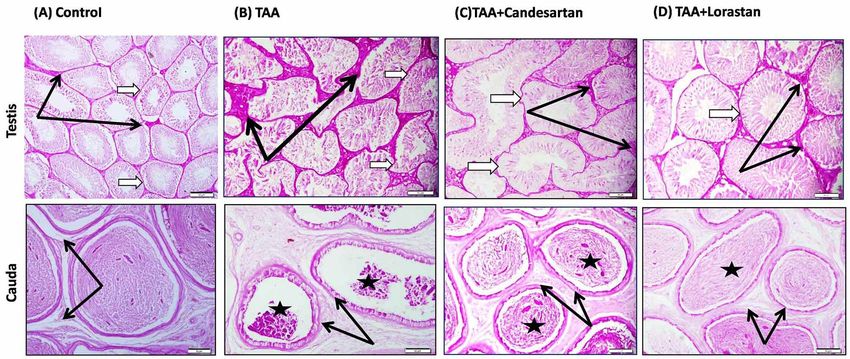
FADDLADDEEN, K. A.; MURAD, H. A. M. & ALI, S. S. Improved histoarchitectural changes with angiotensin receptor blockers in early testicular and cauda toxicity in rats.
                                                               Int. J. Morphol., 37(2):515-521, 2019.

Pereira et al., 2014). Both captopril and telmisartan                                        for collagen, and immunohistochemically for caspase-3
reversed the cadmium-induced testicular damage with non-                                     and proliferating cell nuclear antigen (PCNA).
significant differences in-between. They increased serum                                     Photographs were taken for all groups and compared
testosterone level and the testicular reduced glutathione                                    (Suvarna et al., 2013).
and decreased testicular MDA level and expression of
testicular caspase-3 (Fouad & Jresat, 2013). ACEIs and                                       Assay of serum testosterone. The serum testosterone level
ARBS are used to treat cardiovascular diseases like                                          was measured by an ELISA kit (Sigma, MO, US) as per
hypertension and heart failure, to prevent renal failure in                                  instructed by the manufacturer.
cases of hypertension and/or diabetes, and to decrease the
risk of stroke. Generally ARBs are more tolerated than                                       Measurement of testicular MDA and GSH. The testes
ACEIs.                                                                                       were homogenized in ice-cold medium of 50 mM Tris-
                                                                                             HCl (pH 7.4), the homogenate was centrifuged at 1000 xg
       Taken together, this study was designed to evaluate                                   for 10 min at 4 °C, the supernatant was used for
the morphological changes in testicular histoarchitecture                                    measurement of MDA & GSH. For estimation of MDA,
before and after treatment with angiotensin receptor                                         the testis homogenate was incubated in 10 % trichloroacetic
blockers in early testicular toxicity induced by low dose                                    acid and 0.67 % thiobarbituric acid (1 mL of each) for 30
thioacetamide in rats.                                                                       min (Ohkawa et al., 1979). The GSH was determined by
                                                                                             the reduction of Elman’s reagent (Ellman, 1959).

MATERIAL AND METHOD                                                                          Immunohistochemistry of testis and cauda

                                                                                             Caspase-3. The testicular tissue sections were dewaxed
Animals and Drugs. The study was approved by the                                             and rehydrated. Endogenous peroxidase was blocked using
Institutional Research Ethics Committee. It agreed with                                      0.3 % H2O2 in methanol for 15 min. Then sections were
the International guidelines for use of Laboratory animals.                                  incubated with primary diluted polyclonal antibodies for
Sprague–Dawley male rats (200-250 g) were housed in                                          caspase-3 (Thermo Fisher Scientific Co., USA) for 30 min.
cages at 22 °C room temperature with food and water ad                                       and subsequently with biotinylated polyvalent secondary
libitum. Drugs and chemicals were purchased from Sigma-                                      antibody (Thermo Fisher Scientific Co., USA) for 30 min.
Aldrich Corp. (St. Louis, MO, USA).                                                          Metal Enhanced DAB Substrate Working Solution was
                                                                                             added to the tissue followed by counterstaining with
Induction of low-grade testicular toxicity. Low grade                                        hematoxylin stain (Kim et al., 2001; Elgawish &
testicular toxicity was induced in rats by injection of                                      Abdelrazek, 2014).
thioacetamide (TAA, 50 mg/kg) intraperitoneally once
daily for two weeks. This small dose was used before, to                                     Proliferative cell nuclear antigen (PCNA). After
induce low-grade hepatotoxicity. The rats were randomly                                      deparaffinization and rehydration steps, the testicular tissue
divided into four groups (n = 8): negative control group                                     sections were incubated in methanol containing 0.3 %
(distilled water), TAA (positive control) group,                                             H2O2 and normal goat serum for 15 minutes to decrease
candesartan cilexetil group (0.2 mg/kg/day) (Gaur &                                          non-specific peroxidase reactions. Then, they were
Kumar, 2011), and losartan potassium group (7.5 mg/kg/                                       incubated for 30 minutes with a primary antibody against
day) (Croquet et al., 2002). Treatments were given orally                                    PCNA (Santa Cruz Biotech., Texas, US) and subsequently
for three weeks. At the end of treatment duration, blood                                     with goat anti-rat IgG as a secondary antibody (Sigma,
was collected through the retroorbital plexus, centrifuged                                   MO, USA). After that, tissue sections were incubated with
at 4000 rpm for 5 min, and serum was separated and kept                                      the peroxidase/anti-peroxidase complex for 90 min.
at -80°C for biochemical measurements. Rats were                                             Diaminobenzidine was used as chromogen followed by
sacrificed by cervical dislocation and the male system was                                   counterstaining with hemotoxylin and examination by a
removed as one block for preparation of the testicular                                       light microscope (Sternberger, 1986; Altay et al., 2003;
homogenate,         and       histopathological        and                                   Dkhil et al., 2016).
immunohistochemical examination. For histopathological
and immunohistochemical examination the testis and                                           Statistical analysis. Data was expressed as means ± SEM
cauda were fixed in 10 % neutral buffered formalin then                                      and analyzed using SPSS version 18. One-way ANOVA
processed for paraffin sections. Slides were stained by                                      followed by Tukey's multiple comparison post-hoc test was
hematoxylin and eosin for general structure, periodic acid                                   used to assess differences between groups. P < 0.05 was
Schiff (PAS) for basement membrane, Masson trichrome                                         considered to be statistically significant.
516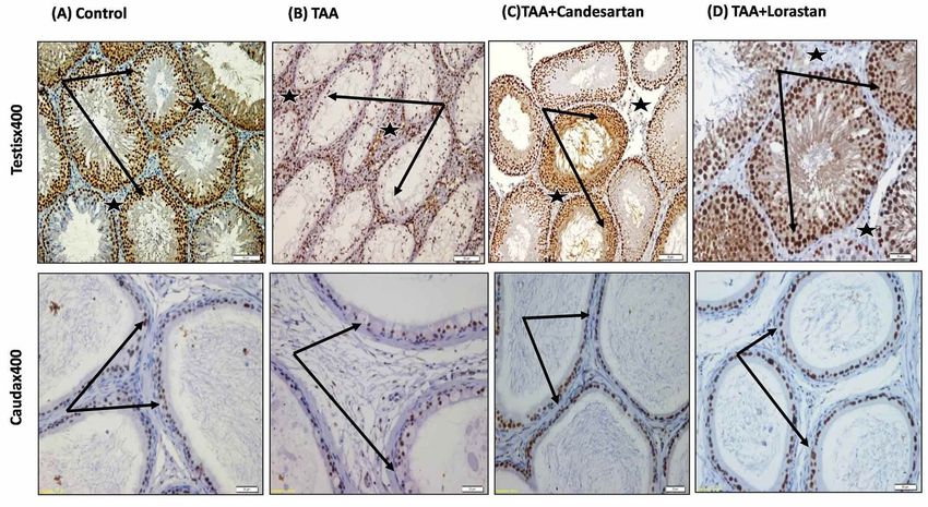
FADDLADDEEN, K. A.; MURAD, H. A. M. & ALI, S. S. Improved histoarchitectural changes with angiotensin receptor blockers in early testicular and cauda toxicity in rats.
Int. J. Morphol., 37(2):515-521, 2019.
RESULTS significantly reversed these change with non-significant
difference in-between (Table I).
Serum testosterone and Testicular MDA and GSH. TAA The histoarchitectural study. Both candesartan and losartan
decreased serum testosterone level, increased testicular MDA protected against TAA-induced testicular and cauda toxicity
level while decreased testicular GSH level compared with as shown using the following stains: Hematoxylin and eosin
the normal control group. Both candesartan and losartan (Figs. 1 and 2), Masson trichrome (Fig. 3), PAS (Fig. 4),
Caspase-3 (Fig. 5), and PCNA (Fig. 6).
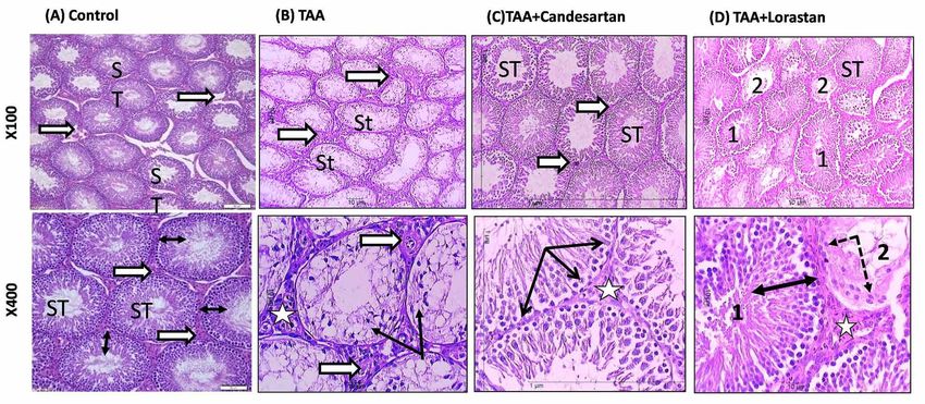
Fig. 1. Sections of rat testis stained by H&E photographed by low power and high power . (A) Normal Control group showing normal rounded seminiferous
tubules (ST) with intact full germ cell layer (double headed arrows), and normal population of interstitial Leydig cells (white arrow). (B) TAA group showing
degeneration and depletion of germ cell layer (black arrows). The interstitial tissue showed blood vessels with thick wall and Leydig cells looked smaller and
degenerated (star). (C) TAA+Candesartan group: Most tubules looked normal with full thickness germ cell layers (double headed arrows). The interstitial spaces
are normal and similar to NC (white stars). (D) TAA+Losartan group: Moderate improvement of TAA induced changes (1 & double arrows). Some tubules
showed degenerative changes (2 & dotted arrows).
Fig 2. Sections of rat cauda epididymis stained by H&E low power and high power. (A) Normal Control group: The tubules (arrows) show variations in
size and plan of cutting. They are separated by scanty connective tissue with few connective tissue cells (black stars). The tubules are lined by simple
columnar ciliated epithelium. The cytoplasm is slightly basophilic and the nuclei are rounded, vesicular, and basal. The lumen is filled with mature sperms
(white stars). (B) TAA group: There is increased interstitial tissue (black stars) with numerous inflammatory cells (dotted arrows). The epithelium showed
degenerated cells and loss of apical cilia (white arrows). The lumen showed absence of mature sperms and is filled with acidophilic debris (white stars).
(C) TAA+Candaesartan group: The tubules are still separated by wide spaces with fine fibrous tissue (black star) and few inflammatory cells (dotted
arrow). The lumina contain mature sperms (black star). (D) TAA+Lorasartan group: The tubules are separated by interstitial spaces containing fibrous
tissue (black stars). The lumina contain few mature sperm (white stars). The lining epithelium is normal with intact apical cilia (white arrows).
517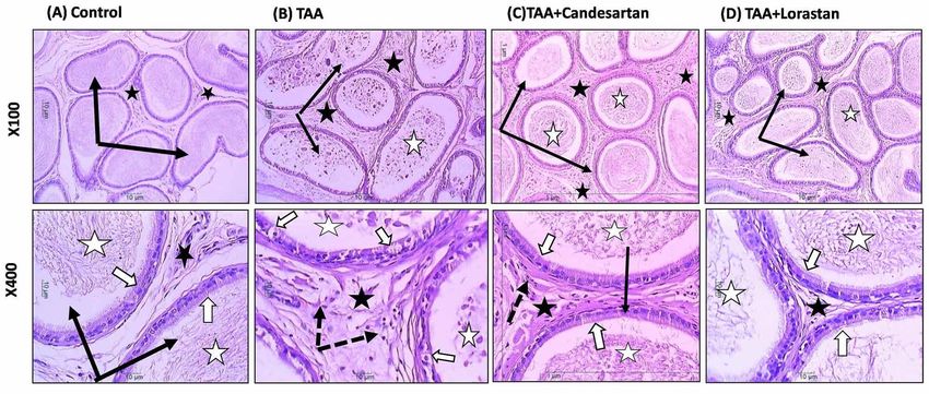
FADDLADDEEN, K. A.; MURAD, H. A. M. & ALI, S. S. Improved histoarchitectural changes with angiotensin receptor blockers in early testicular and cauda toxicity in rats.
                                                                 Int. J. Morphol., 37(2):515-521, 2019.

                         Table I. Effects of treatment with ARBs on parameters of thioacetamide-induced testicular toxicity.
                          Group                       Serum testosterone        Testicular GSH        Testicular MDA
                                                      (mmol/L)                  (µg/g testis)         (mmol/mg protein)
                          Normal control              3.26 ± 0.14               57.63 ± 0.90          16.91 ± 0.45
                          Thioacetamide               1.71 ± 0.11               38.59 ± 1.55          29.61 ± 0.63
                          TAA+Candesartan             2.82 ± 0.18 *             55.31 ± 1.33 *        17.65 ± 0.32 *
                          TAA+Losartan                2.90 ± 0.14 *             53.75 ± 1.09 *        18.14 ± 0.38 *
                          Data was expressed as mean ± SEM. *: P < 0.05 vs. thioactamide (TAA) group.

Fig 3. Sections of rat testis and cauda epididymis stained for collagen by Masson trichrome. (A) Normal Control group: The testis shows normal amount
of collagen fibers separating seminiferous tubules (white arrow). The cauda shows fine collagen fibers along the tubular wall (black stars). (B)TAA group:
The testis shows increases in collagen in the widely-spaced interstitial tissue (white arrows) and around the thickened congested blood vessels (BV). The
cauda shows thickened collagen (black stars) around the tubules. (C) TAA+Candesartan group: The testis shows normal distribution of collagen (white
arrows) between seminiferous tubules (ST) and the cauda shows normal distribution of collagen along and in-between tubular walls (black stars). (D)
TAA+Losartan group: The testis shows moderate improvement with nearly normal distribution of collagen (white arrows) between seminiferous tubules
(ST) and the cauda shows also moderate improvement normal distribution of collagen along tubular walls (black stars).

Fig 4. Sections of rat testis and cauda epididymis stained by PAS: (A) Normal Control group showing PAS positive reaction in the thin basal lamina of
seminiferous tubules (white arrows) and mild positive reaction in interstitial connective tissue (black arrows). (B) TAA group showing increase in the positive
PAS reaction in the thickened basal lamina of seminiferous tubules (white arrow) and degenerated germ cells and interstitial connective tissue (black arrows).
(C) TAA+Candesartan group showing restoration of normal PAS reaction in the basal lamina (white arrow) nearly similar to control. (D) TAA+Losartan group
showing normal PAS positive reaction in the basal lamina with focal increase in reaction in the interstitial tissue (black arrows). The cauda epididymis showed
increase in PAS positive reaction in the basal lamina of tubules (black arrows) and cellular debris in TAA group (black stars) which appeared to be ameliorated
by both candesartan and losartan.

518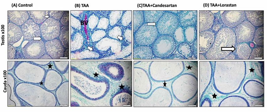
FADDLADDEEN, K. A.; MURAD, H. A. M. & ALI, S. S. Improved histoarchitectural changes with angiotensin receptor blockers in early testicular and cauda toxicity in rats.
                                                                 Int. J. Morphol., 37(2):515-521, 2019.

Fig 5. Sections from rat testis and cauda epididymis immunohistochemically stained for caspase-3. (A) Normal Control group: the testis showed focal few or
mild expression of caspase 3 in germ cells (black arrows) and the interstitial cells showed negative expression (white arrow). Cauda epithelium also showed
negative reaction while few degenerated cells within the lumen showed positive expression. (B)TAA group: the testis showed expression of caspase 3 mainly in
the interstitial cells (white arrows) and also in the left few degenerated germ cells (black arrows) in the seminiferous tubules (ST). The cauda epithelium showed
negative expression (black arrows) except for few degenerated cells in the lumen which showed positive reaction (dotted arrows). (C) TAA+Candesartan group:
the testis showed mild positive caspase 3 expression in few tubules (black arrows) and the interstitial cells showed negative expression (white arrows). The
cauda epithelium also showed negative reaction similar to control with few cells in the lumen showing positive reaction (dotted arrows). (D) TAA+Losartan
group: the testis expression of caspase 3 is similar to control. Few scattered tubules showed expression in germ cells (black arrows). The Interstitial cells showed
negative expression (white arrow). The cauda showed absence of caspase 3 expression in lining cells (black arrows) and few desquamated cells showed positive
reaction in the lumen (dotted arrow).

Fig 6. Sections from rat testis immunohistochemically stained for PCNA. (A) Normal Control group: the testis shows high immunopositive expression for
PCNA in all the spermatogenic intact germ cells of lining the seminiferous tubules (black arrows). Few interstitial cells showed positive reaction (black stars).
The cauda showed that most nuclei of lining epithelial cells of cauda tubules showed positive expression for PCNA (black arrows). (B) TAA group: the testis
showed a decrease in PCNA reaction in the seminiferous tubule germ cells (black arrows) as most germ cells are degenerated. An increase in PCNA expression
was observed in Leydig cells (black stars). The cauda showed negative expression for PCNA in degenerated swollen cells (arrows) while other cells showed
normal expression. (C) TAA+Candesartan group: the testis showed restoration of immunopositive reaction for PCNA in the intact proliferating spermatogenic
germ cells of the seminiferous tubules (black arrows) and few interstitial cells showed positive reaction (black stars). The cauda showed also PCNA expression
in the nuclei of lining cells similar to control (black arrows). (D) TAA+Losartan group: the testis showed immunopositive reaction for PCNA in the intact
proliferating germ cells of seminiferous tubules (black arrows) and few interstitial cells (black stars). The cauda showed also PCNA expression in the nuclei of
lining cells nearly similar to control (black arrows).

                                                                                                                                                                                   519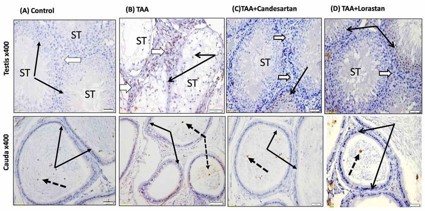
FADDLADDEEN, K. A.; MURAD, H. A. M. & ALI, S. S. Improved histoarchitectural changes with angiotensin receptor blockers in early testicular and cauda toxicity in rats.
                                                               Int. J. Morphol., 37(2):515-521, 2019.

DISCUSSION                                                                                          In conclusion, treatment with ARBs (candesartan and
                                                                                             losartan) significantly reversed thioacetamide-induced low-
                                                                                             grade testicular and cauda toxicity in rats. This could be
         Liver fibrosis could decrease male fertility (Foresta                               potentially useful for early treatment of male patients with
et al.). The increased markers of lipid peroxidation and the                                 occupational toxicant-induced reproductive dysfunction
decreased natural antioxidants in the testicular tissue                                      especially if they are using ARBs for other comorbidities.
supported the postulation of a direct toxic effect of TAA on
testicular tissue via stimulating oxidative mechanisms (Kang
et al.). The effect of TAA may be directly on the                                            ACKNOWLEDGEMENTS. Funding: This project was
spermatogenic cells or via damage of their supporting Sertoli                                funded by the Deanship of Scientific Research (DSR) at King
cells (Lenzi et al., 2002; Cheng, 2014). In the present study,                               Abdulaziz University, Jeddah under grant no. J-735-247-
the decrease in Leydig cells population could be explained                                   38. The authors, therefore, acknowledge with thanks DSR
in view of reported testosterone changes in patients with                                    for technical and financial support. The participation of the
liver fibrosis (Nitsche et al.). It was found that exposure to                               medical students Yasser A. Alkhairy, Abdulrahman M.
flutamide during intrauterine life resulted in a chronic                                     Khafagy, Hassan Gabbani, Mohammed Abdulhamid
apoptotic germ cell death in the adult rat testicular tissue                                 Alfuraydi, Hesham Flemban, and Tarek Abou-Taleb is
correlated with an increase in the expression and activation                                 gratefully acknowledged.
in germ cells of caspases-3 and -6 which are two important
components in the apoptotic machinery (Omezzine et al.,
2003). In rats, lead acetate significantly increased level of                                FADDLADDEEN, K. A.; MURAD, H. A. M. & ALI, S. S.Mejora
testicular         caspase-3         expression.       PCNA                                  de los cambios en la histoarquitectura con bloqueadores de receptores
immunohistochemistry showed three differences between                                        de angiotensina en la toxicidad precoz testicular y de cauda en ratas.
                                                                                             Int. J. Morphol., 37(2):515-521, 2019.
the testes of control and hypothyroid rats indicating its
usefulness as a marker to differentiate between the two                                               RESUMEN: La disfunción reproductiva es una complicación
regarding germ cell kinetics and spermatogenesis (Tousson                                    por muchas enfermedades y toxinas. Su diagnóstico y tratamiento
et al., 2011).                                                                               tempranos son inmensamente importantes. Aquí se evaluaron los cam-
                                                                                             bios morfológicos en la histoarquitectura en la toxicidad precoz
        PAS was used in the present study to demonstrate                                     testicular y cauda antes y después del tratamiento con bloqueadores
                                                                                             de receptores de angiotensina. Se indujo daño testicular de bajo grado
any changes in the tubule basement membrane and the result
                                                                                             usando tioacetamida (TAA, 50 mg / kg / día) por vía intraperitoneal
showed that it was thickened in TAA group which may have                                     durante dos semanas en ratas. Las ratas se dividieron aleatoriamente
a role in impaired spermatogenesis and germ cell                                             en cuatro grupos (n = 8) tratados diariamente por vía oral durante tres
degeneration. Thickening of basement membrane and                                            semanas de la siguiente manera: control normal (agua destilada), TAA
excessive deposition of extracellular matrix proteins are                                    (control positivo), TAA + candesartan (0,2 mg / kg) y TAA + losartán
characteristics of fibrotic disorders of multiple organs                                     (7,5 mg / kg). Se midieron la testosterona sérica, el malondialdehído
including liver and kidney (Rosenbloom et al., 2010). In                                     testicular y el glutatión. Los cambios en la histoarquitectura de los
                                                                                             testículos y la epidermis de la cauda se evaluaron mediante
rats, PAS staining was used as a histomorphological method                                   Hematoxilina y Eosina para determinar la estructura general, con
to detect disorders in seminiferous tubular histoarchitecture                                tricrómicro de Masson para el colágeno, ácido periódico de Schiff para
(Ogedengbe et al., 2016). In the present study, both                                         la membrana basal y la caspasa-3 y el antígeno nuclear de células
candesartan and lorsartan modulated the TAA-induced                                          proliferantes (PCNA) para análisis inmunohistoquímico. Las ratas TAA
testicular toxicity and all histological and                                                 mostraron disminución de la testosterona sérica y glutatión testicular,
immunohistochemical alteration in both testicular tissue and                                 aumentos en el malondialdehído testicular, cambios degenerativos y
                                                                                             apoptosis en células germinales, engrosamiento de la lámina basal
cauda epididymis tubules with non-significant differences
                                                                                             tubular y aumentos en la expresión de la caspasa 3, y disminución en
in-between. Candesartan relieved the cisplatin-induced                                       la expresión de PCNA. Los ARB (candesartán y losartán) revirtieron
testicular damage by modulating the expression patterns of                                   significativamente estos cambios con diferencias no significativas en
the testicular nephrin-podocin complex (Enatsu et al., 2015).                                el medio. El tratamiento con BRA (candesartán y losartán) revirtió
In rats, losartan partially reversed the angiotensin II type 1                               significativamente la toxicidad testicular y cauda inducida por TAA
receptor dependent increase in interstitial fibrosis and                                     en ratas. Esto podría ser potencialmente útil para el tratamiento tem-
impairment of spermatogenesis that occurred post-                                            prano de pacientes con disfunción reproductiva inducida por tóxicos
                                                                                             ocupacionales, especialmente si están usando BRA para otras
vasectomy due to oxidative stress (Shiraishi et al., 2003).                                  comorbilidades.
Losartan protected against testicular germ cell apoptosis
caused by an experimental varicocele (Bolat et al., 2016).                                           PALABRAS CLAVE: Caspasa-3; Masson; Antígeno nu-
Limitations of the current study include lack of quantitative                                clear de células en proliferación; Bloqueadores de receptores de
histological analysis and use of only two ARBs members.                                      angiotensina.

520
FADDLADDEEN, K. A.; MURAD, H. A. M. & ALI, S. S. Improved histoarchitectural changes with angiotensin receptor blockers in early testicular and cauda toxicity in rats.
Int. J. Morphol., 37(2):515-521, 2019.
REFERENCES tissues by thiobarbituric acid reaction. Anal. Biochem., 95(2):351-8,
1979.
Omezzine, A.; Chater, S.; Mauduit, C.; Florin, A.; Tabone, E.; Chuzel, F.;
Bars, R. & Benahmed, M. Long-term apoptotic cell death process with
Altay, B.; Cetinkalp, S.; Doganavsargil, B.; Hekimgil, M. & Semerci, B.
increased expression and activation of caspase-3 and -6 in adult rat
Streptozotocin-induced diabetic effects on spermatogenesis with
germ cells exposed in utero to flutamide. Endocrinology, 144(2):648-
proliferative cell nuclear antigen immunostaining of adult rat testis.
61, 2003.
Fertil. Steril., 80 Suppl. 2:828-31, 2003.
Rosenbloom, J.; Castro, S. V. & Jimenez, S. A. Narrative review: fibrotic
Alves-Pereira, J. L.; Frantz, E. D. & da Fonte Ramos, C. Beneficial effects
diseases: cellular and molecular mechanisms and novel therapies. Ann.
of Renin-Angiotensin system blockers on testicular steroidogenesis. J.
Intern. Med., 152(3):159-66, 2010.
Urol., 192(6):1878-83, 2014.
Shiraishi, K.; Yoshida, K.; Fujimiya, T. & Naito, K. Angiotensin II dependent
Bolat, D.; Oltulu, F.; Uysal, A.; Kose, T.; Gunlusoy, B.; Yigitturk, G.; Turk,
testicular fibrosis and effects on spermatogenesis after vasectomy in
N. S. & Turan, T. Effects of losartan on experimental varicocele-induced
the rat. J. Urol., 170(5):2104-8, 2003.
testicular germ cell apoptosis. Andrologia, 48(7):840-6, 2016.
Sternberger, L. A. Immunocytochemistry. Ann Arbor, John Wiley & Sons,
Cheng, C. Y. Toxicants target cell junctions in the testis: Insights from the
1986.
indazole-carboxylic acid model. Spermatogenesis, 4(2):e981485, 2014.
Suvarna, K. S.; Layton, C. & Bancroft, J. D. Bancroft's Theory and Practice
Croquet, V.; Moal, F.; Veal, N.; Wang, J.; Oberti, F.; Roux, J.; Vuillemin,
of Histological Techniques. 7th ed. Oxford, Churchill Livingstone
E.; Gallois, Y.; Douay, O.; Chappard, D. & Calès, P. Hemodynamic
Elsevier, 2013.
and antifibrotic effects of losartan in rats with liver fibrosis and/or por-
Tousson, E.; Ali, E. M.; Ibrahim, W. & Mansour, M. A. Proliferating cell
tal hypertension. J. Hepatol., 37(6):773-80, 2002.
nuclear antigen as a molecular biomarker for spermatogenesis in PTU-
D'Andrea, M. R.; Lawrence, D.; Nagele, R. G.; Wang, C. Y. & Damiano,
B. P. PCNA indexing as a preclinical immunohistochemical biomarker induced hypothyroidism of rats. Reprod. Sci., 18(7):679-86, 2011.
for testicular toxicity. Biotech. Histochem., 83(5):211-20, 2008.
Dkhil, M. A.; Zrieq, R.; Al-Quraishy, S. & Abdel Moneim, A. E. Selenium
nanoparticles attenuate oxidative stress and testicular damage in
streptozotocin-induced diabetic rats. Molecules, 21(11):E1517, 2016.
Elgawish, R. A. R. & Abdelrazek, H. M. A. Effects of lead acetate on
Corresponding author:
testicular function and caspase-3 expression with respect to the Prof. Hussam Aly Sayed Murad
protective effect of cinnamon in albino rats. Toxicol. Rep., 1:795-801, Department of Pharmacology
2014. Faculty of Medicine Rabigh
Ellman, G. L. Tissue sulfhydryl groups. Arch. Biochem. Biophys., 82(1):70- King Abdulaziz University
7, 1959. Jeddah
Enatsu, N.; Miyake, H.; Chiba, K. & Fujisawa, M. Candesartan mediated SAUDI ARABIA
amelioration of cisplatin-induced testicular damage is associated with
alterations in expression patterns of nephrin and podocin. Biomed Res.
Int., 2015:273784, 2015.
Foresta, C.; Schipilliti, M.; Ciarleglio, F. A.; Lenzi, A. & D’Amico, D. E mail: hamurad@kau.edu.sa
Male hypogonadism in cirrhosis and after liver transplantation. J. HussamMurad@med.asu.edu.eg
Endocrinol. Invest., 31(5):470-8, 2008. muradha2000@yahoo.com
Fouad, A. A. & Jresat, I. Captopril and telmisartan treatments attenuate
cadmium-induced testicular toxicity in rats. Fundam. Clin. Pharmacol.,
27(2):152-60, 2013. Received: 16-10-2018
Gaur, V. & Kumar, A. Neuroprotective potentials of candesartan, atorvastatin
and their combination against stroke induced motor dysfunction.
Accepted: 14-01-2019
Inflammopharmacology, 19(4):205-14, 2011.
Kang, J. S.; Morimura, K.; Toda, C.; Wanibuchi, H.; Wei, M.; Kojima, N.
& Fukushima, S. Testicular toxicity of DEHP, but not DEHA, is elevated
under conditions of thioacetamide-induced liver damage. Reprod.
Toxicol., 21(3):253-9, 2006.
Kim, J. M.; Ghosh, S. R.; Weil, A. C. & Zirkin, B. R. Caspase-3 and caspase-
activated deoxyribonuclease are associated with testicular germ cell
apoptosis resulting from reduced intratesticular testosterone.
Endocrinology, 142(9):3809-16, 2001.
Lenzi, A.; Gandini, L.; Lombardo, F.; Picardo, M.; Maresca, V.; Panfili, E.;
Tramer, F.; Boitani, C. & Dondero, F. Polyunsaturated fatty acids of
germ cell membranes, glutathione and blutathione-dependent enzyme-
PHGPx: from basic to clinic. Contraception, 65(4):301-4, 2002.
Nitsche, R.; Coelho, J. C.; Freitas, A. C.; Zeni Neto, C. & Martins, E.
Testosterone changes in patients with liver cirrhosis before and after
orthotopic liver transplantation and its correlation with MELD. Arq.
Gastroenterol., 51(1):59-63, 2014.
Ogedengbe, O. O.; Jegede, A. I.; Onanuga, I. O.; Offor, U.; Naidu, E. C.;
Peter, A. I. & Azu, O. O. Coconut oil extract mitigates testicular injury
following adjuvant treatment with antiretroviral drugs. Toxicol. Res.,
32(4):317-25, 2016.
Ohkawa, H.; Ohishi, N. & Yagi, K. Assay for lipid peroxides in animal
521You can also read