Syndesmotic malreduction may decrease fixation stability: a biomechanical study
←
→
Page content transcription
If your browser does not render page correctly, please read the page content below
Bai et al. Journal of Orthopaedic Surgery and Research            (2020) 15:64
https://doi.org/10.1186/s13018-020-01584-y

 RESEARCH ARTICLE                                                                                                                               Open Access

Syndesmotic malreduction may decrease
fixation stability: a biomechanical study
Lu Bai1,2, Wentao Zhang1*, Siyao Guan1, Jianxin Liu3 and Peng Chen1

  Abstract
  Background: This study aims to investigate the malreduction of syndesmosis and its effects on stability.
  Methods: The biomechanical tests, including the three-dimensional (3D) displacement of the syndesmotic incisura,
  fibular rotation angle, and torque resistance, were performed on six cadaver legs. These specimens were first tested
  intact (intact group), then cut all the syndesmotic ligaments and fixed in anatomical position (anatomical model
  group) and test again. After that, syndesmosis was fixed in 1 cm malreduction (anterior and posterior displacement
  group) to do the same test.
  Results: In internal or external load, there were significant differences in torque resistance and fibular rotation angle
  (internal t = 2.412, P = 0.036; external t = 2.412, P = 0.039) between the intact and post-malreduction groups. In
  internal rotation load, there were significant differences in sagittal displacement between the intact and post-
  malreduction groups (P = 0.011), and between the anatomical and post-malreduction groups (P = 0.020). In external
  rotation load, significant differences existed between the intact and ant-malreduction group (P = 0.034) in sagittal
  (anterior-posterior) displacement. Significant differences also existed between the intact and post-malreduction
  groups (P = 0.013), and between the anatomical and post-malreduction groups (P = 0.038) in coronal (medial-lateral)
  displacement.
  Conclusions: Malreduction in different conditions does affect the stability of the syndesmotic fixation. The result of
  the study may reveal the biomechanical mechanism of poor clinical outcome in syndesmosis malreduction patients
  and pathological displacement patterns of the ankle under syndesmotic malreduction conditions.
  Level of evidence: III
  Keywords: Syndesmotic separation, Malreduction, Biomechanical stability

Background                                                                           in 2006 [3] after observing the bilateral computed tomog-
Syndesmotic separation is a common injury associated with                            raphy (CT) scans of 25 patients with ankle fractures and
ankle fractures, and its incidence in fracture is approximately                      syndesmotic injury. They proposed that the distance be-
7–20% [1, 2]. Anatomical reduction and rigid fixation of                             tween the anterior and posterior margin of the fibula and
both the fracture and syndesmosis should be performed to                             syndesmosis defines the malreduction. That is, when the
acquire a good clinical result. However, it is difficult to ac-                      measured ipsilateral and contralateral values differ by 2 mm
curately judge the reduction of the syndesmosis during sur-                          (G value), this can be called a malreduction. In 77% (10/13)
gery. Furthermore, due to the improper position of the                               of patients with syndesmotic malreduction, the relative dis-
syndesmotic screw or abnormal placement of the reduction                             placement and external rotation of the fibula relative to the
forceps, syndesmosis may be fixed at the non-anatomical                              tibia were the major persistent deformities. Sagi et al. [4]
positon, which normally leads to malreduction. The concept                           conducted a 2-year postoperative follow-up on syndesmotic
of syndesmotic malreduction was reported by Gardner et al.                           malreduction patients, and the results revealed that the
                                                                                     functional scores in the malreduction group were signifi-
                                                                                     cantly lower than scores in the anatomical reduction group.
* Correspondence: zhangwtshenzhen@163.com
1
 Department of Sports Medicine, Peking University Shenzhen Hospital, #1120           However, the underlying mechanism that makes the differ-
Lianhua Road, Shenzhen, Guangdong Province, China                                    ence remains unclear. Does syndesmotic malreduction cause
Full list of author information is available at the end of the article

                                       © The Author(s). 2020 Open Access This article is distributed under the terms of the Creative Commons Attribution 4.0
                                       International License (http://creativecommons.org/licenses/by/4.0/), which permits unrestricted use, distribution, and
                                       reproduction in any medium, provided you give appropriate credit to the original author(s) and the source, provide a link to
                                       the Creative Commons license, and indicate if changes were made. The Creative Commons Public Domain Dedication waiver
                                       (http://creativecommons.org/publicdomain/zero/1.0/) applies to the data made available in this article, unless otherwise stated.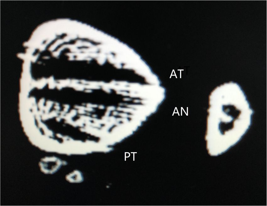
Bai et al. Journal of Orthopaedic Surgery and Research   (2020) 15:64                                                     Page 2 of 8

mechanical instability that might affect ankle function? In                  malreduction and post-malreduction groups, re-
order to further test this question, a biomechanical study                   spectively. Then, a 3D-printed guide plate was used
was performed by directly investigating the mechanical rela-                 to re-fix the specimens, in order to form the malre-
tionship between syndesmotic malreduction and its stability.                 duction model (ant-malreduction and post-
                                                                             malreduction groups). Afterwards, the same bio-
Materials and methods                                                        mechanical tests were performed, and data were
Storage and preparation of bone specimens                                    collected as the ant-malreduction and post-
Six adult (age range 49–67 years old; four males and two                     malreduction groups. Figure 1 presents the experi-
females) legs (including the complete tibia and fibula,                      mental protocol, Fig. 2 shows the experimental
ankle, and feet) were included for this study, with the                      setup, and Fig. 3 shows the CT scan of three bone
permission of the Anatomy Department of the Medical                          tunnels made by template.
College of Shenzhen University. Radiographic examina-
tions were performed to exclude pathological conditions                 Biomechanical testing
(pre-exist trauma, neoplasm, bony deformity). The whole                 Testing apparatus
specimens were stored at − 20 °C prior to the mechanical                The biomechanical test was performed using an MTS
experiments. Before the mechanical test, the entire spec-               simulator. The simulator included different channels, such
imens were thawed at room temperature for 12 h, and                     as axial displacement (proximal-distal), sagittal displace-
saline was sprayed during the thawing process to prevent                ment (anterior-posterior), coronal displacement (medial-
the specimens from drying up.                                           lateral), and torsional rotation. Moreover, the system was
                                                                        equipped with a six degrees of freedom (6DOF) force
The establishment of testable models                                    transducer (ATI-Mini 45-SI-580-20, Schunk GmbH & Co.
Before the biomechanical experiment, all the muscles                    KG, Germany), which could record the Tx (axial), Ty (sa-
and tendons of the leg were removed, while the inter-                   gittal), and Tz (coronal) displacement.
osseous membrane and proximal/distal tibiofibular syn-
desmosis were kept intact (intact model). Soft tissues of               Specimen fixation and marker placement
the foot were left to enhance fixation into the machine.                The proximal tibia of the specimens was fixed on the super-
CT scans were performed to virtually make the 3D-                       ior fixture. Then, the foot was rigidly fixed on the fixture,
printed guide template, and ensure that the bone tunnels                and the fixture was secured to the base via eight screws.
of the syndesmotic fixation, including late anatomical                     The Optotrak Certus motion analysis system (North-
fixation and malreduction fixation, do not interfere with               ern Digital Inc., Ontario, Canada) was applied to capture
each other. Then, all tibiofibular syndesmotic ligaments                the motion of the tibia and fibula. Four active optical
and the interosseous ligament were cut off to establish                 markers were firmly fixed on the middle of the tibia and
the syndesmotic separation model. A suture-button                       fibula, in order to set the base coordinate system. An-
(Arthrex, Naples, USA) was used as the syndesmotic fix-                 other two active optical markers were placed on the inci-
ation (anatomical model). In order to establish the anter-              sura of the syndesmosis on the fibular side and tibial
ior malreduction model, the fibula was moved anteriorly                 side, in order to record the initial coordinates of the two
by 1 cm, relative to the syndesmosis incisura (ant-malre-               bone landmarks. All displacements and relative angles of
duction model), or moved posteriorly by 1 cm to estab-                  the syndesmosis during the test were captured in this
lish the post-malreduction model.                                       system. Before the test, all markers were captured at a
                                                                        sampling rate of 100 Hz for 5 s to record the initial rela-
Experimental protocol and specimen allocation                           tive position between the incisura edge of the syndes-
                                                                        mosis in the tibial and fibular side. The whole set of the
  (1) Biomechanical tests were performed with all                       apparatus and detail were showed in Fig. 2.
      syndesmotic structures intact. These data was
      recorded as the intact group.                                     Biomechanical test
  (2) The ligaments of the syndesmosis were cut off, the                We set the experimental conditions according to the
      3D-printed template was used to make bone tunnels,                method of previous literature [5, 6]. At the beginning of
      and a suture-button was used to anatomically fix the              the test, the specimens were preconditioned for five cycles
      syndesmosis. Afterwards, the same abovementioned                  to ± 7.5 Nm, while a 600-N axial compressive load was ap-
      biomechanical tests were performed, and these data                plied and maintained. Subsequently, the specimens were
      were recorded as the anatomical model group.                      tested in an intact, anatomical, and anterior/posterior mal-
  (3) With the lateral fibular bone tunnels unchanged,                  reduction state in sequence. For each state, the specimens
      the fibula samples were moved anteriorly and                      were first placed in a neutrally positioned orientation
      posteriorly by 1 cm to establish the ant-                         without axial load. Then, a 600-N axial compressive load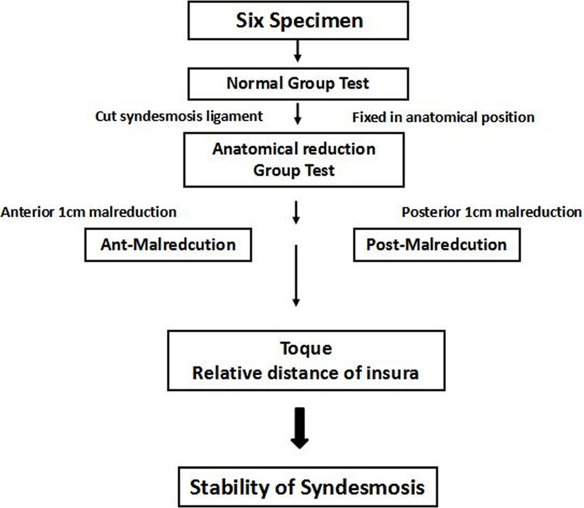
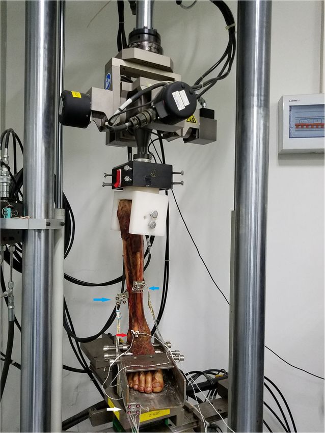
Bai et al. Journal of Orthopaedic Surgery and Research   (2020) 15:64                                                     Page 3 of 8

 Fig. 1 Experimental protocol

was applied to simulate the weight bearing load. While                  malreduction groups, and the foot was under internal
maintaining the axial compressive load, the foot was exter-             load (torque 6.75 ± 1.14 vs. 5.29 ± 0.95 Nm, t = 2.412,
nally rotated to 10°, and internally rotated to 10° (1°/min).           P = 0.036; rotation angle − 1.79 ± 0.21 vs. − 2.27 ± 0.19
During the test, the coordinates of these two rigids,                   degree, t = 4.216, P = 0.002). In addition, a significant
torque, and rotational degrees were continuously re-                    difference in torque resistance was found between the
corded. The Optotrak Certus motion analysis system can                  intact and ant-malreduction groups, while the foot was
catch and record the micro-displacement of the syndes-                  under external load (3.70 ± 0.57 vs. 3.02 ± 0.41 Nm, t =
mosis markers in three-dimensional spaces.                              2.380, P = 0.039). Fibular rotation was also detected to
                                                                        be significantly different between the intact and post-
Statistical analysis                                                    malreduction groups under both internal (− 1.79 ± 0.21
Statistical analysis was conducted in the PASW 18.0                     vs. − 2.27 ± 0.19 degree, t = 4.216, P = 0.002) and external
software (IBM Chicago, USA). The metric data were re-                   (3.57 ± 0.62 vs. 4.84 ± 0.74 Nm, t = − 3.228, P = 0.017)
ported as mean ± standard deviation (x ± SD). One-way                   loads (Table 1).
ANOVA was used to compare the differences among
groups, followed by the post hoc LSD method (homo-
geneity of variance) or Tamhane method (heterogeneity                   The 3D displacement of the syndesmotic incisura in the
of variance). A P value of < 0.05 was considered statisti-              internal/external rotation model
cally significant.                                                      Internal loading model
                                                                        No differences were found among all groups for
Results                                                                 axial displacement (intact vs. anatomical, P = 0.430;
Torque resistance and fibular rotation among the                        intact vs. post-malreduction, P = 0.326; intact vs.
different groups under internal rotation and external                   ant-malreduction, P = 0.391; anatomical vs. post-
rotation force                                                          malreduction, P = 0.842; anatomical vs. ant-malreduction,
There were significant differences in torque resistance                 P = 0.944; post-malreduction vs. ant-malreduction,
and fibular rotation angle between the intact and post-                 P = 0.897).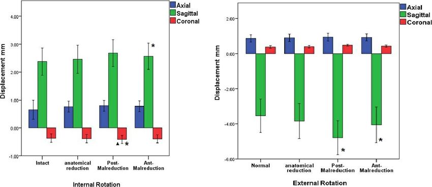
Bai et al. Journal of Orthopaedic Surgery and Research    (2020) 15:64                                                               Page 4 of 8

 Fig. 2 Experimental setup. The specimen was fixed on MTS machine. Blue arrow pointed optical marker of tibia and fibular. Red arrow pointed
 marker of syndesmosis incisura. White arrow pointed to the marker fixed on the base of the system

  There were significant differences in sagittal (anterior-               External loading model
posterior) displacement between the intact and posterior                  No differences were found among all groups for
malreduction group (P = 0.011). Also, there were signifi-                 axial displacement (intact vs. anatomical, P = 0.797;
cant difference between anatomical and posterior malre-                   intact vs. post-malreduction, P = 0.562; intact vs.
duction group (P = 0.020) in sagittal displacement.                       ant-malreduction, P = 0.656; anatomical vs. post-
However, there were no significant differences in the                     malreduction, P = 0.745; anatomical vs. ant-malreduction,
other groups (intact vs. anatomical, P = 0.787; intact                    P = 0.850; post-malreduction vs. ant-malreduction,
vs. ant-malreduction, P = 0.302; anatomical vs. ant-                      P = 0.892).
malreduction, P = 0.441; post-malreduction vs. ant-                         Merely the intact and anterior malreduction groups were
malreduction, P = 0.099) (Fig. 4).                                        significantly different in terms of sagittal displacement (in-
  No differences were found among all groups for                          tact vs. ant-malreduction, P = 0.034) (intact vs. anatomical,
coronal (medial-lateral) displacement (intact vs. ana-                    P = 0.598; intact vs. post-malreduction, P = 0.298; anatom-
tomical, P = 0.844; intact vs. post-malreduction, P =                     ical vs. post-malreduction, P = 0.600; anatomical vs. ant-
0.597; intact vs. ant-malreduction, P = 0.768; anatomical                 malreduction, P = 0.098; post-malreduction vs. ant-
vs. post-malreduction, P = 0.739; anatomical vs. ant-                     malreduction, P = 0.243).
malreduction, P = 0.922; post-malreduction vs. ant-                         For coronal displacement, merely intact and posterior
malreduction, P = 0.814).                                                 malreduction (P = 0.013), and anatomical and posterior
Bai et al. Journal of Orthopaedic Surgery and Research        (2020) 15:64                                                              Page 5 of 8

  Fig. 3 The CT scan of different bony tunnels created by 3D template. AT, anterior malreduction group; AN, anatomical group; PT, posterior
  malreduction group

malreduction (P = 0.038) were significantly different (in-                       group. It refers that malreduction does affect the stability
tact vs. anatomical, P = 0.607; intact vs. ant-malreduction,                     of syndesmosis. Huber et al. [9] confirmed through bio-
P = 0.179; anatomical vs. ant-malreduction, P = 0.394;                           mechanical studies that screw fixation of the tibiofibular
post-malreduction vs. ant-malreduction, P = 0.192)                               syndesmosis still has a joint motion range of 0.5–2.5°.
(Fig. 2).                                                                        Wang et al. [10] found that the fibula had an average rota-
                                                                                 tion of approximately 1° relative to the tibia through 3D
Discussion                                                                       image analysis. In our biomechanical study, syndesmosis
The stabilizing structure of the distal syndesmosis in-                          motion enlarged under malreduction condition that may
cludes the anterior and posterior inferior tibiofibular liga-                    also prove the overactivity had the high correlation of syn-
ment, and interosseous membrane. Distal syndesmosis is                           desmosis instability.
an amphiarthrosis, which has a joint motion range of 2–5°                           Studies have shown that when syndesmotic separation
on the coronal, sagittal, and horizontal planes [7, 8]. Al-                      occurs, its displacement does not only shift in a single
though the distal tibiofibular syndesmosis has micro-                            plane, but in both the sagittal and coronal planes [11]. If
motion, its stability is an important guarantee for normal                       the rotation and anterior/posterior displacement of the
weight bearing and function of the ankle. In our study, the                      fibula are not corrected, the fibula might be fixed in an
rotation angle of syndesmosis (fibular relative to tibia) had                    improper position, leading to syndesmotic malreduction
significant change between intact group and malreduction                         [3, 11, 12]. Moreover, due to the over-compression of the

Table 1 Data of torque (Nm) and fibular rotation (degree) among the different groups under internal/external rotation load
Load model                     Group                 Torque                  t            P            Rotation              t                P
Internal rotation              Intact                6.75 ± 1.14                                       − 1.79 ± 0.21
                               Anatomical            6.61 ± 0.99             0.239        0.816        − 1.87 ± 0.24         0.635            0.540
                               Post-malreduction     5.29 ± 0.95             2.412        0.036*       − 2.27 ± 0.19         4.216            0.002*
                               Ant-malreduction      6.13 ± 0.78             1.111        0.292        − 2.07 ± 0.22         1.111            0.292
External rotation              Intact                3.70 ± 0.57                                       3.57 ± 0.62
                               Anatomical            3.45 ± 0.37             0.818        0.432        3.82 ± 0.71           − 0.635          0.540
                               Post-malreduction     3.22 ± 0.53             1.438        0.181        4.47 ± 0.45           − 2.869          0.017*
                               Ant-malreduction      3.02 ± 0.41             2.380        0.039*       4.84 ± 0.74           − 3.228          0.009*
*Statistical significance: P < 0.05
Bai et al. Journal of Orthopaedic Surgery and Research (2020) 15:64 Page 6 of 8 Fig. 4 Summary of the sagittal, axial, and coronal displacements in the external and internal rotation models. Asterisk denotes the significant difference when compared with the intact group. Triangle denotes significant difference when compared with the anatomical group trans-syndesmotic screw, the tibiofibular space becomes ligaments were cut off, and the stress histogram revealed too small. If judged simply from the position of the fibula that the peak of the contact stress of the talus moved an- and tibia, it is possible to misjudge the restoration distance teriorly and laterally. Moreover, in clinical practice, it was [13, 14]. Through clinical follow-ups, it was found that revealed that the unbalanced stress distribution was the syndesmotic malreduction is a factor that may affect ankle main cause of ankle dysfunction and osteoarthritis [20]. function [4, 15, 16]. However, it remains unclear whether The relative micro motion of syndesmosis was reported this effect was due to the decline in ankle stability or the by many researchers. In Beumer’s biomechanical study [21], change in stress distribution of the contact area. Our study when the ankle is rotated internally or externally, the fibula proved the obvious change when syndesmosis was fixed in has an inward or outward displacement of 0–2 mm relative a non-anatomical position. The torsional stress test under to the tibia. When simulating partial weight bearing, the internal rotation conditions revealed that the torque was tibiofibular syndesmosis has a joint motion range of 3–5°. significantly smaller in the posterior malreduction group Rigby et al. [22] performed a 2-year postoperative follow-up than in the intact group. However, the torque for the an- on patients who had suture-button surgery and measured terior malreduction group was relatively normal. In con- the tibiofibular clear space (TCS), tibiofibular overlap trast, in the external rotation state, the torque was lower (TFO), and medial clear space (MCS). The results revealed in the anterior malreduction group than in the intact that although fixed by suture-button, the relative position of group, while the torque in the posterior malreduction syndesmosis had changed during follow-up time, and it re- group was relatively normal. This also indicates that when vealed a certain trend of displacement after the joint fix- the syndesmosis is not fixed at the anatomical position, its ation, and this kind of displacement was often observed in stability would decrease. Wei’s research revealed that malreduction cases. In our study, regardless of the type of when the syndesmosis was completely separated, the syndesmotic malreduction, its displacement along the axis torque of the ankle increased by more than three times of the lower extremity was not significantly different from [17]. In addition, in 3D measurements, the external rota- that in the intact group. Furthermore, even if the fibula is tion stability of the tibiofibular syndesmosis was relatively fixed to the wrong position relative to the tibia, its axial sta- weak, while the internal rotation was relatively stable [18]. bility is not affected either in the internal rotation stress However, in the external rotation state, the posterior mal- state or external rotational stress state. Therefore, in the reduction (relative external rotation) was more stable than treatment of ankle fractures, it is critical to restore the the anterior malreduction, while in the internal rotation length of the tibia and properly fix it. For sagittal and cor- state, the anterior malreduction was more stable. All these onal stability, the mode of joint motion in different dimen- increasing relative motion of the syndesmosis indicate its sions of the fibula relative to the incisura becomes the key stability decreased under the non-anatomical fixation. to determine the joint stability of the syndesmosis. The fur- LaMothe et al. [19] demonstrated that the contact stress ther multi-dimensional displacement analysis of the syndes- of the ankle joint was significantly increased after the mosis revealed that under internal rotation conditions, the
Bai et al. Journal of Orthopaedic Surgery and Research   (2020) 15:64                                                                       Page 7 of 8

anteroposterior mobility of the posterior malreduction                  Abbreviations
group (external rotation) increased, and the angle of rota-             CT: Computed tomography; MCS: Medial clear space; TCS: Tibiofibular clear
                                                                        space; TFO: Tibiofibular overlap
tion of the fibula with respect to the tibia was larger than
that of the intact group, while under external rotation con-            Acknowledgements
ditions, the position of the fibula relative to the incisura in         Not applicable.
the anterior malreduction group (internal rotation) signifi-            Authors’ contributions
cantly changed. Westermann et al. [23] reported that the                L Bai and Wt Zhang designed/performed most of the biomechanical test,
syndesmosis had a certain degree of displacement after the              and wrote the manuscript; Jx Liu did data analysis. P Chen and Sy Guan
                                                                        contributed to syndesmotic separation model and dynamical analysis. All of
suture-button fixation, and this malreduction had a ten-                the authors have read and approved the final manuscript.
dency to automatically be reduced. Our study also confirms
this finding: the internal rotation stability of the posterior          Funding
malreduction was poor, indicating that the fibula is less re-           This study was supported by a grant from the Foundation of Shenzhen
                                                                        Science and Technology Research (no. JCYJ20160428173808742), the
sistant in the direction of the reversed deforming force, and           Foundation of Shenzhen Health and Family Planning Commission
has a greater degree of motion. This also indicates that the            (SZXJ2018085), and the Sanming Project of Medicine in Shenzhen (No.
instability of syndesmotic malreduction might force the fib-            SZSM201612078).

ula to displace to the anatomic position with overwhelming              Availability of data and materials
deforming force, which could result in the excessive activity           The datasets generated and analyzed during this study are available from
of the tibiofibular syndesmosis after fixation. Similarly, Tera-        the corresponding author on reasonable request.

moto [24] performed a 3D analysis on the joint instability of           Ethics approval and consent to participate
the syndesmotic separation and found that ankle external                This study was approved by the Anatomy Department of the Medical
rotation instability could be caused by simple anterior tibial-         College of Shenzhen University.
fibular ligament damage. The above studies all revealed that            Consent for publication
under the condition that the syndesmosis was unstable, the              Not applicable.
mechanical state of the joint would be significantly changed,
                                                                        Competing interests
and the ankle function might be impaired.
                                                                        The authors declare that they have no competing interests.
   The limitations of the study were as follows: in order
to avoid the effects of multiple drilling holes, a 3D-                  Author details
                                                                        1
                                                                         Department of Sports Medicine, Peking University Shenzhen Hospital, #1120
printed guide plate was used to set different bone canals,
                                                                        Lianhua Road, Shenzhen, Guangdong Province, China. 2National and Local
and single suture-button fixation was used to minimize                  Joint Engineering Research Center of Orthopaedic Biomaterials, Peking
the measurement error between different specimens.                      University Shenzhen Hospital, #1120 Lianhua Road, Shenzhen, Guangdong
                                                                        Province, China. 3Department of Rehabilitation, Peking University Shenzhen
Some studies have shown that using merely a single but-
                                                                        Hospital, #1120 Lianhua Road, Shenzhen, Guangdong Province, China.
ton cannot achieve complete stability of the syndesmosis
[18]. However, in this study, the intact and anatomical                 Received: 21 October 2019 Accepted: 7 February 2020
groups had no significant differences in stability. Fur-
thermore, there were also studies [25, 26] that revealed                References
that the stability of the syndesmosis had no significant                1. Hunt KJ, et al. Epidemiology of syndesmosis injuries in intercollegiate
difference between suture-button fixation and screw fix-                    football: incidence and risk factors from National Collegiate Athletic
                                                                            Association injury surveillance system data from 2004-2005 to 2008-2009.
ation. Due to the limitation in sample size, the anatomy                    Clin J Sport Med. 2013;23(4):278–82.
variation of the tibiofibular syndesmosis incisura was not              2. Carr JC 2nd, Werner BC, Yarboro SR. An update on management of syndesmosis
considered [5].                                                             injury: a national US database study. Am J Orthop. 2016;45(7):E472.
                                                                        3. Gardner MJ, et al. Malreduction of the tibiofibular syndesmosis in ankle
                                                                            fractures. Foot Ankle Int. 2006;27(10):788–92.
                                                                        4. Sagi HC, Shah AR, Sanders RW. The functional consequence of syndesmotic
Conclusions                                                                 joint malreduction at a minimum 2-year follow-up. J Orthop Trauma. 2012;
                                                                            26(7):439–43.
Malreduction of the syndesmosis does affect the stability               5. Tsai J, et al. Strength of syndesmosis fixation: 2 TightRope versus 1 TightRope
of the syndesmotic fixation. No matter anterior or poster-                  with plate-and-screw construction. Foot Ankle Surg. 2017;23(2):143.
ior malreduction of the fibular, the stability of torque re-            6. Che J, et al. Novel anatomical reconstruction of distal tibiofibular ligaments
                                                                            restores syndesmotic biomechanics. Knee Surg Sports Traumatol Arthrosc.
sistance of the system was decreased under non-                             2017;25(6):1866–72.
anatomical fixation. The relative motion of the syndes-                 7. Chi PY, Lui TH. Distal tibiofibular syndesmosis: anatomy, biomechanics,
mosis under three-dimensional space was also signifi-                       injury and management. Open Orthop J. 2017;11(Suppl-4, M7):670.
                                                                        8. Hermans JJ, et al. Anatomy of the distal tibiofibular syndesmosis in adults: a
cantly changed in malreduction group. The results of the                    pictorial essay with a multimodality approach. J Anat. 2010;217(6):633–45.
study reveal the relationship between instability and syn-              9. Huber T, Schmoelz W, Bölderl A. Motion of the fibula relative to the tibia
desmosis malreduction and provide more biomechanical                        and its alterations with syndesmosis screws: a cadaver study. Foot Ankle
                                                                            Surg. 2012;18(3):203–9.
evidence on unsatisfied clinical outcome of ankle fracture              10. Wang C, et al. Three-dimensional motions of distal syndesmosis during
fixation under syndesmotic malreduction conditions.                         walking. J Orthop Surg Res. 2015;10(1):1–8.
Bai et al. Journal of Orthopaedic Surgery and Research (2020) 15:64 Page 8 of 8
11. Ebinger T, et al. 3D model analysis of existing CT syndesmosis
measurements. Iowa Orthop J. 2013;33:40.
12. Knops SP, et al. Rotational malreduction of the syndesmosis. Foot Ankle Int.
2013;34:1403–10.
13. Tornetta P 3rd, et al. Overtightening of the ankle syndesmosis: is it really
possible? J Bone Joint Surg Am. 2001;83-A(4):489.
14. Mahapatra P, Rudge B, Whittinghamjones P. Is it possible to overcompress
the syndesmosis? J Foot Ankle Surg. 2018;57(5):1005–9.
15. Michelson JD, Wright M, Blankstein M. Syndesmotic ankle fractures: a
systematic review. J Orthop Trauma. 2017;32(1):10.
16. Ntalos D, et al. Incidence and severity of malreduction of the tibiofibular
syndesmosis following surgical treatement of displaced ankle fractures and
impact on the function – clinical study and MRI evaluation. Injury. 2018;
49(6):1220–7.
17. Wei F, et al. A biomechanical investigation of ankle injury under excessive
external foot rotation in the human cadaver. J Biomech Eng. 2010;132(9):
427–35.
18. Lamothe JM, et al. Three-dimensional analysis of fibular motion after
fixation of syndesmotic injuries with a screw or suture-button construct.
Foot Ankle Int. 2016;37(12):1350–6.
19. Lamothe J, et al. Effect of complete syndesmotic disruption and deltoid
injuries and different reduction methods on ankle joint contact mechanics.
Foot Ankle Int. 2017;38(3):1071100717696360.
20. Martin RL, Stewart GW, Conti SF. Posttraumatic ankle arthritis: an update on
conservative and surgical management. J Orthop Sports Phys Ther. 2007;
37(5):253–9.
21. Beumer A, et al. Kinematics of the distal tibiofibular syndesmosis. Acta
Orthop Scand. 2003;74(3):337–43.
22. Rigby RB, Cottom JM. Does the Arthrex TightRope (R) provide maintenance
of the distal tibiofibular syndesmosis? A 2-year follow-up of 64 TightRopes
(R) in 37 patients. J Foot Ankle Surg. 2013;52(5):563–7.
23. Westermann RW, et al. The effect of suture-button fixation on simulated
syndesmotic malreduction: a cadaveric study. J Bone Joint Surg (Am Vol).
2014;96(20):1732–8.
24. Teramoto A, et al. Three-dimensional analysis of ankle instability after
tibiofibular syndesmosis injuries: a biomechanical experimental study. Am J
Sports Med. 2008;36(2):348–52.
25. Soin SP, et al. Suture-button versus screw fixation in a syndesmosis rupture
model: a biomechanical comparison. Foot Ankle Int. 2009;30(4):346–52.
26. Kim JH, et al. A comparison of screw fixation and suture-button fixation in a
syndesmosis injury at an ankle fracture. J Foot Ankle Surg. 2016;55(5):985–90.
Publisher’s Note
Springer Nature remains neutral with regard to jurisdictional claims in
published maps and institutional affiliations.You can also read