Heterochronic Phenotype Analysis of Hypodermal Seam Cells in Caenorhabditis elegans
←
→
Page content transcription
If your browser does not render page correctly, please read the page content below
Vol 9, Iss 01, Jan 05, 2019
                                           www.bio-protocol.org/e3132            DOI:10.21769/BioProtoc.3132

    Heterochronic Phenotype Analysis of Hypodermal Seam Cells in Caenorhabditis elegans
                                              Yon Ju Ji and Jiou Wang*

Department of Biochemistry and Molecular Biology, Bloomberg School of Public Health, Johns Hopkins
University, Baltimore, MD, USA
*For correspondence: jiouw@jhu.edu

[Abstract] Heterochrony refers to changes in the timing of developmental events, and it is precisely
regulated in the organisms by the heterochronic genes such as C. elegans lin-4 and let-7. Mutations in
these genes cause precocious or retarded development of certain cell lineages. With well-defined cell
lineages, C. elegans is one of the best model systems to study heterochronic genes, since the subtle
changes in the development of cell lineages can be easily identified. Among the different cell types in C.
elegans, hypodermal seam cells and their lineages are well known to be maintained by lin-14, whose
expression level is regulated by two miRNA genes, lin-4 and let-7, at the larval stages. Therefore,
analyzing the heterochronic phenotype of hypodermal seam cells in C. elegans could yield detailed
insights into the status of the miRNA pathway. Here we describe the assay protocol to analyze the
heterochronic phenotypes of C. elegans hypodermal seam cells, which can be used as a reliable method
to study the miRNA pathway.
Keywords: C. elegans, Hypodermal seam cells, Heterochronic phenotype, miRNA, lin-4, let-7, lin-14

[Background] Caenorhabditis elegans is a transparent nematode which is found in the soil. It was
introduced as a new model organism by Sydney Brenner in the 1960s to study neural development.
Since then, it has been extensively studied because of the simple anatomy, the easy cultivation, and the
rapid growth. The reproductive life cycle of C. elegans, which takes only three days, consists of the
embryonic stage, four larval stages (L1-L4), and the adult stage. After 14 h of embryogenesis, C.
elegans grows in size during the four larval stages which are divided by each molt, then it reaches the
adult stage which can produce the next generation. In unfavorable conditions, C. elegans larvae can
choose the alternative developmental pathway, so-called “dauer”, which can survive a few months in
the adverse conditions. When the environment becomes favorable, C. elegans exits the dauer stage
and develops into the L4 stage.
  In C. elegans, most hypodermal seam cells have the characteristic of stem cells. In each molt, they
divide into two daughter cells; the anterior daughter cell fuses with hyp7, which is the major hypodermis,
and the posterior daughter cell continues to divide like the stem cells until they terminally differentiate at
the L4 stages. Thus, ten hypodermal seam cells at L1 stage generate sixteen hypodermal seam cells at
the end of the L4 stage in each side (Figure 1) (Altun and Hall, 2009).
  Functionally, hypodermal seam cells are important for the formation of stage-specific cuticles that are
composed of various collagen proteins (Thein et al., 2003). They also produce cuticular alae, which are
the protruding ridges extending longitudinally along the two sides of the animal over the seam cells

Copyright © 2019 The Authors; exclusive licensee Bio-protocol LLC.                                        1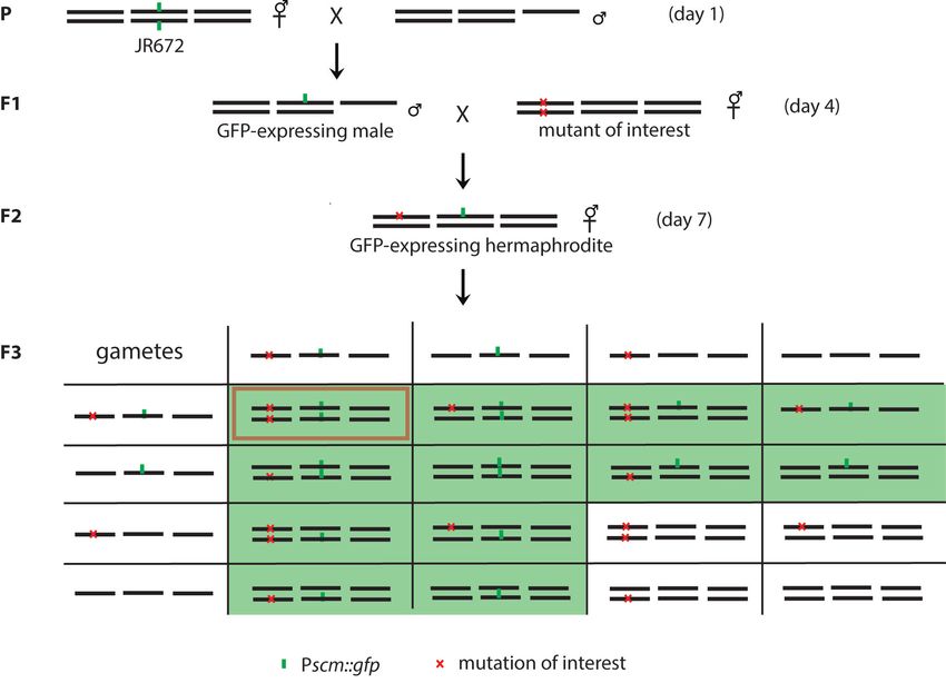
Vol 9, Iss 01, Jan 05, 2019
                                           www.bio-protocol.org/e3132              DOI:10.21769/BioProtoc.3132

(Singh and Sulston, 1978). Alae are produced only in the L1 stage, dauer larva, and adult. Therefore,
adult alae are commonly used as an indicator which shows terminal differentiation of hypodermal seam
cells. For example, lin-4 or let-7 loss-of-function mutant, which shows the retarded development of
hypodermal seam cells, has the alae defects in adult stages, while lin-14 loss-of-function mutant, which
shows the precocious development of hypodermal seam cells, has alae in the L3 stage (Hong et al.,
2000).
  The cell fate of a hypodermal seam cell is regulated by lin-14, whose expression is high in L1 animals,
and decreases by the L2 stage (Ruvkun and Giusto, 1989). The lin-14 expression is regulated by well-
characterized miRNAs lin-4 and let-7 (Ambros, 1989; Reinhart et al., 2000). It is known that lin-4 and
let-7 bind the 3’ untranslated region of lin-14 and downregulate its expression at the larval stages (Lee
et al., 1993, Slack et al., 2000). When the expression level of lin-14 is not decreased in the larval stages,
hypodermal seam cells abnormally proliferate, generating retarded phenotypes in lin-14 gain-of-function
mutants and lin-4 loss-of-function mutants.

    Figure 1. Hypodermal seam cell lineage in C. elegans. A. Ten hypodermal seam cells at the L1
    stage. These cells generate major hypodermis hyp7, hypodermal seam cells, neurons and glia
    throughout the development. B. Cell division patterns of hypodermal seam cells in each molt. Most
    of the hypodermal seam cells generate one terminally differentiated daughter cell and one stem-
    cell-like seam cell in each division. By the L4 stage, sixteen of hypodermal seam cells are generated
    in each lateral side.

  LIN-14 is a transcription factor that regulates its target gene expressions. One of the target genes of
LIN-14 transcription factor is a cell cycle inhibitor cki-1. Inactivation of cki-1 results in the division of the
vulva precursor cell (VPC) during the L2 stage. Therefore, decreasing lin-14 expression via lin-4 miRNA
is important for the development of vulva as well as the hypodermal seam cell.
  miRNAs are small, non-coding RNAs that are produced by a series of RNA-processing steps from
Copyright © 2019 The Authors; exclusive licensee Bio-protocol LLC.                                           2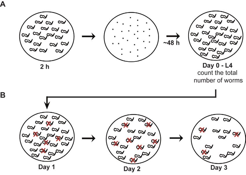
Vol 9, Iss 01, Jan 05, 2019
                                           www.bio-protocol.org/e3132           DOI:10.21769/BioProtoc.3132

their precursors known as pri-miRNAs. The pri-miRNAs are generated by transcription via RNA
polymerase II (Bartel, 2004; Lee et al., 2004) and then cleaved by the RNase III endonuclease Drosha
to produce pre-miRNAs in the nucleus (Bracht et al., 2004; Lee et al., 2002). These pre-miRNAs are
then transported into the cytoplasm by Exportin-5 (Yi et al., 2003). In the cytoplasm, the pre-miRNA is
further processed by Dicer to produce the mature 20-25 nt miRNA (Bernstein et al., 2001; Grishok et al.,
2001). The mature miRNA functions as a guide to recruit RNA-induced silencing complex (RISC), which
is composed of the miRNA:mRNA duplex, Argonaute, and other proteins (Hammond et al., 2001;
Carmell et al., 2002; Caudy et al., 2002; Mourelatos et al., 2002; Caudy et al., 2003). In the RISC
complex, Argonaute cleaves the target mRNA when it is activated. In C. elegans, there are about 24
Argonaute proteins (Bartel, 2004; Carmel et al., 2002). Among them, ALG-1 and AGL-2 are appeared
to be important for downregulating of lin-4 and let-7 targets, because alg-1 and alg-2 mutants share the
phenotypes with lin-4 and let-7 mutants. (Grishok et al., 2001).
  Here we describe the assay protocols to analyze the heterochronic phenotypes of hypodermal seam
cells in C. elegans. These assays are useful for the analysis of the miRNA pathway by taking advantage
of the fact that hypodermal seam cell fate is regulated by the well-characterized miRNAs, lin-4 and let-
7 (Zhang et al., 2018).

Materials and Reagents

    1. 60 mm Petri dishes (Fisher Scientific, FisherbrandTM, catalog number: AS4051)
    2. 100 mm Petri dishes (Fisher Scientific, FisherbrandTM, catalog number: FB0875712)
    3. Syringe Filter Unit, 0.22 μm (Millipore Sigma, Millex®-GV, catalog number: SLGV033RS)
    4. Frosted microscope slides 25 x 75 x 1.0 mm (Fisher Scientific, FisherbrandTM, catalog number:
         12-552-3)
    5. Microscopic cover glass 18 x 18 mm (Fisher Scientific, FisherbrandTM, catalog number: 12-
         542A)
    6. C. elegans wild type: N2 Bristol strain from C. elegans Genetic Center
    7. JR672 wIs54 [Pscm::gfp] V; wIs54 is the integration allele of the seam cell-specific
         transcriptional GFP reporter that is expressed in all of the seam cells at all developmental stages
         (Terns et al., 1997; Koh and Rothman, 2001)
    8. E. coli OP50-1: streptomycin resistant strain from C. elegans Genetic Center
    9. Levamisole (Sigma-Aldrich, catalog number: L9756)
    10. NaCl (Fisher Scientific, Fisherbrand, catalog number: S271, CAS 7647-14-5)
    11. Agar, Bacteriological, Ultrapure (Thermo Scientific, catalog number: J10906, CAS 9002-18-0)
    12. Peptone (BD Bioscience, BD BactoTM, catalog number: 211677)
    13. Calcium Chloride hexahydrate (Sigma-Aldrich, catalog number: 442909)
    14. Magnesium sulfate (Sigma-Aldrich, catalog number: 208094)
    15. Potassium phosphate dibasic (Sigma-Aldrich, catalog number: P3786)
    16. Potassium phosphate monobasic (Sigma-Aldrich, catalog number: P0662)

Copyright © 2019 The Authors; exclusive licensee Bio-protocol LLC.                                       3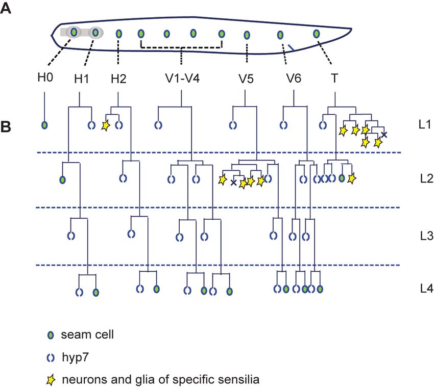
Vol 9, Iss 01, Jan 05, 2019
                                           www.bio-protocol.org/e3132      DOI:10.21769/BioProtoc.3132

    17. Cholesterol (Sigma-Aldrich, catalog number: C75209)
    18. Pure alcohol 200 proof (Pharmco products, catalog number: 111000200)
    19. Tryptone (BD Bioscience, BD BactoTM, catalog number: 211705)
    20. Yeast Extract (BD Bioscience, BD BactoTM, catalog number: 212750)
    21. Nail polish
    22. Nematode Growth Media (NGM) Agar (see Recipes)
    23. 1 M CaCl2 (see Recipes)
    24. 0.5 M MgSO4 (see Recipes)
    25. 1 M Potassium phosphate (pH 6) (see Recipes)
    26. 5 mg/ml cholesterol (see Recipes)
    27. LB agar (see Recipes)
    28. LB (see Recipes)
    29. OP50-1 culture (see Recipes)

Equipment

    1. Amsco® Century SV-120 Scientific Prevacuum Sterilizer (STERIS)
    2. 4 L flask
    3. Stir bar
    4. Stir plate
    5. PourBoy® 4 Sterile Media Dispenser (TritechTM Research)
    6. Home-made cell spreader (made with glass Pasteur pipets by bent by heating, spreader size is
         less than 1 inch.)
    7. 37 °C incubator (VWR, model: 1535)
    8. Incubator Shaker (Eppendorf, New Brunswick Scientific, model: I2500, catalog number: M1284-
         0000)
    9. 20 °C incubator for C. elegans culture (Intellus control system) (PERCIVAL, model: I-36NL)
    10. A home-made worm picker with a 5¾ glass Pasteur pipet (Fisher Scientific, FisherbrandTM,
         catalog number: 13-678-6A) and a platinum wire (Scientific Instrument Services, Inc., catalog
         number: W414)
    11. Fluorescent Stereo Microscope (Leica Microsystems, model: Leica M165FC) with Leica
         PLANAPO 2.0x objective lens
    12. Confocal laser scanning microscopy platform (Leica Microsystems, model: Leica TCS SP8) with
         Leica CTR6500 electronics box

Software

    1. GraphPad Prism 7.0a (GraphPad Software, Inc., www.graphpad.com)
    2. Leica Application Suite Advanced Fluorescence (Leica Microsystems)

Copyright © 2019 The Authors; exclusive licensee Bio-protocol LLC.                                  4
Vol 9, Iss 01, Jan 05, 2019
www.bio-protocol.org/e3132 DOI:10.21769/BioProtoc.3132
Part I: Analysis of vulva phenotypes
Procedure
A. Synchronize the populations and grow the worms (Figure 2A)
1. Transfer twenty one-day-old adult worms, which have approximately ten eggs in the uterus, onto
60 mm NGM plates seeded with E. coli (OP50-1).
Note: If your mutant of interest has smaller brood size or egg-laying defects, you may need to
transfer more worms onto each plate.
2. Make 5 replicates of plates per strain.
3. Let them lay eggs for two hours. More than fifty eggs per plate are needed. If you have less than
fifty eggs per plate, go back to Step A1 and increase the number of adult worms in each plate.
4. Remove the adult worms.
5. Let the offspring grow until they reach the L4 stage (~48 h) at 20 °C. The L4 stage can be
distinguished by a small white crescent spot in the vulva area as well as the size of the worms
(Fielenbach and Antebi, 2008).
B. Analyzing the vulva phenotypes (Figure 2B)
Figure 2. The flow chart of vulva phenotype analysis. A. Synchronization of the worm population.
Eggs are collected from young adult worms for two hours, then they are cultured for approximately
48 h until they reach the L4 stage. B. Analysis of vulva phenotype. Count the total number of worms
on Day 0. Red X indicates the worms that show vulva phenotype. Count the worms that show the
vulva defects and remove them to avoid repeated counting on following days.
1. Count the total number of L4 worms as soon as they reach the L4 stage. Typically, each plate
has approximately one hundred worms.
2. After 24 h, censor and remove the worms that show protruded vulva or bagging. Continue with
Copyright © 2019 The Authors; exclusive licensee Bio-protocol LLC. 5Vol 9, Iss 01, Jan 05, 2019
www.bio-protocol.org/e3132 DOI:10.21769/BioProtoc.3132
counting and censoring for 3 days.
3. Calculate the percentages of vulva defects (Zhang et al., 2018).
Data analysis
1. More than 250 worms should be analyzed for each strain, and three independent experiments
should be performed.
2. Calculate the percentage of worms having vulva defects using the following equation:
The number of worms showing vulva defects
× 100
The total number of worms
3. Enter the value into GraphPad Prism and analyze the mean with SEM and P-value with two-
tailed Student’s t-test.
Part II: Analysis of Alae Phenotypes (Figure 3)
Procedure
1. Synchronize the worms as described above.
2. Transfer either young adult or the L3 animals to a new plate. Place ten animals per plate. Make
five replicates per strain.
3. Analyze the alae phenotype. Under the stereomicroscope, focus on the upper side of each worm
under the 24x objective lens. You will see the protruding ridge of the alae on the upper side of
the worm from the head to the tail. Observe the alae and record whether they are intact,
discontinued, or absent in each individual animal. Refer to Slack et al., 2000 for the alae
phenotypes. In the adult stage, discontinued or absent alae are abnormal, while the presence
of alae is abnormal at the L3 stage.
4. Count the number of worms that have intact, discontinued, or absent alae (Zhang et al., 2018).
Figure 3. The flow chart of alae phenotype analysis. When synchronized worms reach the
specific stage, ten worms are transferred into the new plate and the alae phenotypes are
analyzed under the microscope.
Copyright © 2019 The Authors; exclusive licensee Bio-protocol LLC. 6Vol 9, Iss 01, Jan 05, 2019
www.bio-protocol.org/e3132 DOI:10.21769/BioProtoc.3132
Data analysis
1. For each strain, at least 50 worms are observed and three independent experiments are
performed.
2. Calculate the percentage of worms having abnormal alae.
4. Means with SEM and P-value with two-tailed Student’s t-test are calculated using GraphPad
Prism.
Part III: Analysis of the number of hypodermal seam cells
Procedure
A. Generating the desired strains expressing Pscm::gfp using JR672 (Figure 4)
Figure 4. Schematic diagrams of genetic crosses for Pscm::gfp expressing mutants. JR672
is crossed with wild-type males to get the GFP-expressing heterozygote males, which are mated
with mutants of interests on Day 4. Then, the GFP-expressing hermaphrodites in the F2 generation
are the double heterozygotes. With the Mendelian Segregation, the double heterozygotes from the
F2 generation would give rise to double homozygotes with a 1/16 chance, when two mutations are
not linked. However, since we select only the GFP-expressing worms, the chance for the double
homozygotes is increased to 1/12. The green boxes in F3 indicate the possible genotypes of GFP-
expressing worms and the red box indicates the genotype of the double mutant.
1. Prepare NGM plates for the crosses by seeding 20 μl of OP50 in the middle of 60 mm plates.
By making a small bacterial lawn, the chances of worm mating are increased.
Copyright © 2019 The Authors; exclusive licensee Bio-protocol LLC. 7Vol 9, Iss 01, Jan 05, 2019
www.bio-protocol.org/e3132 DOI:10.21769/BioProtoc.3132
2. Cross hermaphrodite of the strain JR672 with N2 males. Hermaphrodite to male ratio for the
crosses is usually 1:3.
3. On Day 3, remove the parent worms to avoid mixing of two generations.
4. On Day 4, select F1 males, which are GFP-positive heterozygote to set up a cross with
hermaphrodites of your mutant of interest. Keep the hermaphrodite to male ratio 1:3.
5. On Day 6, remove the parent worms from the plate. Now, it is expected to have only the F2
generation as larvae or eggs in the plate.
6. On Day 7, select the hermaphrodites that express GFP in the hypodermal seam cells. There is
a 50% chance to get the GFP-expressing hermaphrodites if the mating is successful. Clone out
a couple of GFP-expressing hermaphrodites, one animal per seeded plate.
7. On Day 9, remove the parent worm from the plate.
8. On Day 10, chose one plate which has a good number of worms. Discard the rest of the plates.
From the chosen plate, single out GFP-expressing worms. The chance to get the double mutant
is one out of twelve if the mutation is not in the same linkage group as the GFP locus, because
only the GFP-expressing worms are selected for analysis. Based on the chance to getting
double mutants, select the appropriate number of F3 worms. To increase probability of getting
double mutants, pick three to four times number of worms. We usually select forty worms if the
mutation of interest is not linked with Pscm::gfp. Note that Pscm::gfp is located on linkage group
V.
9. On Day 12, genotype F3 worms for the mutation of interest and select the plates which have
homozygotes. Check the plates that have homozygous mutants and identify the ones with all
F4 animals positive for GFP expression. If desired, GFP homozygotes can be selected by PCR
in F3 generation.
B. Synchronizing and preparing the worms for microscopy
1. To obtain a synchronized population of animals, allow ten to twenty of fertile adult worms to lay
eggs for two hours and let them grow until they reach the L4 stages at 20 °C as described in
Part I Procedure A.
2. Prepare the 2% agarose pads on the imaging slide as described before (Huynh et al., 2018).
3. Place ten L4 worms in 7 μl of 10 mM levamisole on the 2% agarose pad, put a coverslip and
stabilize the coverslip by painting four corners of coverslip with transparent nail polish.
4. Under the 40x objective lens, check the GFP signals. GFP is expressed in the nuclei of
hypodermal seam cells. Wild-type worms have sixteen hypodermal seam cells on each side at
L4 stage (Figure 5), thus the total number of hypodermal seam cells per worm is 32. By focusing
up and down, count the number of hypodermal seam cells on each side. In the tail, the focal
planes of right and left hypodermal seam cells are closer than others. Thus it is easier to count
the total number of hypodermal seam cells by counting one side of cells from the head to tail
and the other side from tail to head.
Copyright © 2019 The Authors; exclusive licensee Bio-protocol LLC. 8Vol 9, Iss 01, Jan 05, 2019
www.bio-protocol.org/e3132 DOI:10.21769/BioProtoc.3132
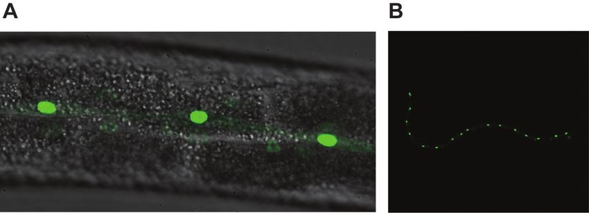
Figure 5. GFP-expression pattern of Pscm::gfp in wild-type C. elegans. A. GFP is
expressed in the nuclei of hypodermal seam cells. A 40x objective lens was used in this image.
B. Sixteen hypodermal seam cells are shown in wild-type C. elegans under the 10x objective
lens. The smear GFP signal is from the other side of hypodermal seam cells (Zhang et al., 2018).
Data analysis
1. Analyze at least 30 worms per strain to count the seam cells, and three independent
experiments are performed.
2. Count the total numbers of hypodermal seam cells per worm and calculate the average, SEM,
and P-value with two-tailed Student’s t-test using GraphPad Prism.
Notes
1. The genotype of JR672 is wIs54 [Pscm::gfp] V. Note that Pscm::gfp transgene is located on
chromosome V.
2. The GFP signal is stronger in the larval stage than in adults. Thus, select the GFP-expressing
worm as soon as they reached the L4 stage.
Recipes
1. Nematode Growth Media (NGM) Agar (2 L)
a. Add the following ingredients in a 4 L flask:
NaCl 6.0 g
Agar, Bacteriological, Ultrapure 34 g
Bacto peptone 5 g
dH2O 1,950 ml
Note: Worms do not grow well on plates prepared with agar of lower grade.
b. Add a stir bar and mix well. Leave the stir bar inside of the flask
c. Prepare 1 L of dH2O to wash the tubes for PourBoy® 4 Sterile Media Dispenser, and wrap
the tubes with aluminum foil
d. Autoclave NGM, dH2O and the tubes for PourBoy® 4 Sterile Media Dispenser using liquid
45 min cycle in Amsco® Century SV-120 Scientific Prevacuum Sterilizer
Copyright © 2019 The Authors; exclusive licensee Bio-protocol LLC. 9Vol 9, Iss 01, Jan 05, 2019
www.bio-protocol.org/e3132 DOI:10.21769/BioProtoc.3132
e. Cool down the NGM on the stir plate until it reaches approximately 55 °C. Meanwhile,
prepare 200 of 60 mm Petri-dishes
f. Once NGM cools down, add the following ingredients:
2 ml of 1 M CaCl2
4 ml of 0.5 M MgSO4
50 ml of Potassium Phosphate (pH 6) and 2 ml Cholesterol (5 mg/ml in ethanol)
g. Mix well by stirring. It is okay that NGM turns cloudy
h. Dispense 10 ml of NGM into 60 mm Petri-dish using PourBoy® 4 Sterile Media Dispenser.
The NGM plates are dried for a couple of days at RT
i. Seed OP50-1 as a food source for C. elegans. Add approximately 100 μl of OP50-1 in each
plate and spread the bacteria using a home-made cell spreader to make a small circle of
bacterial lawn. The bacterial lawn should not touch the edge of the plate. Then, culture the
OP50-1 overnight at RT
2. 1 M CaCl2
a. Dissolve 21.9 g of Calcium chloride hexahydrate (CaCl2·6H2O) in 90 ml dH2O
b. Once it is dissolved, add more dH2O to make 100 ml of 1 M CaCl2
c. Sterilize it by filtering using 0.22 μm filter units
3. 0.5 M MgSO4
a. Dissolve 12.03 g of Magnesium sulfate (MgSO4) in 90 ml dH2O
b. Once it is dissolved, add more dH2O to make 100 ml of 0.5 M MgSO4
c. Autoclave it with a liquid 45 min cycle in Amsco® Century SV-120 Scientific Prevacuum
Sterilizer
4. 1 M Potassium phosphate (pH 6)
a. Prepare 100 ml of 1 M K2HPO4 by dissolving 17.42 g of potassium phosphate dibasic in
dH2O
b. Prepare 100 ml of 1 M KH2PO4 by dissolving 13.61 g of potassium phosphate monobasic
in dH2O
c. Take 13.2 ml of 1 M K2HPO4 and add 86.8 ml of 1 M KH2PO4 to make 1 M Potassium
phosphate
d. Autoclave 1 M potassium phosphate with a liquid 45 min cycle in Amsco® Century SV-120
Scientific Prevacuum Sterilizer
5. 5 mg/ml cholesterol
Dissolve 0.5 g of cholesterol in 100 ml of ethanol
6. LB agar
a. Add the following ingredients:
NaCl 2.5 g
Tryptone 2.5 g
Yeast Extract 1.25 g
Agar 3.75 g
Copyright © 2019 The Authors; exclusive licensee Bio-protocol LLC. 10Vol 9, Iss 01, Jan 05, 2019
www.bio-protocol.org/e3132 DOI:10.21769/BioProtoc.3132
dH2O up to 250 ml
b. Mix well with a stir bar and autoclave with a liquid 45 min cycle
c. Cool it down to 55 °C and add 250 μl of 50 mg/ml streptomycin
d. Stir it on the stir plate and pour into 100 mm Petri-dish
e. Dry the plates on the bench overnight
7. LB
a. Add the following ingredients:
NaCl 10 g
Tryptone 10 g
Yeast Extract 5 g
dH2O up to 1 L
b. Dissolve by stirring and autoclave with a liquid 45 min cycle
8. OP50-1 culture
a. Streak OP50-1 on the LB agar plate containing streptomycin
b. Culture OP50-1 in 37 °C incubator overnight
c. Pick one colony of OP50-1 from the LB agar plate and culture in 20 ml of LB containing
streptomycin at the final concentration of 50 μg/ml at 37 °C shaking incubator overnight
Acknowledgments
This work was supported by grants from NIH (NS074324, NS089616), The Robert Packard Center
for ALS Research at Johns Hopkins, The ALS Association, and The Muscular Dystrophy Association.
Competing interests
The authors have no conflicts of interest or competing interests to declare.
References
1. Altun, Z. F. and Hall, D. H. (2009). Epithelial system, hypodermis. In: WormAtlas.
Doi:10.3098/wormatlas.1.13.
2. Ambros, V. (1989). A hierarchy of regulatory genes controls a larva-to-adult developmental
switch in C. elegans. Cell 57(1): 49-57.
3. Bartel, D. P. (2004). MicroRNAs: genomics, biogenesis, mechanism, and function. Cell 116:
281-297.
4. Bernstein, E., Caudy, A. A., Hammond, S. M. and Hannon, G. J. (2001). Role for a bidentate
ribonuclease in the initiation step of RNA interference. Nature 409(6818): 363-366.
5. Bracht, J., Hunter, S., Eachus, R., Weeks, P. and Pasquinelli, A. E. (2004). Trans-splicing and
polyadenylation of let-7 microRNA primary transcripts. RNA 10(10): 1586-1594.
Copyright © 2019 The Authors; exclusive licensee Bio-protocol LLC. 11Vol 9, Iss 01, Jan 05, 2019
www.bio-protocol.org/e3132 DOI:10.21769/BioProtoc.3132
6. Carmell, M. A., Xuan, Z., Zhang, M. Q. and Hannon, G. J. (2002). The Argonaute family:
tentacles that reach into RNAi, developmental control, stem cell maintenance, and
tumorigenesis. Genes Dev 16(21): 2733-2742.
7. Caudy, A. A., Myers, M., Hannon, G. J. and Hammond, S. M. (2002). Fragile X-related protein
and VIG associate with the RNA interference machinery. Genes Dev 16(19): 2491-2496.
8. Caudy, A. A., Ketting, R. F., Hammond, S. M., Denli, A. M., Bathoorn, A. M., Tops, B. B., Silva,
J. M., Myers, M. M., Hannon, G. J. and Plasterk, R. H. (2003). A micrococcal nuclease
homologue in RNAi effector complexes. Nature 425(6956): 411-414.
9. Fielenbach, N. and Antebi, A. (2008). C. elegans dauer formation and the molecular basis of
plasticity. Genes Dev 22(16): 2149-2165.
10. Grishok, A., Pasquinelli, A. E., Conte, D., Li, N., Parrish, S., Ha, I., Baillie, D. L., Fire, A., Ruvkun,
G. and Mello, C. C. (2001). Genes and mechanisms related to RNA interference regulate
expression of the small temporal RNAs that control C. elegans developmental timing. Cell
106(1): 23-34.
11. Hammond, S. M., Boettcher, S., Caudy, A. A., Kobayashi, R. and Hannon, G. J. (2001).
Argonaute2, a link between genetic and biochemical analyses of RNAi. Science 293(5532):
1146-1150.
12. Hong, Y., Lee, R. C. and Ambros, V. (2000). Structure and function analysis of LIN-14, a
temporal regulator of postembryonic developmental events in Caenorhabditis elegans. Mol Cell
Biol 20(6): 2285-2295.
13. Huynh, J. M., Dang, H. and Fares, H. (2018). Measurement of lysosomal size and lysosomal
marker intensities in adult Caenorhabditis elegans. Bio-protocol 8 (3): e2724.
14. Koh, K. and Rothman, J. H. (2001). ELT-5 and ELT-6 are required continuously to regulate
epidermal seam cell differentiation and cell fusion in C. elegans. Development 128:2867-2880.
15. Lee, R. C., Feinbaum, R. L. and Ambros, V. (1993). The C. elegans heterochronic gene lin-4
encodes small RNAs with antisense complementarity to lin-14. Cell 75(5): 843-854.
16. Lee, Y., Jeon, K., Lee, J. T., Kim, S. and Kim, V. N. (2002). MicroRNA maturation: stepwise
processing and subcellular localization. EMBO J 21(17): 4663-4670.
17. Lee, Y., Kim, M., Han, J., Yeom, K. H., Lee, S., Baek, S. H. and Kim, V. N. (2004). MicroRNA
genes are transcribed by RNA polymerase II. EMBO J 23(20): 4051-4060.
18. Mourelatos, Z., Dostie, J., Paushkin, S., Sharma, A., Charroux, B., Abel, L., Rappsilber, J., Mann,
M. and Dreyfuss, G. (2002). miRNPs: a novel class of ribonucleoproteins containing numerous
microRNAs. Genes Dev 16(6): 720-728.
19. Reinhart, B. J., Slack, F. J., Basson, M., Pasquinelli, A. E., Bettinger, J. C., Rougvie, A. E.,
Horvitz, H. R. and Ruvkun, G. (2000). The 21-nucleotide let-7 RNA regulates developmental
timing in Caenorhabditis elegans. Nature 403(6772): 901-906.
20. Ruvkun, G. and Giusto, J. (1989). The Caenorhabditis elegans heterochronic gene lin-14
encodes a nuclear protein that forms a temporal developmental switch. Nature 338(6213): 313-
319.
Copyright © 2019 The Authors; exclusive licensee Bio-protocol LLC. 12Vol 9, Iss 01, Jan 05, 2019
www.bio-protocol.org/e3132 DOI:10.21769/BioProtoc.3132
21. Singh, R. N. and Sulston, J. E. (1978). Some observations on moulting in Caenorhabditis
elegans. Nematologica 24: 63-71.
22. Slack, F. J., Basson, M., Liu, Z., Ambros, V., Horvitz, H. R. and Ruvkun, G. (2000). The lin-41
RBCC gene acts in the C. elegans heterochronic pathway between the let-7 regulatory RNA
and the LIN-29 transcription factor. Mol Cell 5(4): 659-669.
23. Terns, R. M., Kroll-Conner, P., Zhu, J., Chung, S and Rothman, J. H. (1997). A seficiency screen
for zygotic Loci required for establishment and patterning of the epidermis in Caenorhabditis
elegans. Genetics 146(1):185-206.
24. Thein, M. C., McCormack, G., Winter, A. D., Johnstone, I. L., Shoemaker, C. B. and Page, A.
P. (2003). Caenorhabditis elegans exoskeleton collagen COL-19: an adult-specific marker for
collagen modification and assembly, and the analysis of organismal morphology. Dev Dyn
226(3): 523-539.
25. Yi, R., Qin, Y., Macara, I. G. and Cullen, B. R. (2003). Exportin-5 mediates the nuclear export
of pre-microRNAs and short hairpin RNAs. Genes Dev 17(24): 3011-3016.
26. Zhang, T., Wu, Y. C., Mullane, P., Ji, Y. J., Liu, H., He, L., Arora, A., Hwang, H. Y., Alessi, A. F.,
Niaki, A. G., Periz, G., Guo, L., Wang, H., Elkayam, E., Joshua-Tor, L., Myong, S., Kim, J. K.,
Shorter, J., Ong, S. E., Leung, A. K. L. and Wang, J. (2018). FUS regulates activity of microRNA-
mediated gene silencing. Mol Cell 69(5): 787-801 e788.
Copyright © 2019 The Authors; exclusive licensee Bio-protocol LLC. 13You can also read


