Empty fetal renal fossa results of a tertiary center - Perinatal Journal
←
→
Page content transcription
If your browser does not render page correctly, please read the page content below
AL JO
          AT           U
                           R
     IN

                           N
 R
PE

                               AL
                                                                                                                                                   Original Article
                                                                                                                           Perinatal Journal 2021;29(2):148–154
                               L
PE

     IN                        A
                           N
 R

                           R
          AT       U
             AL JO
                                                                                                                                    ©2021 Perinatal Medicine Foundation

               Empty fetal renal fossa results of a tertiary center
                                                                Erzat Toprak1         İD   , fiadan Tutufl2        İD
                                    1Division   of Perinatology, Department of Obstetrics and Gynecology, Kayseri City Hospital, Kayseri, Turkey
                                                          2Department of Radiology, Kayseri City Hospital, Kayseri, Turkey

  Abstract                                                                                     Özet: Üçüncü basamak bir merkezin bofl fetal renal
                                                                                               fossa sonuçlar›
  Objective: Our aim is to present the anomalies included empty                                Amaç: Amac›m›z, 2020 y›l›nda hastanemizin perinatoloji polikli-
  renal fossa (ERF) in the ultrasonographic examination performed                              ni¤inde gerçeklefltirilen ultrason muayenelerinde bofl renal fossa-
  in the perinatology outpatient clinic of our hospital in 2020.                               n›n (BRF) yer ald›¤› anomalileri sunmakt›r.
  Methods: Ultrasonography requests were made in Kayseri City                                  Yöntem: Kayseri fiehir Hastanesi Perinatoloji Poliklini¤inde 1
  Hospital Perinatology Outpatient Clinic between January 1 and                                Ocak – 31 Aral›k 2020 tarihleri aras›nda yap›lan ultrason muaye-
  December 31, 2020, and 2405 ultrasonographic examinations were                               nesi talepleri sonucunda 1961 gebede 2405 ultrason muayenesi
  performed on 1961 pregnant women. The reports of the patients                                gerçeklefltirildi. Hastanemizden al›nan etik kurul onay› sonras›nda
  were analyzed retrospectively after the ethics committee approval of                         hastalar›n raporlar› retrospektif olarak analiz edildi. Tüm ultrason
  our hospital was obtained. All ultrasonographic examinations were                            muayeneleri, Samsung HS70A (Hampshire, Birleflik Krall›k), prob
  performed by an experienced perinatologist using Samsung HS70A                               CA 1-7A kullanan deneyimli bir perinatolog taraf›ndan gerçeklefl-
  (Hampshire, UK), probe CA 1-7A. The fetuses diagnosed with                                   tirildi. Bofl renal fossa tan›s› alan fetüsler efllik eden anomaliler için
  empty renal fossa were screened in terms of accompanying anomalies                           tarand› ve tespit edilen anomaliler kaydedildi.
  and the detected anomalies were noted.                                                       Bulgular: Gebeler 13 ile 34 (ortalama 23.9) yafl aras›ndayd› ve ge-
  Results: Pregnant women were between 13 and 34 (mean 23.9) years                             belik haftalar› 17 ile 34 hafta aras›ndayd›. Bofl renal fossa insidan-
  old and their gestational ages were between 17 and 34 weeks. The                             s› tüm gebeler için %0.75 olarak bulundu. Bofl renal fossa bulgusu
  incidence of empty renal fossa was found to be 0.75% for all pregnant                        toplam 15 fetüste gözlemlendi. Dört fetüste (%0.20) at nal› böb-
  women. An empty renal fossa finding was observed in a total of 15                            rek anomalisi, befl fetüste (%0.25) bilateral renal agenezi, iki fetüs-
  fetuses. Horseshoe kidneys were present in four fetuses (0.20%),                             te (%0.10) sol renal agenezi, üç fetüste (%0.15) pelvik böbrek (sa¤-
  bilateral renal agenesis was present in five fetuses (0.25%), left renal                     da bir ve solda iki tane) ve bir fetüste (%0.05) çapraz kaynaflm›fl ek-
  agenesis was present in two fetuses (0.10%), pelvic kidneys (one on                          topik böbrek anomalisi mevcuttu.
  the right and two on the left) were present in three fetuses (0.15%),                        Sonuç: Anhidramniyoz ile birlikte görülen BRF’nin nedeni bilate-
  and crossed fused ectopic kidney was present in one fetus (0.05%).                           ral renal agenezidir. Normal amniyotik s›v› varl›¤›nda çeflitli ne-
  Conclusion: The cause of ERF seen with anhydramnios is bilat-                                denlerden dolay› BRF düflünülmelidir ve prognoz efllik eden ano-
  eral renal agenesis. In the presence of normal amniotic fluid, it                            malilere göre de¤iflebildi¤inden, prenatal tan› do¤rulu¤unu art›r-
  should be considered that ERF may be due to various reasons and                              mam›z gerekmektedir.
  since the prognosis may change according to the accompanying
  anomalies, we must increase the accuracy of prenatal diagnosis.
  Keywords: Empty renal fossa, renal agenesis, renal ectopia,                                  Anahtar sözcükler: Bofl renal fossa, renal agenezi, renal ektopi, at
  horseshoe kidney, lying down adrenal sign.                                                   nal› böbrek, düzleflmifl adrenal bez belirtisi.

Introduction                                                                                  in the prenatal period.[1] The fetal urinary system can be
                                                                                              evaluated ultrasonographically after 11th week of preg-
Urinary system anomalies including the morphological
                                                                                              nancy. Most of the renal anomalies can be detected in the
and functional pathologies of the fetal kidney, ureter,                                       second trimester. Standard anatomical examination of
bladder, and urethra constitute 15–20% of all anomalies                                       fetal kidneys includes evaluation of bilateral renal fossa,

Correspondence: fiadan Tutufl, MD. Department of Radiology, Kayseri City Hospital, Kayseri, Turkey.
e-mail: sadantutus35@yahoo.com.tr / Received: April 11, 2021; Accepted: July 3, 2021
How to cite this article: Toprak E, Tutufl fi. Empty fetal renal fossa results of a tertiary center. Perinatal Journal 2021;29(2):148–154.
doi:10.2399/prn.21.0292005

ORCID ID: E. Toprak 0000-0002-2877-1232; fi. Tutufl 0000-0001-5936-5643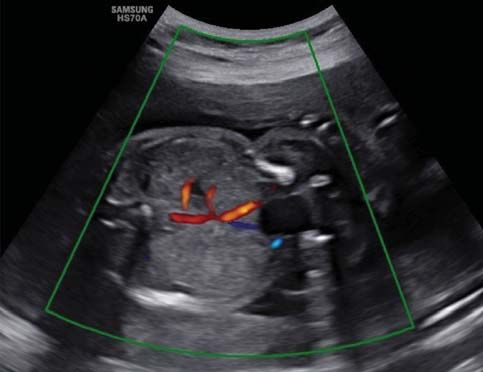
Empty fetal renal fossa results of a tertiary center

kidney size, the morphology of cortex, and collecting sys-      the biparietal diameter (BPD), head circumference (HC),
tem. The fetal kidneys are located paravertebrally, just        abdominal circumference (AC), and femur length (FL) of
below the fetal stomach level, and their pelvis faces the       the fetuses were measured. Biometry, amniotic fluid and
midline. In the third trimester, cortex-medulla distinc-        fetal anatomy were evaluated according to the
tion can be made. There are adrenal glands on top of            International Obstetrics and Gynecology Ultrasound
both kidneys.[2] Renal blood flow is evaluated with             Association (ISUOG).[6] After the “lying down adrenal
Doppler examination. When the renal anomaly is unilat-          sign” is detected in at least one renal fossa, the whole
eral, the amount of amniotic fluid is generally normal.         abdomen and thorax were scanned axially, coronally, and
Therefore, it should not be allowed to miss the diagnosis       sagittally in terms of the ectopic kidney. If the kidney is
in cases of possible unilateral renal agenesis or renal         found at the pelvic region, near the bladder, the diagno-
ectopia. It should be careful with the examination of the       sis is “pelvic kidney”. When both kidneys are fused at any
bilateral renal fossa and pelvic region.[3] When fetal kid-     point (upper or lower pole) we diagnosed a “horseshoe
neys are not observed in their normal anatomical local-         kidney”. “Crossed fused ectopic kidney” was diagnosed
ization in the ultrasonographic examination, this situa-        when both kidneys were fused end-to-end. “Renal agen-
tion is called “empty renal fossa” (ERF). The frequency         esis” was diagnosed when renal artery could not be visu-
of ERF in fetuses has been reported as 0.29% in preg-           alized in Doppler ultrasonography and kidney could not
                                                                be observed in other possible regions. Fetuses diagnosed
nancies with normal amniotic fluid, and the most com-
                                                                with ERF were also screened in terms of accompanying
mon cause is renal ectopia or unilateral renal agenesis.[3]
                                                                anomalies and the detected anomalies were noted.
Empty renal fossa pathologies include renal agenesis,
                                                                Diagnostic weeks and gender of fetuses were specified.
pelvic kidney, horseshoe kidney, and crossed fused
ectopic kidney. The “lying down adrenal sign” defined               Karyotype analysis was not recommended for cases
by Hoffman et al. in 1992 is observed in ultrasonogra-          with a diagnosis of a pelvic kidney with ERF and crossed
phy.[4] Defected migration during early embryological           fused ectopic kidney because it would not affect the fetal
development causes renal ectopia, and failure in the onset      prognosis. Karyotype analysis was recommended for all
                                                                other cases.
of pronephros-mesonephros causes the absence of kid-
neys and ureters.[3] If ERF is present, careful evaluation of      Prenatal diagnoses were confirmed by the radiologist
the fetal anatomy is required to assess for associated com-     (S.T.) in pregnancies with delivery by postnatal ultra-
plex anomalies including chromosomal disorders and              sonographic evaluation (Fig. 1).
syndromes such as VACTERL syndrome.[5]
   Our aim is to present the anomalies included empty           Results
renal fossa (ERF) in the ultrasonographic examination           Ultrasonographic examination was performed on 1961
performed in the perinatology outpatient clinic of our          pregnant women who were referred to the perinatology
hospital in 2020.                                               outpatient clinic with consultation or the suspicion of
                                                                anomaly in 2020. Racial origin of 170 pregnant women
                                                                were Asian immigrants. Of the total pregnancies, 34
Methods
                                                                were twin pregnancies. Empty renal fossa findings were
Obstetric anomaly screening, obstetric ultrasonography,         observed in a total of 15 fetuses. Pregnant women were
and obstetric Doppler ultrasonography requests were             between 13 and 34 (mean 23.9) years old and their ges-
made in Kayseri City Hospital Perinatology Outpatient           tational ages were between 17 and 34 weeks. Twelve
Clinic between January 1 and December 31, 2020, and             pregnancies were diagnosed at the second trimester
2405 ultrasonographic examinations were performed on            (between 17 and 23 weeks of gestation) and three preg-
1961 pregnant women, 34 of whom were twin pregnan-              nancies were diagnosed at the third trimester (between
cy and the reports of the patients were analyzed retro-         27 and 36 weeks of gestation). Of the 15 fetuses, 10 were
spectively after the ethics committee approval of our hos-      female and 5 were male. The incidence of ERF was
pital was obtained (decision number: 314, 18.02.2021).          found to be 0.75% for all pregnant women, 0.60% for
All ultrasonographic examinations were performed by an          Caucasian, and 1.76% for Asian immigrants. Horseshoe
experienced perinatologist (E.T.) using Samsung HS70A           kidneys was found in four fetuses (0.20%), bilateral renal
(Hampshire, UK), probe CA 1-7A. For fetal biometry,             agenesis in five (0.25%), left renal agenesis in two

                                                                                     Volume 29 | Issue 2 | August 2021   149
Toprak E, Tutufl fi
(0.10%), pelvic kidneys in three (one on the right and
two on the left) (0.15%), and crossed fused ectopic kid-
ney (0.05%) in one.
The number of unilateral ERF cases was 6 (two left
renal agenesis, three pelvic kidneys, one crossed fused
ectopic kidney) and it was observed at a rate of 40%.
Bilateral renal agenesis was observed in nine cases (60%)
with five bilateral renal agenesis, four horseshoe kidneys.
One of four fetuses with horseshoe kidneys was
found no additional anomaly and was male. One of
other fetuses with horseshoe kidneys had anencephaly,
the other two had coexistence of neural tube defects
and hydrocephalus (Chiari 2); Also, two of them had
additional lower extremity anomaly and one had major
cardiac anomaly; all of these fetuses were female. In a
fetus with a neural tube defect, kidneys were fused
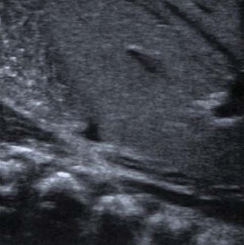
from the lower pole. Fusion was observed in the upper
pole of the remaining three fetuses with horseshoe kid-
neys (Fig. 2). In fetus number four mentioned in
Table 1, lower pole fusion was observed, and upper Fig. 1. In sonographic image, “lying down adrenal sign” of a newborn
with a pelvic kidney is seen.
pole fusion was observed in fetuses number one, two
and three. There were complex anomalies in fetuses
number one, two and four. While only the number one were evacuated with perinatologist recommendation
fetus with anencephaly was evacuated, other fetuses and family decision. The number of cases with unilat-
were delivered. The diagnosis week of fourth fetus was eral renal agenesis was two and none had a left kidney.
realized at 36th week due to the pregnancy without fol- They were both males. A persistent right umbilical
low-up. Also, in our horseshoe kidney cases, only the vein was detected in fetus number five and mild
number three fetus was male, the others were female. hydronephrosis in the right kidney in fetus number six.
One of the fetuses with bilateral renal agenesis had Both fetuses were born healthy.
dextrocardia, one had occipital encephalocele, and the There was no left kidney in all fetuses with unilat-
other had scoliosis and an echogenic focus in the heart. eral renal agenesis, hydronephrosis in the other kidney
No additional anomaly was observed in the other two in one and persistent right umbilical vein in another
fetuses. All fetuses with bilateral renal agenesis had
anhydramnios. The amniotic fluid amount was suffi-
cient in other fetuses. Four fetuses were female, only
one was male. In our cases, the number of bilateral
renal agenesis was five, and it was observed at a fre-
quency of 33.3%. In Table 1, dextrocardia in fetus
number 8, scoliosis and echogenic focus in the heart in
fetus number 9, and occipital encephalocele in fetus
number ten were detected. No additional anomalies
were found in the others. Four were females and one
was male. Fetus number seven, male, was not evacuat-
ed by family decision, Cesarean delivery occurred
when he was 35 weeks old according to the last men-
strual period. Fetus number 10 was an intrauterine exi-
tus around four weeks after diagnosis and delivery was
immediately performed. Fetus numbers eight and nine Fig. 2. The horseshoe kidney fused from the upper pole is seen.
150 Perinatal JournalEmpty fetal renal fossa results of a tertiary center
Table 1. General characteristics of fetuses with empty renal fossa (ERF).
Fetus no Maternal age Diagnosis (Week) ERF reason Accompanying anomaly Result Gender
1 (FN) 27 19 Horseshoe kidney Anencephaly Evacuation F
(upper pole fusion)
2 (FN) 13 22 Horseshoe kidney Hydrocephalus, NTD, hypoplastic Delivery F
(upper pole fusion) right ventricle, VSD, tricuspid valve (33 weeks C/S)
dysplasia, right foot talipes
3 24 21 Horseshoe kidney None Delivery M
(upper pole fusion)
4 23 36 Horseshoe kidney NTD, hydrocephalus, Delivery F
(lower pole fusion) food talipes
5 (FN) 31 34 Left renal agenesis Persistent right umbilical vein Delivery (C/S) M
6 21 21 Left renal agenesis Right hydronephrosis Delivery M
7 22 23 Bilateral renal agenesis None Evacuation not accepted; M
delivery with C/S at 35
weeks according to the
last menstrual period
8 31 22 Bilateral renal agenesis Dextrocardia Evacuation F
9 20 18 Bilateral renal agenesis Scoliosis, echogenic focus Evacuation F
10 17 17 Bilateral renal agenesis Occipital encephalocele Evacuation after F
intrauterine exitus
11 21 20 Bilateral renal agenesis None Evacuation F
12 29 21 Left pelvic kidney Echogenic focus in the left ventricle Delivery M
13 34 27 Left pelvic kidney None Delivery F
14 26 23 Right pelvic kidney None Delivery F
15 20 21 Crossed fused None Delivery (C/S) F
ectopic kidney
C/S: cesarean section; ERF: empty renal fossa; F: female; FN: foreign national; M: male; NTD: neural tube defect; VSD: ventricular septal defect.
fetus were accompanying, renal artery outflows were trimester during the investigation of the cause of anhy-
not observed on the side of agenesis in Doppler. dramnios, seven in the second-trimester screening, and
There were three pelvic kidneys in our cases, and they the remaining three in the sonographic examinations at
were observed in the left lateral in fetuses number twelve
and thirteen shown in Table 1, and in the right lateral
pelvic in fetus number 14. Fetus number 12 was male and
also echogenic focus was observed in the left ventricle of
the heart. The fetuses number 13 and 14 were female and
no additional anomalies were found in both.
Among our cases, fetus number fifteen in Table 1
had a crossed fused ectopic kidney and it was a twin, no
extrarenal anomaly detected. The left renal fossa was
observed empty (Fig. 3). It was a female and was deliv-
ered healthily. No anomaly was found in the other twin.
Horseshoe kidney anomaly with anencephaly and
Chiari 2 anomaly was found in two of the Asian immi-
grants and left renal agenesis with persistent right
umbilical vein was detected in the third pregnant.
Among the 15 fetuses with ERF, five fetuses with Fig. 3. In the case of cross-fused ectopic kidney on Doppler ultraso-
bilateral renal agenesis were diagnosed in the second nography, both renal arteries are observed on the same side.
Volume 29 | Issue 2 | August 2021 151Toprak E, Tutufl fi
27, 34, and 36 weeks of gestation. A total of five fetus- horseshoe kidneys in their study. In addition, the gen-
es were evacuated. Four of them were fetuses with der of the patients with accompanying anomalies were
bilateral renal agenesis and the other with anencephaly 17 male and 15 female.[10] The four horseshoe kidneys
with horseshoe kidney. detected in our study were found with a rate of 26.6%
Fetal karyotyping was offered in 11 cases and only among all ERF cases. Central nervous system, cardiac
was performed in one. Fetal karyotype of fetus number and skeletal system anomalies were seen together in
three with isolated horseshoe was normal. Genetic 75% of our horseshoe kidney cases. Since the prenatal
analysis could not be done in other cases as their fami- diagnosis of isolated horseshoe kidney is more difficult,
lies did not want fetal karyotype. we think that we encountered horseshoe kidney more
Fetuses number one, two, and five of our 15 cases frequently in fetuses with complex anomalies in our
with ERF belonged to Asian immigrants. a foreign cases. We hypothesize that it is due to the fact that
family. The first two had horseshoe kidneys and other most of the pregnant women were sent to our clinic
accompanying system anomalies were present. Fetus with suspected anomaly. Contrary to the literature,
number five had left renal agenesis and persistent right horseshoe kidney anomaly was observed three times
umbilical vein. While fetus number one was evacuated, more frequently in female fetuses. This might be due
other fetuses were delivered. to the small sample size.
General characteristics of fetuses with ERF are Renal agenesis is characterized by a congenital
summarized in Table 1. absence of the kidney. Bilateral renal agenesis is
incompatible with life. Unilateral renal agenesis is 3–4
times more common than bilateral renal agenesis and
Discussion is three times more common in males.[11] Unilateral
An ideal method for evaluating the presence and renal agenesis can be diagnosed after ectopic kidney
growth of the fetal kidneys is prenatal sonography. and renal hypoplasia have been ruled out. Cho et al.[12]
Kidneys can be seen at 12 weeks of gestation. They are measured and compared the anteroposterior and trans-
located on either side of the spine in the posterior verse diameter ratios of unilateral renal agenesis,
abdomen. When no kidney is visualized in its normal ectopic multicystic dysplastic kidney, pelvic kidney and
paravertebral location, the diagnosis of an empty renal healthy fetus kidneys in the second and third
fossa is made on prenatal ultrasonography, adrenal trimesters. Unilateral renal agenesis and ectopic multi-
gland is oriented longitudinally in the renal fossa. cystic dysplastic kidney were observed with compensa-
The most common renal fusion anomaly of the kid- tory hypertrophy in the third trimester compared to
ney is the horseshoe kidney occurring in 1 in 400 normal kidneys, while compensatory hypertrophy was
births, with twice as many in males.[7,8] Although it is not observed in the pelvic kidneys. In bilateral renal
common in the postnatal period, prenatal diagnosis is agenesis, anhydramnios accompanies after the 16th
made less frequently.[9] Kidneys are fused on both sides gestational week, the fetus bladder cannot be visualized
of the midline to contain equal amounts of renal tissue. even if the examination is repeated. Since adrenal
Kidneys frequently undergo horseshoe fusion from the glands extend towards the renal fossa in cases of agen-
lower poles. The kidneys appear medially or anteriorly esis, it should not be considered as a kidney by mistake.
rotated. The horseshoe kidney shape can be traced in The normal sonographic appearance of the adrenals is
the coronal plane. Upper pole, both upper and lower observed as a small inverted “Y” or “V”, they have a
pole, or alternative fusions are less common.[8] central hyperechogenic medulla and peripheral hypoe-
Although the horseshoe kidney clinic is silent in the choic cortex, and they do not have a pelvis.[13] The renal
adult population and there is no increase in the risk of artery cannot be visualized in color Doppler examina-
tumors or urinary system stones, the risk of anomalies tion in agenesis. Since the adrenal gland artery is sig-
is three times higher in these fetuses and is also associ- nificantly smaller than the renal artery, it can be distin-
ated with chromosomal syndromes such as Turner or guished by careful examination.[5] Retroperitoneal
trisomy 18.[5] Boatman et al.[10] reported that complex colon segment filled with hypoechoic content filling
anomalies (mainly skeletal, cardiovascular, central ERF in the third trimester may also be mistaken for a
nervous system, and anorectal malformations) were kidney. Careful examination reveals that the corti-
observed in almost one third of the patients with comedullary separation is not noticed and the renal
152 Perinatal JournalEmpty fetal renal fossa results of a tertiary center
artery is not observed in the Doppler.[5] Since the rate fetuses with crossed fused ectopic kidneys, they found
of occurrence with chromosomal anomalies and genet- a double collecting system in two cases, bilateral
ic syndromes in bilateral or unilateral renal agenesis hydronephrosis in one, a single umbilical artery in
can reach up to 30%, the fetus should be examined sep- four, ventricular septal defect in one, and persistent left
arately in terms of accompanying anomalies.[11] In a superior vena cava in one.
study by Tutufl it was reported that Blake pouch’s cyst Chow et al.[15] retrospectively examined unilateral
and also corpus callosum agenesis were observed in a ERF cases between 1989 and 2003 and identified 93 ERF
patient with bilateral renal agenesis.[14] Unlike the liter- cases. The most common cases were renal agenesis
ature, the number of bilateral renal agenesis was high- (47%), ectopic kidney (42%), normal localization (11%),
er in our cases than unilateral agenesis. We think that but no dysplastic kidney cases. They pointed out that
it is due to the fact that most of the pregnant women 42% of the cases were accompanied by multisystemic
were referred with the suspicion of anomaly. The fact anomalies. Markov et al.[24] prospectively investigated
that both fetuses with unilateral renal agenesis were ERF cases between 2007 and 2010 other than bilateral
male was consistent with the literature.[11] renal agenesis. They identified 9 cases of ectopic kidney
Unilateral ERF is caused by the pelvic kidney at a (seven of them pelvic kidney, one iliac settlement and one
rate of approximately 42%[15] and its frequency is 1 in 500 crossed fused ectopic kidney) and 8 cases of unilateral
to 3000 live births.[16,17] It occurs when the upward migra- renal agenesis. While 16 of the 17 cases were isolated
tion of the kidney towards the lumbar area is impaired anomalies, they pointed out that there was tetralogy of
between the sixth and the tenth gestational weeks.[18] A Fallot and a single umbilical artery association in only
pelvic kidney can mimic renal agenesis in prenatal ultra- one case that resulted in evacuation. They noted that a
sonography. When an empty renal fossa and lying down case with unilateral renal agenesis was diagnosed after
adrenal sign are detected, possible renal locations should birth. The number of unilateral ERF cases in our study
be screened before the diagnosis of renal agenesis is was 6 and the number of bilateral ERF cases was 9, and
made. Also, in unilateral renal agenesis, hypertrophy can complex multisystem anomalies were observed in bilater-
be observed in the contralateral kidney even in the al ERF cases. However, due to the small number of cases,
intrauterine 22nd week, while the contralateral kidney it may not be very accurate to compare.
size in the pelvic kidney is within normal limits.[12] Most As it is known, the frequency of congenital anom-
ectopic kidneys are located within the bony pelvis. Hill alies may differ depending on race, socioeconomic
et al.[19] defined the pelvic kidney as ipsilateral or midline, level, geographical regions, and environmental factors,
but Meizner et al.[20] found that postnatally opposite the so the rate of ERF in Asian immigrants in our study
sacrum and below the aortic bifurcation. In the study was approximately three times higher than Caucasians.
conducted by Yüksel and Batukan, 24 of 40 ERF cases However, this excess should not be generalized due to
with at least one ERF and normal amniotic fluid amount the small number of our cases.
were found to have pelvic kidney and 13 had unilateral
The low number of cases and the families not want-
renal agenesis.[3] In the cases in our study, our rate of a
ing fetal karyotype are the limitations of our study.
pelvic kidney was 20% among all ERFs and its incidence
Diversity with a higher number of cases and associa-
among unilateral ERFs was 50% and it was consistent
tion with the genetic study will shed more light on the
with the literature.[3,15]
subject we touched upon.
A crossed fused ectopic kidney is defined as the
fusion of one kidney with its ureter crossing the mid-
line to the other kidney. Its frequency is reported as 7.5 Conclusion
per 10,000 newborns.[21] There are several subtypes of The cause of ERF seen with anhydramnios is bilateral
the crossed fused ectopic kidney. It can be seen as sig- renal agenesis. In the presence of normal amniotic fluid,
moid or “S” shaped, lump shaped, “L” shaped dis- it should be considered that ERF may be due to various
coid.[22] A crossed fused ectopic kidney can be isolated, reasons and since the prognosis may change according to
or it can be seen especially with uterine anomalies, the accompanying anomalies, we must increase the accu-
imperforate anus, or skeletal anomalies.[5] Jazicek et racy of prenatal diagnosis. In our cases, the incidence of
al.[23] retrospectively analyzed 185 fetuses with ERF ERF was higher according to the total number of preg-
between 2005 and 2015 and when they examined 10 nant women examined. This was attributed to the spe-
Volume 29 | Issue 2 | August 2021 153Toprak E, Tutufl fi
cial-problematic cases being sent to the perinatology 11. Deshpande C, Hennkam RCM. Genetic syndromes and prena-
outpatient clinic. tally detected renal anomalies. Semin Fetal Neonate Med 2008;
13:171–80. [PubMed] [CrossRef]
Funding: This work did not receive any specific grant from 12. Cho JY, Moon MH, Lee YH, Kim KW, Kim SH. Measurement
funding agencies in the public, commercial, or not-for-prof- of compensatory hyperplasia of the contralateral kidney: use-
it sectors. fulness for differential diagnosis of fetal unilateral empty renal
fossa. Ultrasound Obstet Gynecol 2009;34:515–20. [PubMed]
Compliance with Ethical Standards: The authors stated [CrossRef]
that the standards regarding research and publication ethics,
13. Majmudar A, Cohen HL. “Lying-down” adrenal sign: there
the Personal Data Protection Law and the copyright regula-
are exceptions to the rule among fetuses and neonates. J
tions applicable to intellectual and artistic works are com-
Ultrasound Med 2017;36:2599–603. [PubMed] [CrossRef]
plied with and there is no conflict of interest.
14. Tutufl fi. The incidence and distribution of anomalies found in
the pregnant women applied to Kayseri City Hospital for obstet-
References ric ultrasound in 2019: a retrospective analysis. Perinatal Journal
1. Rodriguez MM. Congenital anomalies of the kidney and the 2021;29:54–62. [CrossRef]
urinary tract (CAKUT). Fetal Pediatr Pathol 2014;33:293– 15. Chow JS, Benson CB, Lebowitz RL. The clinical significance
320. [PubMed] [CrossRef] of an empty renal fossa on prenatal sonography. J Ultrasound
2. Dias T, Sairam S, Kumarasiri S. Ultrasound diagnosis of fetal Med 2005;24:1049–54; quiz 1055–7. [PubMed] [CrossRef]
renal abnormalities. Best Pract Res Clin Obstet Gynaecol 16. Cinman NM, Okeke Z, Smith AD. Pelvic kidney: associated
2014;28:403–15. [PubMed] [CrossRef] diseases and treatment [review]. J Endourol 2007;21:836–42.
3. Yüksel A, Batukan C. Sonographic findings of fetuses with an [PubMed] [CrossRef]
empty renal fossa and normal amniotic fluid volume. Fetal 17. Bader AA, Tamussino KF, Winter R. Ectopic (pelvic) kidney
Diagn Ther 2004;19:525–32. [PubMed] [CrossRef] mimicking bulky lymph nodes at pelvic lymphadenectomy.
4. Hoffman CK, Filly RA, Callen PW. The “lying down” adrenal Gynecol Oncol 2005;96:873–75. [PubMed] [CrossRef]
sign: a sonographic indicator of renal agenesis or ectopia in fetus- 18. Moore KL, Persaud TVN. The developing human. Clinically
es and neonates. J Ultrasound Med 1992;11:533–6. [PubMed] oriented embryology. 6th ed. Philadelphia, PA: WB Saunders;
[CrossRef] 1998. p. 305.
5. Oh KY, Holznagel DE, Ameli JR, Sohaey R. Prenatal diagnosis 19. Hill LM, Grzybek P, Mills A, Hogge WA. Antenatal diagno-
of renal developmental anomalies associated with an empty renal sis of fetal pelvic kidneys. Obstet Gynecol 1994;83:333–6.
fossa. Ultrasound Q 2010;26:233–40. [PubMed] [CrossRef] [PubMed]
6. Salomon LJ, Alfirevic Z, Da Silva Costa F, Deter RL, Figueras 20. Meizner I, Yitzhak M, Levi A, Barki Y, Barnhard Y, Glezerman
F, Ghi T, et al. ISUOG Practice Guidelines: ultrasound assess- M. Fetal pelvic kidney: a challenge in prenatal diagnosis?
ment of fetal biometry and growth. Ultrasound Obstet Ultrasound Obstet Gynecol 1995;5:391–3. [PubMed] [CrossRef]
Gynecol 2019;53:715–23. [PubMed] [CrossRef] 21. Arena F, Arena S, Paolata A, Campenni A, Zuccarello B,
7. Strauss S, Dushnitsky T, Peer A, Manor H, Libson E, Romeo G. Is a complete urological evaluation necessary in all
Lebensart PD. Sonographic features of horseshoe kidney: review newborns with asymptomatic renal ectopia? Int J Urol 2007;14:
of 34 patients. J Ultrasound Med 2000;19:27–31. [PubMed] 491–5. [PubMed] [CrossRef]
[CrossRef]
22. Glodny B, Petersen J, Hofmann KJ, Schenk C, Herwig R, Trieb
8. Glodny B, Petersen J, Hofmann KJ, Schenk C, Herwig R, Trieb
T, et al. Kidney fusion anomalies revisited: clinical and radiolog-
T, et al. Kidney fusion anomalies revisited: clinical and radiolog-
ical analysis of 209 cases of crossed fused ectopia and horseshoe
ical analysis of 209 cases of crossed fused ectopia and horseshoe
kidney. BJU Int 2009;103:224–35. [PubMed] [CrossRef]
kidney. BJU Int 2009;103:224–35. [PubMed] [CrossRef]
9. Kovo-Hasharoni M, Mashiach R, Levy S, Meizner I. Prenatal 23. Zajicek M, Perlman S, Dekel B, Lahav E, Lotan D, Lotan D,
sonographic diagnosis of horseshoe kidney. J Clin Ultrasound et al. Crossed ectopic kidney: prenatal diagnosis and postnatal
1997;25:405–7. [PubMed] [CrossRef] follow-up. Prenat Diagn 2017;37:712–5. [PubMed] [CrossRef]
10. Boatman DL, Kolln CP, Flocks RH. Congenital anomalies asso- 24. Markov D, Atanassova D, Pavlova E, Markov P. Empty renal
ciated with horseshoe kidney. J Urol 1972;107:205–7. [PubMed] fossa – a prenatal diagnostic dilemma. [Article in Bulgarian]
[CrossRef] Akush Ginekol (Sofiia) 2010;49(5):13–9. [PubMed]
This work is licensed under the Creative Commons Attribution-NonCommercial-NoDerivs 4.0 Unported (CC BY-NC-ND4.0) License. To view a
copy of this license, visit http://creativecommons.org/licenses/by-nc-nd/4.0/ or send a letter to Creative Commons, PO Box 1866, Mountain View, CA
94042, USA.
Publisher’s Note: The content of this publication does not necessarily reflect the views or policies of the publisher, nor does any mention of trade names, commercial products, or
organizations imply endorsement by the publisher. Scientific and legal responsibilities of published manuscript belong to their author(s). The publisher remains neutral with regard
to jurisdictional claims in published maps and institutional affiliations.
154 Perinatal JournalYou can also read