Diagnostics in Trauma: Looking into the Shadows - Daria C. Ruffolo Loyola University Medical Center
←
→
Page content transcription
If your browser does not render page correctly, please read the page content below
Diagnostics in Trauma:
Looking into the Shadows

            Daria C. Ruffolo
    Loyola University Medical Center-
                Chicago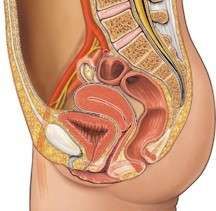
druffol@lumc.edu
 708-216-4541
At the end of this session the
participant will be able to:
Describe the impact radiographic
diagnostic therapies have on the
trauma population
Discuss the indications for studies for
major body systems and injuries
Identify the most current treatment
radiographic diagnostic and
modalities in trauma care
Take a napBasic Concepts
> 1/3 are emergent
They are sick
Will require either GETA or conscious
sedation
Be PREPARED for any and everything
*integrity of access
*check labs
*bring meds with you if needed
*check suction and 02Keep the patient SAFE *informed *warm *monitored *comfortable *breaks in technique *positioning *secure connections
Check site and distal
 pulse over and over and
 over again!
   Feel under the
 patient!
X-ray is shortwave of
electromagnetic radiation that
penetrates matter.
Medically useful and can be harmful
The radiation waves are absorbed in
black and white hues---the more
dense the lighter the color on film
Brightest to darkest, most dense to
least dense
Metal>bone>blood>brain>soft
tissue>water>fat>airDensities
Structure Color Example
Gas/air black pneumo
Fat grey tissue
Fluid white +/- vessels
heart/muscle
Bone/metal white bone/foreign
objectiveTricks
RIGHT PATIENT RIGHT FILM RIGHT TIME
Work around the patient NOT he around
you
Positioning is KEY
Symmetry is your friend
Look again and again and again
Look at ALL films then abnormal will
become more obvious
Look at all films in the same orderThere is NO substitute for looking at, touching, and speaking to the patient!!
Common Associated Injuries
Maxillofacial             Cervical spine
Ribs 6-12                  Liver/spleen
AP compression pelvic fx IP bladder
Lateral compression pelvic EP bladder
Lap belt        Mesentery, bowel,
                    Chance injury
Shoulder belt      Carotid, C spine
                    injury
THINK ENERGY
Overview of Chest X-ray Positioning Technique Indication Full inspiration Structures reviewed systematically
Order to Review Structures Side to side Top to bottom Structures
Structures Lung fields Trachea Mediastinum Bony structures/ribs Diaphragms Structures below diaphragms Intercostal spaces Soft tissues Tubes
The Radiologic ABC’s
GOAL: To detect correctable
  problems that cause hypoperfusion
  or hypoxia
AIRWAY:
-ETT position is unreliable on PE
  ~75%
-~10% are malpositioned on CXR
-~3 cm above the carina
-Remember neck excursion is ~ 4cm
Thoracic Aortic Disruption
Defined
• MOI deceleration
• Typically patients will survive the initial injury insult
30% mortality in 6 hrs
50% mortality in 24 hrs
70% mortality in 1 week
• Injury may be confined to areas of aorta attachment
• Signs & Symptoms
Rapid and deterioration of vitals
Pulse deficit between right and left upper or lower
extremitiesSite of Disruption • Locations Ligamentun arteriosum – 85% Ascending root Descending diaphragm attachment
Avulsion Sites
           Ligamentum arteriosum
Why Are These Injuries So
Morbid (5-28%)?
Left thoracotomy
Single-lung ventilation
Aortic cross-clamping
Interposition grafting
Systemic heparinizationThoracic Aortic Disruption
Aortography
• *Gold Standard*
CT
• Spiral
• Specificity comparable to A-gram
• Utilized on stable patients
TEE
• Utilized on unstable patients to be “lost” to the
OR
• Positive OR
• Negative A-gramThoracic Aortic Disruption
Management
• ABC’s
• Maintain normotension P/ T - -
blockade < 135/70
• Operative management
• Post-operative care
“Clamp-time sequelae”
Maintain normotensionThoracic Aortic Disruption
• THINK MOI
• Radiographic studies
Plain film
•Wide mediastinum*
•Loss of aortic knob*
•Tracheal deviation
•Left pleural cap
•Thickening/deviation
tracheal stripe
*Watch Quality of Film*Thoracic Aortic Disruption
Aortography
• *Gold Standard*
CT
• Spiral
• Specificity comparable to A-gram
• Utilized on stable patients
TEE
• Utilized on unstable patients to be “lost”
to the ORCT Scan Evaluation of the TAI What is a “Multi-detector scanner”? --A rapid scanner --Multi-slicer thin sections (2.5 mm) --Power contrast injectors --Multiplannar reformats
Is CT the Answer?
Multi-center trials
Results > 95% sensitivity/>93%
specificity in evaluating the TAI
Branch vessels, unstable patients in need
of potential embolization; angiography
remains the diagnostic of choice
Miller, et al JOT 4/05 Maturen et al JOT 3/10If They Need Intervention Does
Everyone Get Their Chest
Cracked?
Endoluminal grafting initiated in the
1980’s for AAA
Application to the TAI
Multiple centers have initiated this
approach
Improved outcome
--less morbid
--less bleeding
--earlier mobilizationTreatment Options
Surgical repair:
• The treatment of choice is surgical
repair.
• Two options are “clamp and sew” and
distal aortic perfusion techniques.
Endovascular stent-graft placement:
• A minimally invasive approach to aortic
repair.
• A small incision is made in the femoral
artery and with the aid of angiography
and fluoroscopy, a self-expanding wall
stent is placed in the region of traumaTraumatic Brain Injury
And Imaging
1. In acute head injury the CT is
done without contrast
2. Review the brain, blood, and
bone images
3. Clinical exam is VITAL
4. Always compare studies
5. Watch for mass effectCerebral Contusion
Mechanism can be coup/contre-
coup
Pathologically consists of small
hemorrhagic areas with necrotic
core
Mostly involves poles
“Blossoms”
Defects are determined by the
locationEpidural Hematoma
Classic lucid interval seen 30%
Often caused from middle meningeal
artery bleed
Biconvex finding with sharply
demarcated against the calvarium.
Skull fracture seen ~90%Poor prognosis: -EDH > 2cm -midline shift > 1.5 cm -brainstem deformity -extensive associated injuries
Acute Subdural Hematoma
Below the dura and superficial to the
arachnoid space
“Smudge” appearance
Tearing of bridging veins
SDH with contusion has 2X mortality
of SDH alone
Better visualized in blood windows
Often associated with midline shiftChronic Subdural Hematoma
SDH is > 3 weeks in age
~ 60 yo is peak age
50% report no injury
Bleed with osmotic expansion into
the SH space
Low density extra axial collection
that conforms to the
skull(hypodense)
May be better visualized on MRDepressed Skull Fracture
DSF are elevated if the inner and
outer tables are compromised, if
there is an open wound, cosmesis is
an issue, or the is a deficit
Utilize the bone windows to get a
good interpretationFirst Things First—Can You
Clinically Clear Them?
NEXUS study says YES! (>90%)
--Are they drunk, head injured, obtunded,
have distracting pain or neck pain? NO!
Clear them!
Current information is a little unclear
Recent publication of a large randomized,
prospective study found ~ 70% sensitivity
(few needed tx)
How is the clinical exam???
(Duane, Ivatury, et al JOT 6/08)Cervical Spine If you cannot clear them clinically how do you study them? Plain film: 45% sens 97% spec CT c-spine: 98% sens 99.5% spec High powered, prospective trials Plain film can miss ~55% of injuries that are clinically significant
Blunt Cerebrovascular Injury of the
Neck (BCVI)
The incidence is < 1%
Associated stroke when untreated
~30%
Risk factors: Clinical
--Hemorrhage nose/mouth
--Expanding neck hematoma
--Wounds over neck region
--Unexplained lateralizing signs Risk factors: Mechanism
Hyperflexion/hyperextension/rotation
Complex midface or mandibular
injury
Near-hanging
Seat-belt sign or swelling over
anterior neck
Biffl et al JOT 6/10CT for Evaluation of Abdominal
Injuries
Computed tomography (CT) can
evaluate solid organ injury,
intra-abdominal fluid/blood/air,
and retroperitioneal organ injury
in hemodynamically stable
patients using both oral and
intravenous contrast.CT
Contraindications:
i) hemodynamically
unstable patient
ii) obvious need for
laparotomyCT
Can miss pancreatic and
diaphragmatic injuries-especially
early on
Excellent at diagnosis and evaluation
of solid organs
Helical brings evaluation of hollow
viscous injury to 94%
Debate the need for oral contrastHepatic injuries
Due to size and location
commonly injured in trauma
70% blunt
30% penetrating
Unique because of its
regeneration capabilitiesAAST Organ Injury
Grade Hematoma
Laceration
I 3cm
AB
IV Ruptured parenchyma
Lobe fx
V Ruptured parenchyma
VC/Hep
disruptionNon-Operative Management
(NOM)
80% are NOM (AAST, 2001)
Much less likely to have delayed
bleeding < 10% (Knudson, 2004)
Undetermined which grades may be
treated NOM
REQUIRES SERIAL CT EXAM AND
EXPECTANT CARESplenic Injuries Single most injured organ in blunt trauma 10% mortality S & S: -Profound shock from blood loss -Positive Kehr’s (50%) -Left rib fractures -Generalized abdominal pain (30%) -Palpable mass in left flank
How is the Spleen Damaged?
Side impact (particularly smaller
cars)
Restraint devices
Falls (usually striking stationary
object with height/weight/velocity)
Contact sportsAAST Organ Injury
Grade Hematoma Laceration
I 3 cm AB
IV Ruptured Parenchyma Hilar lac/
Devitalized
V SHATTERED/DEVITALIZED“Why Save the Bloody Thing?”
Overwhelming Post-Splenectomy
Infection (OPSI)
-first identified in the 1950’s
-incidence ~.05-2 %
-death rate in presence of OPSI ~
50%Splenic Function
Opsonin production
~25% of reticuloendothelial cell
mass
Clearance of blood borne bacteria
Reservoir of ~ 200 cc bloodPseudoanuerysm (PS)
There is no one single finding more
predictive of DSR then that of a PS
The process was first described in
1995 by Sugg et al
The work of Davis, 1998, 2009 et al
reported the presence of a “blush”
on CT“The Blush”
Def. A well circumscribed,
intraparenchymal contrast collection
that represents the formation of a
pseudoanuerysm of the splenic
artery branches.What Then Shall We do?
Shurr, Fabian et al, JOT 1995 the
blush needs angiography and
embolization
Davis, JOT 2009 decreased the DSR
rate from 13% to 6% with this
practice
Bee, JOT 2006 presence of large
volume of hemoperitoneum and
blush there was > 24x DSR rateHemodynamically Stable
|
CT with contrast
Blush | No Blush
| Worsens |
OR
Arteriogram Observe
| | |
Successful Unsucc. CT 24-48 hrs.
|
| | |
Observe OR Blush No Blush
(Bee, 2005 JOT)Focused Abdominal Sonography
for Trauma (FAST)
Ultrasound has been used
extensively in Europe and Japan
for 20 years and is now being
used increasingly in North
America, primarily as a rapid
noninvasive means of identifying
hemoperitoneum in the trauma
resuscitation area Advantages --quick --non-invasive --requires no contrast (full bladder) --reproducible Disadvantages --learning curve --artifact DPL fluid, bladder injury, women --? accuracy in obese patients
FAST
Indications: are likely similar to the
  indications for DPL. Its major role may be
  in the rapid diagnosis of hemoperitoneum.

Contraindications: to the use of ultrasound
 are an obvious need for laparotomy and
 inadequate training or experience of the
 trauma team member performing the
 examination.

Accuracy: Reports of sensitivity (>90%) and
  specificity vary.
Pelvic Fractures
Epidemiology
Pelvic fractures account 1-3% of all
fxs
60% Male
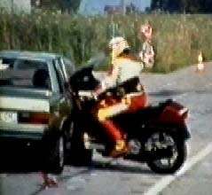
Mechanism
• MVC (57-71%)
• Collision w/ pedestrian (13-18%)
• Motorcycle accident (5-9%)
• Falls (4-9%)
• Crush injury (4-5%)Key Point
Presence of a pelvic fracture
indicates the profound magnitude of
disruptive energy at the time of
injury
• Alerts to likelihood of major injury to
other body systemsEpidemiology
Overall reported mortality figures for
pelvic injuries range from 8%-13%
• Higher energy injuries greater mortality
• Peds vs car (23%)
Poole GV, Ward EF: Causes of mortality in patients with pelvic fractures, Orthop
17:691, 2008.
Pohlemann T et al: Pelvic fractures: epidemiology, therapy and long term
outcome. Overview of the multicenter studey of the pelvis study group,
Unfallchirurg 99:160, 2006.Pelvic Anatomy
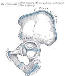
Inominate bones
(2)
• ilium, ischium &
pubis
Sacrum
CoccyxQuestion?
Name the 2 major complications that
are directly related to pelvic fracture.
Hemorrhage
Urologic injuriesWhy is this important?
Why is this important?
It underscores the
importance of
anatomic
relationships and
injuries associated
with pelvic
fracturesWhy is this important?
It underscores the
importance of
anatomic
relationships and
injuries associated
with pelvic
fractures
Pelvic fractures bad, associated injuries very bad!Pelvic Radiography
Pelvic Radiography
Unique skeletal evaluation in trauma
setting
• Only one view is obtained
AP Pelvis
Most injuries can be identified
More commonly missed
• Acetabulum, sacroiliac joints, sacrum
May not define the extent of the injuryAP Pelvis
Adequacy:
• Both iliac crests
• Proximal femurs
• Lower lumbar spine
• No rotation
• Pubic symphysis
aligns midline with
sacral spinous
processesAP Pelvis - Reevaluate
Pelvic CT
CT has replaced
supplementary plain-
films
Greater anatomic
detail
The best study for
acetabular & sacral fxs
Assesses extent of
instability
Evaluates
retroperitoneal
hematomaPelvic CT
In a study comparing plain films
& CT scans in patients with
sacral trauma
Plain films were not diagnostic…
• 29% of SI diastasis
• 57% of acetabular rim fractures
• 34% of vertical shear fractures
All Diagnosed by CT
Montana MA, Richardson ML, Kilcoyne RF, et al. CT of sacral
injury. Radiology. 2006;161:499-503Pelvic CT
Specific indications for pelvic CT
• Acetabular fractures
• Dislocations of the hip
• All potential or recognized sacral
fractures
• All potential or recognized SI
injuries
• Question of instability
Patient must be
hemodynamically stable
Hunter JC, Brandser EA, Tran KA. Pelvic and acetabular trauma.
Radiol Clin North Am. 2007;35:559-590.Question?
Can anyone identify
the following
equation?
V = 4/3 Π r³ Can anyone identify
the following
equation?
V = 4/3 Π r³
Volume of a sphere
What is the significance of this equation? Pelvic volume is
approximated by
volume of a sphere
Control of pelvic
radius is critical as
doubling the radius
theoretically leads
to an 8-fold
increase in volumeWhat Bleeds? Arteries Veins (90%) Bone
The Sad Facts
Remember of those that are
embolized > 95% STOP BLEEDING
< 14% of patient with significant
pelvic arterial bleeding are in the
angio suite within 90 minutes
Veebek, et al. World Journal of Surgery, 2008Pelvic Packing RARE Only in skilled hands Pack the pre-peritoneal space Return to fight another day
Hemorrhage
Primary cause of death
• Retroperitoneal space can accommodate 6 L
Three sources
• Arterial
• Venous
• Osseous
Life-threatening hemorrhage is most
commonly associated with venous source
• Lack muscular wall for post-traumatic
constriction
• Rely on intact peritoneum to contain &
tamponadeAngiography
Method of diagnosing &
controlling life-threatening
arterial hemorrhage in
pelvic fractures
Indicated in hemodynamic
instability when…
• Thoracic source r/o
• External source r/o
• Negative DPL
• Presence of pelvic fx
Use in conjunction with
mechanical fracture
stabilization (Ex-Fix)Pelvic Fractures
Several classification systems exist
• Young & Burgess
Based on mechanism of injury & direction of
force
“most clinically useful” – Tintinalli 6th Ed.
• Kane Modification of the Key & Caldwell
Morphological, based on xray & number of
breaks
• Tile Classification
3 groups based on energy, mechanism &
direction
Does not include acetabular fracturesYoung & Burgess Classification Lateral Compression AP Compression Vertical Shear
Lateral Compression
Laterally directed force
Inward rotation of the
hemipelvis
Coronal fxs of pubic rami
Impaction fx of ipsilateral
sacral wing posteriorly
Compression of ipsilateral
sacrotuberous &
sacrospinous ligaments
Three grades of LC injuryYou can also read