Breast Fibromatosis Case Series and Literature Review
←
→
Page content transcription
If your browser does not render page correctly, please read the page content below
ISSN: 2378-3397
                                                                                                 Yazidi et al. Int J Surg Res Pract 2021, 8:123
                                                                                                           DOI: 10.23937/2378-3397/1410123
                                                                                                                             Volume 8 | Issue 1
                    International Journal of                                                                                      Open Access

                    Surgery Research and Practice
Case Report

Breast Fibromatosis Case Series and Literature Review
Thuraya Al Yazidi1*, Suad Al Aghbari1, Badryia Al Qassabi2 and Marwa Al Riyami3
1
 Department of Surgery, Sultan Qaboos University Hospital, Muscat, Oman
2
 Department of Radiology, Sultan Qaboos University Hospital, Muscat, Oman                                                        Check for
                                                                                                                                 updates
3
 Department of Pathology, Sultan Qaboos University Hospital, Muscat, Oman

*Corresponding author: Thuraya Al Yazidi, Department of Surgery, Sultan Qaboos University Hospital, Muscat, Oman,
Tel: +968-97805052

    Abstract                                                             spite its rarity, this condition may mimic primary breast
                                                                         malignancy. Fibromatosis of the breast can occur either
    Fibromatosis or desmoid tumor is a benign tumor that rarely
    affects the breast. It represents 0.2% of all breast tumors
                                                                         sporadically or genetically like in familial adenomatous
    and 4% of all extra-abdominal desmoid tumors. Wide lo-               polyposis (FAP) and Gardner’s syndrome (GS) [7]. The
    cal excision with adequate safety margins is considered              definite etiology is unclear; however, it was reported as
    the standard of care. We report three cases of breast fibro-         a consequent of surgical trauma or silicone implant as
    matosis who were presented to and operated in the sultan
    Qaboos University. All of these cases underwent wide local
                                                                         well as being an association with Gardener’s syndrome
    excision. After close regular follow up now for six months no        [8,9]. The common presentation is a unilateral, solitary
    local recurrence was reported.                                       mass that is sometimes associated with skin retraction
    Keywords                                                             or fixation to the underlying pectoralis major muscle.
                                                                         Bilateral desmoid tumors have been reported in up to
    Brest fibromatosis, Desmoid tumor, Rare breast disease
                                                                         4% of patients [2], and multicentric lesions have also
                                                                         been found [10]. Wide local excision with adequate
Introduction                                                             safety margins is considered the standard of care. We
    Fibromatosis or desmoid tumor is known as a benign                   report three cases of breast fibromatosis in our surgical
tumor that originates mainly from the fascia or aponeu-                  breast at Sultan Qaboos University Hospital.
roses of the abdominal wall muscles or from the mus-
                                                                         Case One
cles of the shoulders and pelvic girdles. Fibromatosis of
the breast is an unusual site for its occurrence with re-                   An 18-year-old woman presented to our breast sur-
ported incidence of less than 0.2% of all primary breast                 gical clinic with a palpable right breast mass, which she
tumors [1,2]. It may arise in the breast parenchyma or                   had noticed a few days earlier. She denied any history
represent extension of a lesion arising deep in the apo-                 of nipple discharge and skin changes. She had no family
neurosis of chest wall or shoulder girdle [3,4]. It is char-             history of breast cancer. She had no other illness and
acterized by being locally aggressive with significant risk              no history of oral contraceptive use. There was no pre-
for local recurrence rate even with adequate surgical                    vious breast surgery or trauma to the breast or chest
resection but no metastatic potential [5]. By definition,                wall. Clinical breast examination revealed ill-defined
fibromatosis is non-encapsulated well-differentiated                     mass at 9 o’clock along the right mid-axillary line. It was
fibroblastic lesion composed of relatively uniform fi-                   hard in consistency, fixed and mobile. It measures 2 ×
broblasts and collagen and forming a firm, solitary, or                  2 cm in size. The axillary lymph nodes were not palpa-
multinodular mass with an infiltrative growth pattern. It                ble. The sonographic evaluation of the mass revealed
occurs mainly in women, typically between the ages of                    an irregular hypoechoic mass with speculated margins
25 and 45 years and it is extremely rare in men [6]. De-                 and associated with mixed posterior acoustic features.

                                        Citation: Yazidi TA, Aghbari SA, Qassabi BA, Riyami MA (2021) Breast Fibromatosis Case Series and
                                        Literature Review. Int J Surg Res Pract 8:123. doi.org/10.23937/2378-3397/1410123
                                        Accepted: January 28, 2021; Published: January 30, 2021
                                        Copyright: © 2021 Yazidi TA, et al. This is an open-access article distributed under the terms of the
                                        Creative Commons Attribution License, which permits unrestricted use, distribution, and reproduction
                                        in any medium, provided the original author and source are credited.

Yazidi et al. Int J Surg Res Pract 2021, 8:123                                                                                  • Page 1 of 6 •
DOI: 10.23937/2378-3397/1410123                                                                                 ISSN: 2378-3397

   Figure 1: Ultrasound of the right breast showing an irregular hypoechoic mass with speculated margins seen at the axillary
   tail. There associated mixed posterior features.

There was no internal vascularity. It measured 2.7 ×             bilateral mammogram. It was further assessed with ul-
1.7 × 4 cm (Figure 1). The axillary lymph nodes were             trasound breast that showed an irregular hypoechoic
sonographically normal. The ultrasound findings were             mass with speculated borders seen in the subcutaneous
suspicious for malignancy and were characterized, ac-            tissue of the right inframammary region at 5 o’clock. It
cording to the Breast Imaging Reporting and Data Sys-            measured 3 × 1.2 cm (Figure 2). There was a thick right
tem (BI-RADS), as BI-RADS 4C. This was followed with             lymph node with a cortical thickness of 4.7 mm. The giv-
ultrasound guided True cut biopsy that showed benign             en BI-RDAS assessment was 5. Tru cut biopsy was done
myofibroblastic lesion.                                          which showed a spindle cell lesion with dense fibrosis.
                                                                 Wide local excision was performed and histopathologi-
    The patient subsequently underwent ultrasound
                                                                 cal examination showed an irregular spindle cell lesion
guided wire localization followed by surgical lumpecto-
                                                                 formed of fairly uniform cells having plump vesicular
my of the lesion. The postoperative course was uncom-            nuclei with pale eosinophilic cytoplasm, infrequent mi-
plicated. The final histopathologic findings were sugges-        totic figures and no atypia or necrosis. The stroma was
tive. The final histopathologic findings were consistent         variable from edematous to densely collagenous. A mild
with fibromatosis extending to the medial, anterior,             mononuclear infiltrate was present around some blood
lateral and posterior margin. The patient was followed           vessels. Red blood cell extravasation was seen. In some
up and at the time of this report, the disease had not           areas, there were thin walled dilated stag-horn like ves-
recurred.                                                        sels. No epithelial component identified. No normal
Case Two                                                         breast tissue seen. The lesion involved the anterior mar-
                                                                 gin and was 7 mm from the deep margin. Other margins
    A 46-year-old woman presented with a palpable                were clear. With immunohistochemistry, the spindle
right breast mass and breast pain. There was no history          cells were positive for SMA, beta catenin and focally
of nipple discharge or skin changes. She had no fami-            for desmin. They were negative for epithelial markers
ly history of breast cancer but had a history of taking          (CK-AE1/AE3 and CK-MNF-116), CD-34 and s-100. The
oral contraceptive pills for more than five years. She is        final impression was of desmoid type fibromatosis. The
known to have hypertension and diabetic mellitus on              patient was followed up clinically every 6 months, and
medications. There was no previous breast surgery or             at the time of this report, the disease had not recurred.
trauma to the breast or chest wall. Clinical breast exam-
ination revealed a well-defined mass at 5 o’clock on the         Case Three
right breast which was of 2 × 3 cm in size, firm in consis-         A 24-year-old woman presented with a palpable left
tency, mobile and not fixed to skin. There were no skin          breast mass that started 2 months before she present-
changes. The axillary lymph nodes were not palpable.             ed to our breast surgical clinic. There was no history of
    The mass could not be included in the diagnostic             nipple discharge or skin changes. She had family history

Yazidi et al. Int J Surg Res Pract 2021, 8:123                                                                     • Page 2 of 6 •
DOI: 10.23937/2378-3397/1410123                                                                                ISSN: 2378-3397

    Figure 2: Ultrasound of the right breast showing an irregular hypoechoic mass with speculated margins seen at the right
    infra mammary fold. There associated mixed posterior features.

   Figure 3: Ultrasound of left breast showing an irregular hypoechoic mass with indistinct margins seen at 2 o’clock. There
   associated mixed posterior features with peripheral vascularity.

of breast cancer (paternal cousin diagnosed with breast         2.3 × 0.4 × 2.2 cm mass at 2 o’clock (Figure 3). It was
cancer at age of 27 year). She had no other illnesses and       irregular with heterogeneous echotexture. Some of the
no history of oral contraceptive use. There was no previ-       borders were indistinct. It has peripheral vascularity.
ous breast surgery or trauma to the breast or chest wall.       The axillary lymph nodes were sonographically normal.
                                                                The given BI-RADS were 5. For which ultrasound guid-
   Clinical breast examination revealed a well-defined
                                                                ed biopsy was done. The pathology revealed spindle
mass at 2 o’clock on the left breast which was of 2 × 1
                                                                cell lesion suspicious for fibromatosis with additional
cm in size, firm in consistency, mobile and not fixed to
                                                                presence of rare atypical epithelial cells (Figure 4). The
skin. There were no skin changes and the axillary lymph
                                                                patient subsequently underwent wide local excision of
nodes were not palpable.
                                                                the lesion with uneventful postoperative course. The
    The ultrasound assessment of left breast showed             patient subsequently underwent wide local excision of

Yazidi et al. Int J Surg Res Pract 2021, 8:123                                                                    • Page 3 of 6 •
DOI: 10.23937/2378-3397/1410123 ISSN: 2378-3397
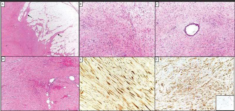
Figure 4: A) Representative photomicrograph of the tumors showing poorly circumscribed spindle cell neoplasm with finger
like extensions into surrounding fat (A, arrows). The spindle cells are fairly uniform and exhibit fascicular arrangement; B)
They surround epithelial structures and C) Fat; D) Tumor cells showed diffuse nuclear and cytoplasmic staining for beta cat-
enin; E) They were also positive for smooth muscle actin; F) However desmin was negative (inset).
the lesion with uneventful postoperative course. Final frequently locally aggressive and is prone to recur (up to
histopathology was reported as Fibromatosis. The pa- 35%), even after complete surgical excision with clear
tient has been on regular follow up now for six months margins [1]. Multicentric and bilateral disease and re-
with no apparent local recurrence. currence at sites other than the primary have been re-
ported [2,7,10,14]. Multicentricity in fibromatosis has
Discussion been reported in 10% of cases [14]. Bilateral lesions
Fibromatosis or desmoid tumor is a rare disease, are extremely rare, found in only 4% of patients [10].
comprising of only 3% of soft tissue tumors and < 0.03% Whether breast involvement is an extension from a
of all neoplasms [10,11]. It is clonal proliferation of fi- primary site within the fibroaponeurotic fascia or the
broblasts and myofibroblasts, typically arising from the pectoral muscle or whether it results from fibroblasts
muscle, the fascia, and aponeurosis. It has aggressive originating from within the breast parenchyma is un-
behavior locally and high incidence of local recurrence determined. However, fibromatosis arising within the
[12]. These tumors do not metastasize. They have no breast parenchyma appears to represent a separate
capsule and tend to infiltrate into local structures. They entity from extramammary fibromatosis, although both
have normal mitotic characteristics [3-5]. lesions may display a similar morphology. The extrama-
mmary lesions display a higher propensity for local re-
The most common extra-abdominal lesions are
currence compared to mammary fibromatosis [4,11].
found in the shoulder girdle, pelvis, and thigh [7,11].
The breast is an unusual location for the development Although the exact etiology of mammary fibromato-
of this tumor. It represents 0.2% of all breast tumors and sis remains unknown. Most of them occur sporadically
4% of all extra-abdominal desmoid tumors. Bilaterality but at least 30% of patients recount history of significant
has been reported in 4% of cases [1,3,4]. All racial and trauma to the involved area [4]. Antecedent trauma
ethnic groups are affected and no specific predilection is has been described at the site of fibromatosis in some
seen. Most of these cases were reported in young fertile patients and in association with saline-filled breast im-
females & rarely also occurs in men based on few study plants [4,15,16]. Few cases have been associated with
reports [13]. Mostly it occurs in the breasts of women Gardner’s Syndrome, familial multicentric fibromatosis
aged between 13 and 80 years. and familial adenomatous polyposis which suggests a
genetic predisposition and probably alteration of the
Fibromatosis of the breast may arise from the pec-
APC/beta-catenin pathway [1,4,15,17]. Some cases
toralis muscle or fascia or the mammary tissue. Mam-
have been associated with sex steroid hormones, mainly
mary fibromatosis appear to originate from fibroblasts
estrogens (during childbearing age, the disease tends to
and myofibroblasts within the breast parenchyma [8].
be more “cellular,” more mitotically active, with a larger
Although it does not metastasize, breast fibromatosis is
Yazidi et al. Int J Surg Res Pract 2021, 8:123 • Page 4 of 6 •DOI: 10.23937/2378-3397/1410123 ISSN: 2378-3397
amount of mild cellular atypia), suggesting a hormonal neoplasms of the breast might be helpful in excluding
correlation [7]. It is important to recognize mammary fi- fibromatosis from its differential diagnoses [20]. Anoth-
bromatosis as it presents a big dilemma for the clinician, er major concern about mammary fibromatosis is the
because it mimics cancer clinically, radiologically and exclusion of the diagnosis of metaplastic carcinoma, be-
sometimes cytologically [4]. cause spindle cell tumors with a myoepithelial immuno-
phenotype may be diagnosed as metaplastic carcinoma
Clinically, breast fibromatosis almost always pres-
even with weak or absent cytokeratin expression. In
ents with a painless, solitary, firm or hard tumor that
the present report, both epithelial (AE1/AE3) and myo-
suggests carcinoma on clinical examination as the case
epithelial markers (CD14 and p63) were negative [15].
with our patients. Rarely, the tumor is non-palpable and
Mammary fibromatosis presents macroscopically as a
detected initially by mammography [4]. Skin dimpling
dense, poorly vascularized, hard, rubbery, grayish-white
and retraction are relatively common signs of breast fi-
mass. The cut-surface usually shows an nonencapsulat-
bromatosis & is caused by fibrous tissue contraction vs.
ed mass with fibrous, gray and white nodular parenchy-
desmoplastic reaction, which is similar to tethering as-
ma [21]. Microscopically, fibromatosis is composed of
sociated with malignancy [2]. The tumor size may range
uniform, spindle shaped fibroblasts forming sweeping
from a few millimeters to 10 cm (the average size being
or interlacing fascicles entrapping ducts and lobules
2.5 cm). Small-sized tumors may be asymptomatic and
with an infiltrative edge. The degree of cellularity var-
show no signs and symptoms. Neither nipple discharge
ies, ranging from relatively cellular to predominantly
nor axillary lymphadenopathies commonly occur with it
collagenized lesions. Devouassoux-Shisheboran, et al.
[8,9]. In our three cases, there was no skin involvement.
studied the morphofunctional features of 33 cases of fi-
On mammography, breast fibromatosis often ap- bromatosis and showed that the cellularity of the lesion
pears as an irregularly shaped, noncalcified, high-densi- varied with the patient’s age. Lesions in the younger pa-
ty mass with spiculatedmargins [18]. Radiologic evalua- tients (childbearing age) were significantly more cellular
tion of our second and the third cases were classified as than those of the perimenopausal and postmenopausal
BIRADS 5 and the first case as BIRADS 4C. On ultrasound, groups, it displayed larger proportion of cells with mild
breast fibromatosis frequently appears as a poorly de- atypia and were mitotically more active. Compared to
fined, hypoechoic mass with a thick echogenic rim and a lesions in the childbearing group, 15 lesions in the peri-
posterior attenuation. The clinical presentation and the menopausal and postmenopausal patients were signifi-
radiological appearance of breast fibromatosis are high- cantly more fibrous and presented with prominent in-
ly suspicious for breast carcinoma. There are 2 cases re- flammatory cells. Immunohistochemically, fibromatosis
ported in the literature: Both patients underwent radi- exhibits positivity for smooth muscle actin and vimentin
cal mastectomy because of an erroneous clinical diagno- and negativity for cytokeratin, estrogen, progesterone
sis of breast carcinoma [19]. Pre-operative diagnosis of and androgen receptors (ER, PR and AR) [22]. Numerous
fibromatosis by FNAC is rare. Nevertheless, fine needle reports showed that even though extramammary fibro-
aspiration cytology, although not entirely specific, may matosis usually exhibit positivity for ER and PR, mamma-
be a source of important information in patients with ry fibromatosis is consistently devoid of such receptors
breast fibromatosis. In particular, it confidently allows [4,22], desmin is rarely positive, whereas S100 and CD34
the exclusion of breast cancer and other more common are usually negative. Immunohistochemical staining
diseases and is useful in planning a surgical approach to usually shows positivity for B-catenin in 70-80% of cases
the lesion [16]. McKinnon, et al. [12] noted that the cor- few studies have shown that many sporadic fibromato-
rect diagnosis of desmoid tumors was made preopera- sis cases have CTNNB1 mutation.
tively in only 50% of cases and that, even after biopsy,
Management remains controversial because of the
the diagnosis was often confused with low-grade fibro-
limited data due to its low incidence. Wide local exci-
sarcoma, confirming the difficulty of diagnosis for this
sion with clear margins is considered the standard of
pathology*.
care. Because of the stellate configurations and grossly
Since, fibromatosis is an infiltrative proliferation of inapparent extensions of most lesions. None of the lit-
fibroblastic and myofibroblastic cells, the positivity for erature we reviewed reported any mammary fibroma-
vimentin and smooth muscle actin was not surprising. tosis metastasis or death of patients from their disease
Curiously, fibromatosis in the breast differs from fibro- [1,4]. However, malignant transformation of fibroma-
matosis arising in other parts of the body due to its hor- tosis following radiation therapy was reported [17]. As
mone receptor profile. Although 30% of extramammary breast fibromatosis do not demonstrate metastatic ca-
fibromatosis are positive for estrogen receptors, only pabilities, axillary dissection is not performed [17]. The
one of the previously reported cases of mammary fibro- positive margin seems to be associated with recurrent
matosis expressed hormonal receptors [20]. Because of disease [3], but not all positive margins were recurrent
the consistent absence of immunoreactivity for estrogen [4]. Positive excision margins and intralesional excisions
and progesterone receptors in mammary fibromatosis, are associated with a greater rate of recurrence. Young-
a positive reaction for these receptors in spindle cell er age and larger tumor size are also associated with
Yazidi et al. Int J Surg Res Pract 2021, 8:123 • Page 5 of 6 •DOI: 10.23937/2378-3397/1410123 ISSN: 2378-3397
an increased risk of recurrence [23]. A literature review 12. Eastley N, Mcculloch T, Esler C, Hennig I, Fairbairn J, et al.
conducted by Trey Thomas, et al. suggests that patients (2016) Extra-abdominal desmoid fibromatosis: A review of
management, current guidance and unanswered questions.
should undergo quarterly clinical examination for a min- Eur J Surg Oncol 42: 1071-1083.
imum of three years; as available literature suggests the
13. Erguvan-Dogan B, Dempsey PJ, Ayyar G, Gilcrease MZ
majority of local recurrences manifest within this time (2005) Primary desmoid tumor (extraabdominal fibromato-
frame [17]. In this time frame, close follow-up is import- sis) of the breast. AJR Am J Roentgenol 185: 488-489.
ant [7]. Because fibromatosis is not cancer, it has a 100%
14. Wongmaneerung P, Somwangprasert A, Watcharachan K,
survival rate [24]. Ditsatham C (2016) Bilateral desmoid tumor of the breast:
Case series and literature review. Int Med Case Rep J 9:
References 247-251.
1. Bhat D, Wear V, Weisenberg E, Alvarado R (2016) Des-
15. Reis-Filho JS, Milanezi F, Pope LZ, Fillus-Neto J, Schmitt
moid-type fibromatosis of the breast. A case report. Breast
FC (2001) Primary fibromatosis of the breast in a patient
Dis 36: 149-152.
with multiple desmoid tumors. Report of a case with eval-
2. Taylor TV, Sosa J (2011) Bilateral breast fibromatosis: Case uation of estrogen and progesterone receptors. Pathol Res
report and review of the literature. J Surg Edu 68: 320-325. Pract 197: 775-759.
3. Wargotz ES, Norris HJ, Austin RM, Enzinger FM (1987) Fi- 16. Ng WH, Lee JS, Poh WT, Wong CY (1997) Desmoid tumor
bromatosis of the breast. A clinical and pathological study of (fibromatosis) of the breast. A clinician’s dilemma-a case
28 cases. Am J Surg Pathol 11: 38-45. report and review. Arch Surg 132: 444-446.
4. Rosen PP, Ernsberger D (1989) Mammary fibromatosis. A 17. Thomas T, Lorino C, John J (1987) Fibromatosis of the
benign spindle-cell tumor with significant risk for local recur- breast: A case report and literature review. J Surg Oncol
rence. Cancer 63: 1363-1369. 35: 70-74.
5. Brennan MF, Alektiar KM, Maki RG. Sarcomas of the soft 18. Cederlund CG, Gustavsson S, Linell F, Moquist-Olsson I,
tissue. In: DeVita VT, Hellman S, Rosenberg SA, Cancer: Andersson I (1984) Fibromatosis of the breast mimicking
Principles and Practice of Oncolog. carcinoma at mammography. Br J Radiol 57: 98-101.
6. Grimaldia MC, Trentinb C, Gullob RL, Cassano E (2018) 19. Rosen Y, Papasozomenos SC, Gardner B (1978) Fibroma-
Fibromatosis of the breast mimicking cancer: A case report. tosis of the breast. Cancer 41: 1409-1413.
Radiol Case Rep 13: 1-5.
20. Rasbridge SA, Gillett CE, Millis RR (1993) Oestrogen and
7. Chummun S, McLean NR, Abraham S, Youseff M (2010) progesterone receptor expression in mammary fibromato-
Desmoid tumour of the breast. J Plast Reconstr Aesthet sis. J Clin Pathol 46: 349-351.
Surg 63: 339-345.
21. Dwyer JB, Clark BZ (2015) Low-grade fibromatosis-like
8. Kumari P, Kapoor A, Kumar V, Kumari S, Kumar HS (2015) spindle cell carcinoma of the breast. Arch Pathol Lab Med
Extra abdominal mammary fibromatosis (desmoid tumor) of 139: 552-557.
breast in an elderly female. Int J Adv Med 2: 56-59.
22. Magro G, Michal M, Bisceglia M (2001) Benign spindle cell
9. Al-Khyatt W, Goyal A, Mansel RE (2010) Nipple-sparing tumors of the mammary stroma: Diagnostic criteria, classi-
skin-sparing mastectomy and vertical latissimus dorsi flap fication, and histogenesis. Pathol Res Pract 197: 453-466.
reconstruction for bilateral fibromatosis of the breast. Clin
23. Neuman HB, Brogi E, Ebrahim A, Brennan MF, Van Zee KJ
Breast Cancer 10: E1-E2.
(2008) Desmoid tumors (fibromatoses) of the breast: A 25-
10. Garg P, Chufal SS, Gupta N, Pant P, Thapliyal NC (2014) year experience. Ann Surg Oncol 15: 274-280.
Multicentric aggressive mammary fibromatosis with cyto-
24. Matherne TH, Green A Jr, Tucker JA, Dyess DL (2004) Fi-
logical features and review of literature. J Clin Diagn Res 8.
bromatosis: The breast cancer imitator. South Med J 97:
11. Greenberg D, McIntyre H, Ramsaroop R, Arthur J, Harman 1100-1103.
J (2002) Aggressive fibromatosis of the breast: A case re-
port and literature review. Breast J 8: 55-57.
Yazidi et al. Int J Surg Res Pract 2021, 8:123 • Page 6 of 6 •You can also read