Liposarcoma of the Oral Cavity - Case Reports of the Pleomorphic and the Dedifferentiated Variants and a Review of the Literature
←
→
Page content transcription
If your browser does not render page correctly, please read the page content below
ANTICANCER RESEARCH 26: 4857-4868 (2006)

                   Liposarcoma of the Oral Cavity – Case Reports
                    of the Pleomorphic and the Dedifferentiated
                      Variants and a Review of the Literature
                        FRANCESCA ANGIERO1, ANGELO SIDONI2 and MICHELE STEFANI1

                              1Institute   of Pathological Anatomy, Oral Pathology Section, Milan;
                                           2Institute
                                                    of Pathological Anatomy, Perugia, Italy

Abstract. Liposarcoma is one of the commonest soft-tissue             accounting for only 0.3% of all liposarcomas (3-6). A review
sarcomas, but very rare in the oral cavity. We present two cases of   of the literature regarding liposarcomas with strictly
liposarcoma of the oral cavity, together with the related clinical,   intraoral location reveals fewer than 100 case reports (3).
histopathological and immunohistochemical findings: one                  Histologically, the World Health Organization (WHO)
affecting the cheek of a 62-year-old man and the other the            classification distinguishes 5 LS subtypes: well-differentiated
gingival maxillary tuber of a 41-year-old woman. At histological      with its variants, myxoid, round cell, pleomorphic and
examination a diagnosis of liposarcoma was made in both cases.        dedifferentiated (2). The commonest subtype is the myxoid
In the first case, immunohistochemical analysis revealed intense      variant, followed by the well-differentiated, round cell,
positivity for p53, MIB-1, MDM2, and focal positivity for S100        dedifferentiated and pleomorphic variants, in that order. Of
protein and CD34, but was negative for alpha-smooth muscle            the pleomorphic variant only five cases primarily of the oral
actin, desmin and CD68. The second case it was intensely              cavity have been reported in the English language literature
positive for p53, MIB-1, S-100, and focal positive for MDM2, but      (6-8) (Table I) and of the dedifferentiated variant in the oral
negative for alpha smooth muscle actin, CD34, CD68 and                cavity our case is the seventh report (3, 9-12) (Table II).
desmin. Histological examination and immunohistochemical                 The peak incidence of all liposarcomas occurs between
profiles in the first case were consistent with pleomorphic           40 and 60 years of age, with men more frequently affected
liposarcoma, whilst that in the second case with dedifferentiated     than women (9). Of intraoral cases, the tongue is the
liposarcoma. Both patients were subjected to surgical treatment       primary site of incidence (9, 13), followed by the cheek and
with wide surgical margins, without adjuvant radio- or                the floor of the mouth (9, 13, 14).
chemotherapy. The first case was lost at follow-up one year after        The gross appearance of liposarcoma may be well-
surgery, while the second case has not undergone relapse after        circumscribed, encapsulated, or both, usually showing a
seven years. We discuss differential diagnosis, examining the         multilobular pattern with occasional satellite nodules. The
histopathological and immunohistochemical features that are           tumor may appear mucinous, gelatinous, or more fibrous,
potentially useful for distinguishing this tumor from other           soft or firm in consistency. The color is pale yellow. Areas
malignant adipose tissue tumors.                                      of necrosis or hemorrhage may be present, either
                                                                      superficially or at a depth, and are frequent in deep soft
While liposarcoma (LS) is considered to be the commonest              tissue. The morphology varies with histological type; in this
soft-tissue sarcoma in adults (9.8%-16% of all cases) (1, 2),         report only the pleomorphic and the dedifferentiated
generally arising in the thighs, buttocks, or the                     variants were considered.
retroperitoneum, intraoral liposarcomas are uncommon,                    The pleomorphic (PL) variant is defined as a high-grade
                                                                      sarcoma, first recognized by Enzinger and Wislow (15), and is
                                                                      considered to be the least common variant of liposarcoma,
                                                                      accounting for approximately 5% of all liposarcomas (16).
Correspondence to: Prof. Michele Stefani, Istituto di Anatomia        Clinically it is characterized by the tendency to occur in the
Patologica sez. Patologia Orale, Via della Commenda 19, 20122
                                                                      limbs of older adults and, less frequently, in the trunk and
Milan, Italy. Tel: +390 250320807 Fax: +390 2799007, e-mail:
michele.stefani@unimi.it                                              retroperitoneum. Histologically, it shows multivacuolated
                                                                      lipoblasts (16) and is further subdivided into two forms, the
Key Words: Liposarcoma, mesenchymal tumor, oral cavity,               malignant fibrous histocytoma-like form and the pleomorphic
dedifferentiated, pleomorphic, MDM2, p53.                             giant-cell-rich form. Recently, Miettinen and Enzinger have

0250-7005/2007 $2.00+.40                                                                                                        4857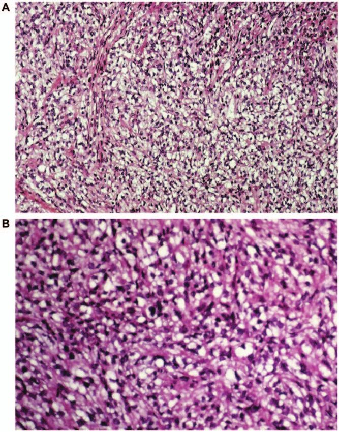
ANTICANCER RESEARCH 26: 4857-4868 (2006)

Table I. Cases of pleomorphic liposarcoma of the oral cavity reported in the literature and the present case.

Case reported (reference/years)           Age (years)/gender            Anatomic location                            Size in cm         Histology

Adkins et al. (5)/1978                    24/male                       Right posterior maxillary gingiva            N/A                Pleomorphic
Eidinger et al. (6)/1990                  80/male                       Cheek                                        8x8x5cm            Pleomorphic
Mc Culloch et al. (7)/1992                N/A                           Cheek                                        8x8x5cm            Pleomorphic
Friedman et al. (8)/1995                  18/male                       Pterymandybular space                        8x7x4 cm           Pleomorphic
Ogawa et al. (9)/1996                     29/male                       Cheek                                        N/A                Pleomorphic
Angiero et al. (pc)/2006                  41/female                     Right posterior maxillary gingiva            1.0 cm             Pleomorphic

N/A, not available; pc: present case.

Table II Cases of dedifferentiated liposarcoma of the oral cavity reported in the literature and the present case.

Case reported                       Age (years)/ Anatomic                  Size in      Types of                                  Follow-up
(reference/years)                   gender       location                  cm           dedifferentiation                         (recurrence)

Diamond et al. (10) /2002           57/male        Cheek                   8.0 cm       Spindle cell pattern with areas           12 months without
                                                                                        showing multinucleated giant cells        recurrence
Fanburg-Smith et al. (11)/2002      39/male        Tongue                  6.0 cm       Bland spindle proliferation and           6 years follow-up
                                                                                        scattered floret cells                    without recurrence
Fanburg-Smith et al. (11) /2002     56/male        Left buccal mucosa      5.0 cm       Spindled pleomorphic                      26-year follow-up,
                                                                                        liposarcoma-like areas                    six recurrences

Nascimento et al. (9) /2002         83/ female     Tongue                  2.5 cm       Non lipogenic rounded tumors cells        N/A
Gustavo de la Roza et al.           61/ male       Cheek                   5.0 cm       Non lipogenic spindle cells with focal    Lost to follow up
(12)/2004                                                                               rhabdomyoblastic differentiation          Recurrence 5 month
                                                                                                                                  after surgery
Werneck da Cunha I et al.           42/women       Cheek                   6.0 cm       Spindle cells with nuclear atypias        12 months
(3)/2005                                                                                arranged in sheets or forming             without
                                                                                        rudimental vascular channels              recurrence
                                                                                        angiosarcomatous dedifferention
Angiero F et al. (pc)/2006          62/male        Cheek                   3.0 cm       Spindle cells with nuclear atypias        7 years follow-up
                                                                                        arranged in sheets                        without recurrence

N/A, not available; pc, present case.

described 12 cases of a new variant of PL with an epithelioid                  sarcoma that constitutes the dedifferentiated component. This
morphology (17). These tumors exhibit focal adipocytic                         pattern may show a transition from a low-grade to a high-
differentiation, but appear to be composed predominantly of                    grade non-lipogenic morphology, within a well-differentiated
sheets of epithelioid-like cells separated by a minimal amount                 liposarcoma; the transition generally occurs abruptly, but can
of intercellular matrix.                                                       be gradual or intermediate (2, 22). Recently, Nascimento et
   The concept of dedifferentiated liposarcoma (DL) was                        al. described a peculiar new variant of DL in which the
introduced by Evans in 1979 (18) and is now widely                             formation of ‘neural-like’ or ‘meningothelial-like’ whorls of
recognized. The dedifferentiated liposarcomas were defined                     spindle cells is seen (23) often in association with metaplastic
as tumors containing distinct areas of well-differentiated                     bone formation. Dedifferentiation in well-differentiated
liposarcoma, and of non-lipogenic cells or pleomorphic                         liposarcoma occurs more frequently in the primary tumor
sarcoma. This was simply a fresh work-up of the earlier                        (90%) than in recurrences (10%) (24).
description of dedifferentiation introduced by David Dhalin
in 1977 in the context of the description of tumor                             Cases
progression in chondrosarcoma (19). The commonest site of
DL is the retroperitoneum, followed by the limbs, accounting                   Case 1. A 62-year-old man was referred for evaluation of a
for approximately 10% of all liposarcoma (20, 21).                             swelling in the cheek, present for about 12 months. The
   Histologically it is a biphasic neoplasm in which one                       swelling was asymptomatic and had increased in size. At
component is an atypical lipomatous tumor or well-                             extraoral examination there was an enlargement of the right
differentiated, and the other is a cellular, non-lipogenic                     cheek, which was of firm consistency. Intraorally, the lesion

4858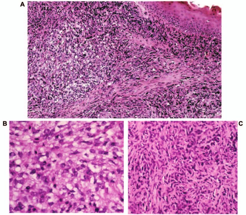
Angiero et al: Pleomorphic and Dedifferentiated Liposarcoma of the Oral Cavity

Table III. Immunohistochemical profile of current cases of liposarcoma.

Antibody                                   Supplier                   Dilution               Reactivity Case 1               Reactivity Case 2
                                                                                             (dedifferentiated)              (pleomorphic)

Mib-1                                      Dako                       1: 200                 ++                              ++
Alfa-Smooth Muscle Actin (Sma)             Sigma                      1: 400                 –                               –
Desmin                                     Dako                       1: 200                 –                               –
S100                                       Dako                       1: 200                 +                               ++
Mdm2                                       Calbioch 5M                1: 25                  ++                              +
Cd34                                       Novocastra                 1: 50                  +                               –
Cd68                                       Dako                       1: 500                 –                               –
p53                                        Novocastra                 1:100                  ++                              ++

– (negative), no staining; + (positive), focally positive for a limited number of cells; and ++ (intensely positive), focally or diffusely positive for
numerous cells.

appeared as a smooth, firm submucosal mass in the right                        Results
cheek extending from the right buccal mucosa toward the
zygomatic area, measuring 3.0 x 2 cm at its greatest                           Microscopic examination of Case 1 (Figure 1) revealed a
dimension. The overlying mucosa was non-ulcerated and                          pleomorphic adipose tissue neoplasm with areas of
normal in color. The patient underwent incisional biopsy                       mature lipocytes and lipoblasts with atypical and
under local anesthesia and a diagnosis of liposarcoma was                      hyperchromatic nuclei. Round cells were also present, and
rendered; surgical treatment with wide surgical margins                        rare mitotic figures. The diagnosis was pleomorphic
followed, without any adjuvant radio- or chemotherapy.                         liposarcoma with rounded cells. Histological examination
There is no evidence of recurrence seven years after surgery.                  of Case 2 showed that the tumor was characterized by a
                                                                               solid non-lipogenic sheet and areas of well-differentiated
Case 2. A 41-year-old woman presented with a history of a                      lipoblasts arranged in lobules interspersed with dense
small painless mass of 1-year duration on the right maxillary                  fibrous bundles. Individual cells were vacuolated,
gingiva. At extraoral examination there was an enlargement of                  containing varying numbers of lipid droplets, and
the right maxillary gingiva. At intraoral examination, the                     displaying hyperchromatic, peripheral and pleomorphic
submucosal lesion appeared firm, nodular, slightly tender,                     nuclei. Small, scattered nests of multi-vacuolated
measuring approximately 1 cm in diameter. An incisional                        lipoblasts were also visible. Signet-ring cells were present,
biopsy was performed and a diagnosis of liposarcoma was                        filled with a single large lipid globule, and showing a
rendered. The patient underwent surgical treatment with wide                   lateral displacement of the nucleus; however, lipoblasts in
surgical margins, without any adjuvant radio- or chemotherapy,                 several stages of differentiation were abundant. The
but was lost at follow-up one year after surgery.                              dedifferentiated areas consisted mostly of spindle or
                                                                               stellate fibroblastic cells (Figure 2).
Materials and Methods                                                             Immunohistochemical studies using the avidin-biotin-
                                                                               peroxidase technique were performed in both cases and are
Excised biopsy specimens were fixed in 10% formalin-buffered and               summarized in Table III.
paraffin-embedded. Sections of 5-Ì were stained with hematoxylin
and eosin. For immunohistochemistry, the avidin-biotin complex
                                                                               Case 1. The tumor cells were intensely positive for p53,
(ABC) method was applied (25). A panel of monoclonal antibodies
was used for the following markers (Table III): Desmin (1: 200,
                                                                               (Figure 3A), focal positive for S-100 (Figure 4A) and for
DAKO), Ki-67 antigen (MIB-1 1: 200, DAKO), p53 (1: 100,                        CD34, strongly positive for MIB-1 and MDM2 (Figures 5A,
NOVOCASTRA) MDM2 (1: 25, CALBIOCH 5M), alpha-smooth                            6A), and negative for desmin, alpha-smooth muscle actin
muscle actin SMA (1: 400, SIGMA), S100 protein (1: 100, DAKO),                 and CD68.
CD34 (1: 100, NOVOCASTRA), CD68 (1: 500, DAKO). The
immunohistochemical antibodies, their sources and dilutions, are               Case 2. Intense positive reactivity was seen for p53,
listed in Table III. Appropriate controls were tested
                                                                               (Figure 3B), S-100 (Figure 4B), MIB-1 (Figure 5B) and
simultaneously. The immunohistochemical reactivity was evaluated
and graded as follows: – (negative), no staining; + (positive),
                                                                               focal positive for MDM2 (Figure 6B); the reaction was
focally positive for a limited number of cells; and ++ (intensely or           negative for alpha-smooth muscle actin SMA, CD34 and
strong positive), focally or diffusely positive for numerous cells.            desmin.

                                                                                                                                                 4859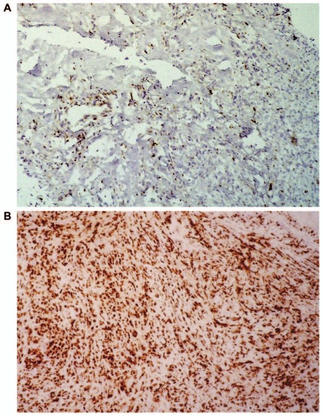
ANTICANCER RESEARCH 26: 4857-4868 (2006)
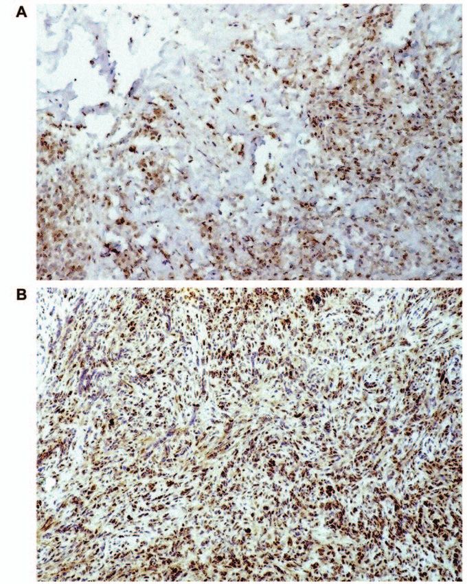
Figure 1. A-B) Pleomorphic liposarcoma (Case 1). A) Low-power view, B) high-power view with pleomorphic lipoblasts and round cells. Histology
disclosed hypercellular areas of neoplastic cells with ill-defined cytoplasm and round nuclei (hematoxylin and eosin staining; original magnification, A
x150 and B x200).
Discussion considered are, above all, those in which evidence of non-
adipose cellular differentiation must be carefully evaluated;
The histopathological diagnosis of intraoral liposarcoma differential diagnosis includes high-grade variants of
presents a challenge to the oral pathologist, because of the myxofibrosarcoma, malignant fibrous histiocytoma,
rarity of this tumor in the oral cavity. Differential diagnosis fibrosarcoma, leiomyosarcoma and rhabdomyosarcoma,
of dedifferentiated liposarcoma and pleomorphic and also towards some liposarcomas containing adipose
liposarcoma in the oral cavity includes a vast panorama of differentiation, such as spindle cell liposarcoma and the
malignant tumors. Malignant tumors that must be well-differentiated sclerosing liposarcoma.
4860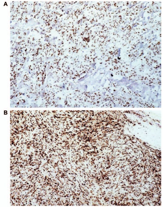
Angiero et al: Pleomorphic and Dedifferentiated Liposarcoma of the Oral Cavity

Figure 2. Dedifferentiated liposarcoma (Case 2). A) Low-power view with both components, B) high-power view, with areas of well-differentiated
lipoblasts, C) areas resembling a high-grade fibrosarcoma with storiform pattern (hematoxylin and eosin staining; original magnification, A x150 and B,
C x200).

   In many cases, the histopathology of pleomorphic                          or pleomorphic sarcoma may lead to incorrect
liposarcoma may be indistinguishable from that of                            interpretation and, thus, to an inaccurate diagnosis.
dedifferentiated liposarcoma. Beyond that, it has been                          It has also been observed that about 5-10% of cases, the
observed that the well-differentiated and high-grade                         dedifferentiated component may exhibit heterologous
components in the dedifferentiated variant may be                            differentiation, most often myogenic or osteo-
intermingled (26). However, in that case the                                 chondrosarcomatous and more rarely angiosarcomatous
dedifferentiated component is non-lipogenic, in contrast to                  (27), and may have either low-grade or high-grade
the pleomorphic form.                                                        differentiation. Such cases, thus, enter into differential
   In a series of 155 cases of dedifferentiated liposarcoma                  diagnosis with the above-mentioned tumor types.
reported by Henricks et al. (20), the majority displayed areas               Furthermore, from the morphological standpoint the
of high-grade dedifferentiation, resembling fibrosarcoma or                  dedifferentiated areas overlap with the storiform and
pleomorphic sarcomas. It is, thus, clear that the presence of                pleomorphic variant of malignant fibrous histiocytoma, or
large dedifferentiated areas similar to that of fibrosarcoma                 less frequently with myxofibrosarcoma. A difficulty consists

                                                                                                                                                 4861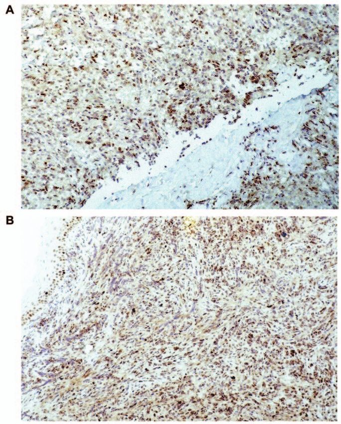
ANTICANCER RESEARCH 26: 4857-4868 (2006)

Figure 3. p53 immunoreactivity can be seen in both components in (Case 1, A) and (Case 2, B) components (original magnification, x150).

in differentiating between dedifferentiated liposarcoma and               dedifferentiation"; however, spindle cell liposarcoma is a
malignant fibrous histiocytoma, and it has been observed                  lipogenic lesion, whereas both low- and high-grade
that some cases reported as the latter may in fact have been              dedifferentiated areas are generally non-lipogenic.
dedifferentiated liposarcomas (28).                                          In PL, an acute inflammatory infiltrate may be present,
   Sometimes dedifferentiated liposarcoma exhibits the                    though rarely, that may cause diagnostic confusion with
presence of fascicles of bland spindle cells with a cellularity           well-differentiated inflammatory liposarcoma.
intermediate between well-differentiated sclerosing                          The histological pattern in the first of our cases, with the
liposarcoma and usual high-grade areas (29, 30). The term                 atypical population of bizarre and pleomorphic cells and the
proposed to describe these areas is "low-grade                            presence of round cells allowed the diagnosis of PL with

4862
Angiero et al: Pleomorphic and Dedifferentiated Liposarcoma of the Oral Cavity

Figure 4. Positive cytoplasmatic S-100 immunopositivity in both (Case 1, A) and (Case 2, B) (original magnification, x150).

rounded cells. In the second case, the dedifferentiated areas               histiocytoma, although it has been noted that poorly-
consisted mostly of spindle or stellate fibroblastic cell in a              differentiated liposarcomas may stain negatively for S-100
storiform pattern intermingled with area of well-differentiated             and vimentin (2).
LS, and was indicative of a dedifferentiated liposarcoma.                      Both of our cases stained positive for the S-100 protein,
   Immunohistochemistry was carried out to achieve a                        the dedifferentiated variant more markedly than the
correct diagnosis. From the immunohistochemical                             pleomorphic variant. In both variants, positivity for p53
standpoint, adipocytes and lipoblasts are known to stain                    and MDM2 (protein frequently expressed in several
positively for S-100 and vimentin. Hashimoto et al. (31)                    sarcoma types) was found (19, 20, 32); the concomitant
suggested that S-100 protein positivity is useful for                       positive expression for MDM2 protein and p53 is thought
distinguishing liposarcoma from myxoid malignant fibrous                    to indicate a form of tumor progression, suggesting a key

                                                                                                                                   4863
ANTICANCER RESEARCH 26: 4857-4868 (2006) Figure 5. The pleomorphic component showing a high MIB-1 immunopositivity (Case 1, A), the dedifferentiated component (Case 2, B) also shows a high MIB-1 immunopositivity (original magnification, x150). role for the p53 gene, from well-differentiated liposarcoma The prognosis of liposarcoma is influenced by several to a high-grade neoplasm (29, 33). Moreover the factors: histopathological variant, location, tumor size, pleomorphic variant was strongly positive for MIB-1, focal adequacy of surgical treatment and distant metastases (7, positive for CD34 and negative for CD 68, alpha-smooth- 34). With regard to the histopathological variant of muscle actin and desmin; the dedifferentiated variant was liposarcoma, it has been observed that the various subtypes strongly positive for MIB-1, but mostly negative for alpha- identified by the WHO have a different biological behavior. smooth-muscle actin and desmin. In the light of these Pleomorphic and dedifferentiated liposarcomas located results, the initial diagnosis was confirmed with H&E outside the oral cavity are reported to have more aggressive staining with the first case classified as pleomorphic biological behavior, and are associated with a significantly liposarcoma with round cells, and the second case as higher rate of mortality and metastasis than myxoid and dedifferentiated liposarcoma. well-differentiated liposarcomas (23, 35), and in some cases 4864
Angiero et al: Pleomorphic and Dedifferentiated Liposarcoma of the Oral Cavity

Figure 6. Positive immunoreactivity for MDM2 (Case 1, A) and (Case 2, B) expression is strong whereas it is reduced in the dedifferentiated variant
(original magnification, x150).

a metastatic liposarcoma may be of a more aggressive                       as has also been reported by Enzinger and Wislow (15),
histological type than the original tumor (3). The                         different locations may correspond to different prognoses.
pleomorphic variant is reported to have a 30% metastasis                   In a recent study, Fanburg- Smith et al. (38) have reported
rate and an overall mortality of at least 40% (36).                        patients with liposarcomas in the oral cavity appear to have
Regarding the dedifferentiated variant, there are limited                  a better prognosis than their soft-tissue counterparts, and
data available on the prognosis, but Mc Cormick et al. (37)                that tumor size irrespective of the histological subtype may
have reported that they have a better prognosis than                       be the best predictor of recurrence.
pleomorphic sarcomas, and are associated with a 15-20%                        In terms of adequacy of surgical treatment and distant
metastatis rate.                                                           metastases, in general, the treatment of choice for
   As far as location concerned, malignant forms tend to be                liposarcomas is surgical excision (38, 39) and the frequent
located in deep soft tissue, whereas less aggressive or benign             presence of satellite nodules means that wide surgical
forms tend to be observed in superficial adipose tissue; thus,             excision is necessary for adequate removal of the tumor. It

                                                                                                                                             4865
ANTICANCER RESEARCH 26: 4857-4868 (2006)
has also been reported that non-surgical treatments have Acknowledgements
limited value, and the role of adjuvant chemotherapy or
radiotherapy is controversial (40-42). We would like to thank B. Vergani for her assistance with the
Cytogenetic and molecular analyses (not done in our immunohistochemistry from the National Tumour Institute of
cases) are considered very important prognostic factors; Milan.
they are helpful in identifying the histological subtype, as
well as in elucidating pathogenesis. Cytogenetic analysis References
is useful in that it provides accurate classification of types
of liposarcoma and enables liposarcoma to be 1 Hashimoto H and Enjoij M: Liposarcoma clinicopathologic
subtyping of 52 cases. Acta Pathol Jpn 32: 933-948, 1982.
distinguished from benign adipose tumors. It has been
2 Enzinger MF and Weiss SW: Liposarcoma. In: Soft Tissue
observed that the dedifferentiated form frequently retains
Tumors. Gay SM, Gery L (eds.). 3rd ed. St Louis (MO): Mosby-
some of the cytogenetic changes present in well- Year Book, pp. 431-446, 1995.
differentiated liposarcoma, including ring and marker 3 Cunha I W, Kolwaski LP and Soares FA: Dedifferentiated
chromosomes composed of amplified material from liposarcoma of the oral cavity with angiosarcomatous
12q13-15 in about 50% of cases (43, 44). There are also dedifferentiation. Virkow Archiv 446: 456-459, 2005.
gene abnormalities in MDM2 in over half of all cases. 4 Adkins WY, Putney FJ, Kreutner A and Cunningham C:
In contrast, pleomorphic liposarcoma frequently has a Liposarcoma of the maxilla. Otolaryngology 86: 710-713, 1978.
5 Eidinger G, Katsikeris N, and Gullane P: Liposarcoma: report
highly abnormal and complex karyotype without specific
of a case and review of the literature. J Oral Maxillofac Surg
abnormalities (45, 46) and does not appear to arise from 48(9): 984-988, 1990.
a low-grade precursor. Recent microarray data using 6 McCulloch TM, Makielski KH and McNutt MA: Head and neck
comparative genomic hybridization and expression liposarcoma. A histopathologic reevaluation of reported cases.
profiling also suggest that the molecular signatures of Arch Otolaryngol Head Neck Surg 118(10): 1045-1049, 1992.
dedifferentiated and pleomorphic liposarcomas are 7 Friedman JL, Bistritz JI and Robinson MJ: Pleomorphic
different (47, 48). liposarcoma of the pterygomandibular space involving the
maxilla. Oral Surg Oral Med Oral Pathol Oral Radiol Endod
79(4): 488-491, 1995.
Conclusion
8 Ogawa T, Kawakami T, Naito M, Yamamoto Y, Moriwaki S
and Sadamoto M: Multidisciplinary treatment of liposarcoma
We have added two more cases of this rare tumor located in the maxillary region. Practica Otologica 79: 1455-1459, 1996.
in the oral cavity, one being the dedifferentiated and the 9 Nascimento AF, Mc Menamin ME and Fletcher CDM:
other the pleomorphic variant. The first type, of which only Liposarcomas/atypical lipomatous tumors of the oral cavity:
six other cases have been reported in the literature, was a clinicopathologic study of 23 cases. Ann Diagn Pathol 6: 83-
located at the cheek. The second is the sixth case reported 93, 2002.
to date involving the gingival maxillary tuber. 10 Diamond C, Prince ME, Covert AA and Morris SF:
Dedifferentiated liposarcoma of the cheek: case report and
Histological classification and differential diagnosis are
literature review. J Otolaryngol 31: 125-128, 2002.
indispensable not only in discriminating the variant, but also
11 Fanburg-Smith JC and Miettinen M: Liposarcoma with
in predicting clinical behavior and prognosis: clinical course meningothelial like whorls: a study of 17 cases of a distinctive
is directly related to aggressiveness, which can only be histological pattern associated with dedifferentiated
determined by histology. Immunohistochemistry helps in liposarcoma. Histopathology 33: 414-424, 1998.
this, and provides the key to discriminate between the 12 De la Roza G, Baredes S, and Aisner SC: Dedifferentiated
various forms. It is probable that the prognosis of the liposarcoma of the cheek. Annals of Diagnostic Pathology 8:
dedifferentiated form in the oral cavity is similar to that 352-357, 2004.
13 Gagari E, Kabani S and Callagher GT: Intraoral liposarcoma:
observed in other parts of the body, although the degree of
case report and review of the literature. Oral Surg Oral Med
aggressiveness has also been reported to depend on Oral Pathol Oral Radiol Endod 89: 66-72, 2000.
location. Case reports to date are very few for proper 14 Allon I, Vered M and Dayan D: Liposarcoma of the tongue:
evaluation; in particular, the case of the pleomorphic form clinicopathologic correlations of a possible underdiagnosed
reported here, only the sixth such case to date, is now lost at entity. Oral Oncology 41: 657-665, 2005.
follow-up and further data are not available. 15 Enzinger FM and Winslow DJ: Liposarcoma: a study of 103
It cannot be ruled out that, in the oral cavity, many more cases. Virkows Arch 335: 367-388, 1962.
cases occur than are reported, due to the difficulty of 16 Fletcher CDM, Unni KK and Mertens F Pathology and genetics.
Tumors of soft tissue and bone. IARC Press, Lyon, 2002.
diagnosis and the rarity of these tumors in this location. For
17 Miettinen M and Enzinger FM: Epithelioid variant of
this reason it is important to enhance diagnostic accuracy, by pleomorphic liposarcoma: a study of 12 cases of a distinctive
using, alongside with the routine histological tests, variant of high-grade liposarcoma. Mod Pathol 12(7): 722-
immunohistochemical exams, such as MDM2, MIB-1 and p53. 728, 1999.
4866Angiero et al: Pleomorphic and Dedifferentiated Liposarcoma of the Oral Cavity
18 Evans HL: Liposarcoma: a study of 55 cases with reassessment 34 O’Connor M and Snover DC: Liposarcoma: a review of factors
of its classification. Am J Surg Pathol 3: 507-523, 1979. influencing prognosis. Am Surg 49: 379-384, 1983.
19 Dhalin DD, Unni KK and Matsuno T: Malignant (fibrous) 35 Weiss SW and Rao VK: Well-differentiated liposarcoma
histiocytoma of bone: fact or fancy? Cancer 39: 1509-1516, 1977. (atypical lipoma) of deep soft tissues of the extremities,
20 Henricks WH, Chu YC, Goldblum JR and Weiss SW: retroperitoneum and miscellaneous sites: a follow-up study of
Dedifferentiated liposarcoma. A clinicopathological analysis of 92 cases with analysis of the incidence of "dedifferentiation".
155 cases with a proposal for an expanded definition of Am J Surg Pathol 16: 1051-1058, 1992.
dedifferentiation. Am J Surg Pathol 21: 271-281, 1997. 36 Mentzel T, Bosemberg M and Fletcher CD: Pleomorphic
21 Nascimento AG: Dedifferentiated liposarcoma. Semin Diagn liposarcoma: clinicopathologic and prognostic analysis of
Pathos 18: 263-266, 2001. 31cases. [abstract] Mod Pathol 12: 13A, 1999.
22 Dei Tos AP, Doglioni C, Piccinin S, Sciot R, Furlanetto A, 37 McCormick D, Mentzel T, Behem A and Fletcher CD:
Boiocchi M, Dal Cin P, Maestro R, Fletcher CD and Tallini G: Dedifferentiated liposarcoma. Clinicopathologic analysis of 32
Coordinated expression and amplification of the mdm2, cdk4 cases suggesting a better prognostic subgroup among
and hmgi-c genes in atypical lipomatous tumors.J Pathol 190: pleomorphic sarcomas. Am J Surg Pathol 18: 1213-1223, 1994.
531-536, 2000. 38 Fanburg-Smith JC, Furlong MA and Childers ELB: Liposarcoma
23 Nascimento AG, Kurtin PJ, Guillou L and Fletcher CDM: of the oral and salivary gland region: a clinicopathologic study of
Dedifferentiated liposarcoma. A report of nine cases with a 18 cases with emphasis on specific sites, morphologic subtypes
peculiar neurallike whorling pattern associated with metaplastic and clinical outcome. Mod Pathol 15: 1020-1031, 2002.
bone formation. Am J Surg Pathol 22: 945-955, 1988. 39 Stout AP: Liposarcoma: malignant tumor of lipoblasts. Ann
24 Dei Tos AP, Doglioni C, Piccinin S, Maestro R, Mentzel T, Surg 19: 86-107, 1994.
Barbareschi M, Boiocchi M and Fletcher CD: Molecular 40 Baden E and Newman R: Liposarcoma of the oropharyngeal
abnormalities of the p53 pathway in dedifferentiated region. Review of the literature and report of two cases. Oral
liposarcoma, J Pathol 181: 8-13, 1997. Surg Oral Med Oral Pathol Endod 44: 889-902, 1977.
25 Hsu SM, Raine L and Fanger H: Use of avidin-biotin- 41 Ham SJ, van der Graaf WT, Pras E, Molenar WM, van den
peroxidase complex (ABC) in immunoperoxidase techniques: a Berg E and Hoekstra HJ: Soft tissue sarcomas of the
comparison between ABC and unlabeled antibody (PAP) extremities: A multimodality diagnostic and therapeutic
procedures. J Histochem Cytochem 29: 577-580, 1981. approach. Cancer Treat Rev 24: 373-391, 1998.
26 Sadeghi EM and Sauk J Jr: Liposarcoma of the oral cavity. 42 Seynaeve C and Verweij J: High-dose chemotherapy in adult
Clinical, tissue culture, and ultrastructure study of a case. J Oral sarcomas: No standard yet. Semin Oncol 26: 119-133, 1999.
Pathol 11: 263-275, 1982. 43 Minic AJ: Liposarcomas of the oral tissues: a clinicopathologic
27 Evans HL, Khurana KK, Kemp BL and Ayala AG: "Heterologous study of four tumors. J Oral Pathol Med 24: 180-184, 1995.
elements in the dedifferentiated component of dedifferentiated 44 O’Connor M and Snover DC: Liposarcoma: A review of factors
liposarcoma". Am J Surg Pathol 18: 1150-1157, 1994. influencing prognosis. Am Surg 49: 379-384, 1983.
28 Coindre JM, Mariani O, Chibon F, Mairal A, Somerhausen 45 Rosai J, Akerman M, Dal Cin P, De Wever I , Fletcher CD,
NSA, Favre-Guillevin E, Bui NB, Stoeckle E, Hostein I and Mandahl N, Mertens F, Mitelman F, Rydholm A, Sciot R, Tallini
Aurias A: Most malignant fibrous histiocytomas developed in G, Van den Berghe H, Vanni R and Willen H: Combined
the retroperitoneum are dedifferentiated liposarcomas: a review morphologic and karyotypic study of 59 atypical lipomatous
of 25 cases initially diagnosed as malignant fibrous tumors: evaluation of their relationship and differential diagnosis
histiocytoma. Hum Pathol 16: 256-262, 2003. with other adipose tissue tumors (a report of the CHAMP study
29 Schneider-Stock R, Radig K, Oda Y, Mellin W, Rys A, group). Am J Surg Pathol 20: 1182-1189, 1996.
Niezabitowski A and Roessner A: p53 gene mutations in soft- 46 Rubin BP and Fletcher CD: The cytogenetics of lipomatous
tissue sarcomas-correlations with p53 immunohistochemistry tumours. Histopathology 30: 507-511, 1997.
and DNA ploidy. J Cancer Res Clin Oncol 123: 211-218, 1997. 47 Mertens F, Fletcher CD, Dal Cin P, De Wever I, Mandahl N,
30 Boltze C, Schneider-Stock R, Jager Vand Roessner A: Mitelman F, Rosai J, Rydholm A, Sciot R, Tallini G, Van den
Distinction between lipoma and liposarcoma by MDM2: a case Berghe H, Vanni R and Willen H: Cytogenetic analysis of 46
report of simultaneously occurring tumours and review of the pleomorphic soft tissue sarcomas and correlation with
literature. Pathol Res Prac 197: 563-568, 2001. morphologic and clinical features: a report of the CHAMP
31 Hashimoto H, Daimaru Y and Enjoji M: S-100 protein study group. Genes Chromosomes Cancer 22: 16-25, 1998.
distribution in liposarcoma: an immunoperoxidase study with 48 Fritz B, Schubert F, Wrobel G, Schwaenen C, Wessendorf S,
special reference to the distinction of liposarcoma from myxoid Nessling M, Korz C, Rieker RJ, Montgomery K and
malignant fibrous histiocytoma. Virkows Arch A Pathol Anat Kucherlapati R: Microarray-based copy number and expression
Histopathol 405: 1-10, 1984. profiling in dedifferentiated and pleomorphic liposarcoma.
32 Cordon-Cardo C, Latres E and Drobnjak M: Molecular Cancer Res 62: 2993-2998, 2002.
abnormalities of mdm2 and p53 genes in adult soft tissue
tumors. Cancer Res 54: 794-799, 1994.
33 Hasegawa T, Seki K, Hasegawa F, Matsuno Y, Shimodo T,
Hirose T, Sano T and Hirohashi S: Dedifferentiated liposarcoma
of retroperitoneum and mesentery: varied growth patterns and
histological grades-a clinicopathologic study of 32 cases. Hum Received August 8, 2006
Pathol 31(6): 717-727, 2000. Accepted √ctober 25, 2006
4867You can also read