In vivo and in vitro impact of miR-31 and miR-143 on the suppression of metastasis and invasion in breast cancer - JBUON
←
→
Page content transcription
If your browser does not render page correctly, please read the page content below
JBUON 2018; 23(5): 1290-1296
ISSN: 1107-0625, online ISSN: 2241-6293 • www.jbuon.com
E-mail: editorial_office@jbuon.com

ORIGINAL ARTICLE

In vivo and in vitro impact of miR-31 and miR-143 on the
suppression of metastasis and invasion in breast cancer
Sorour Soheilyfar1, Zahra Velashjerdi2, Yasamin Sayed Hajizadeh3, Nazila Fathi Maroufi4,5,
Zahra Amini6, Afshin Khorrami7, Saba Haj Azimian8, Alireza Isazadeh8, Sina Taefehshokr9,
Nima Taefehshokr10
1
 Department of Genetics, Ashkezar Branch, Islamic Azad University, Ashkezar, Iran; 2Department of Biochemistry, Science and
Research Branch, Islamic Azad University, Tehran, Iran; 3Department of Microbiology, Urmia Branch, Islamic Azad University,
Urmia, Iran; 4Department of Clinical Biochemistry and Laboratory Medicine, Tabriz University of Medical Sciences, Tabriz,
Iran; 5Student Research Committee, Tabriz University of Medical Sciences, Tabriz, Iran; 6Department of Marine Biology, Khor-
ramshahr Marine Science and Technology University, Khorramshahr, Iran; 7Department of Biology, School of Science, Yazd
University, Yazd, Iran; 8Department of Genetics, Tabriz Branch, Islamic Azad University, Tabriz, Iran; 9Department of Veterinary
Medicine, Tabriz Branch, Islamic Azad University, Tabriz, Iran; 10Division of Biosciences, Department of Life Sciences, College
of Health and Life Sciences, Brunel University London, Uxbridge, Middlesex, United Kingdom

Summary
Purpose: The microRNA (miR)-31 and miR-143 are plei-          the transfection of miR-31 construct was decreased 4, 70 and
otropic anti-metastatic miRs, with an expression that de-     100 times in MCF-7, MDA-MB468 and MDA-MB231 cell
creases significantly in metastatic breast cancer cells. The  lines, respectively, in comparison to normal breast cells; but
aim of this study was to investigate the effect of miR-31     after the transfection of miR-31 construct, the expression
and miR-143 inhibition on metastasis and invasion in both     of miR-31 increased 80 times. Additionally, invasion and
MDA-MB231, MDA-MB468 as well as the MCF-7 breast              migration decreased by 15 and 10 times in MDAMB-468.
cancer cell lines and 5-week old female mice.                 All of the modifications in miR-143 were low in comparison
                                                              to miR-31. The results of the in vivo experiments were ap-
Methods: Following the cloning of miR-31 and miR-143
                                                              proximately the same as in the in vitro experiments.
into vectors, their expressions were determined before treat-
ment with constructs of miR-31 and miR-143 in cancer cell Conclusions: IIt appears that the use of miR-31 is highly
lines and normal breast cells. Then miR-31 and miR-143 efficient than miR-143 in the inhibition of invasion and me-
were transfected to the cell lines and the expression was as- tastasis in breast cancer. Our study improved our conception
sessed after 48 hrs. Moreover, the levels of migration and about miR-31 and miR-143 and their roles in the identifica-
invasion were determined in cell lines. These experiments tion and therapy of breast cancer.
were performed in 5-week old female mice.
Results: The results showed that miR-31 expression before Key words: breast cancer, invasion, metastasis, microRNA

Introduction
    Breast cancer (BC) is the most common cancer velopments, the mortality of BC decreased, how-
in women and the leading cause of cancer-related ever it is estimated that about 1.3 million women
deaths in females worldwide [1]. Due to recent de- are affected by BC every year [2,3]. The current

Correspondence to: Zahra Velashjerdi, MSc. Department of Biochemistry, Faculty of Basic Sciences, Science and Research Branch,
Islamic Azad University, Shahid Hesarak Blvd, Daneshgah Crossroad, Shahid Sattari Highway, Tehran, Iran.
Tel/Fax: +98 2177182530, E-mail: zahravelashjerdi2020@gmail.com
Received: 11/02/2018; Accepted: 12/03/2018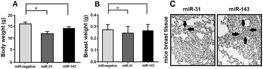
Role of miR-31 and miR-143 in breast cancer                                    1291

treatment methods include chemotherapy, sur-              (Worthington Biochemical Corporation, Lakewood/USA),
gery, radiotherapy, hormonal treatment and tar-           hyaluronidase (Sigma Aldrich, Saint Louis/USA) for 24
geted therapy [4]. Over the past decade, the studies      hrs at 37°C. Then, the cells were centrifuged and super-
largely focused on miRs as new biomarkers in the          natants were stored in pellet. Three ml warm Trypsin/
                                                          EDTA were added and re-suspended in the pellet. Ten ml
diagnosis and treatment of BC [5,6].
                                                          of Hank’s buffer and 2% cold FBS were added to neutral-
     Metastasis is a process in which a tumor cell        ize trypsin and centrifuged for 5 min. Warm Dispose 2Ml
leaves the primary tumor, travels to a distant site       (Stemcell Technologies, Vancouver/Canada, 5mg/ml)
via the circulatory system and establishes a sec-         and 200μl DNase I (Thermo Fisher Scientific, Waltham/
ondary tumor [7]. Tumor metastasis is a key event         USA, 1mg/ml) were added to the pellet and re-suspended
in the progression of BC and is mainly responsible        for 1 min. The suspension was diluted with 10ml cold
for BC-associated mortality [8].                          Hank’s buffer solution and passed through 40-μm cell
     miRs are small noncoding RNAs, which are             strainer. The suspension was centrifuged for 5 min and
                                                          1 ml medium was added to the pellet and cell counting
encoded in the genome of many species. These
                                                          was performed.
molecules are involved in translation, RNA stabil-
ity and gene expression [9]. Increasing evidence          Cloning by vector
indicates that dysfunction of miRs are involved in
                                                               The miR-31-mimic and miR-143-mimic oligom-
the development of cancer, suggesting that miRs           ers were designed and synthesized according to pcDNA
can function as classical oncogenes or tumor sup-         6.2gw/EmGFP vector kits protocol. The double-stranded
pressor genes [10]. The expression of miRs exerts         oligomers hybridization was performed according to
an effect on processes associated with cancer pro-        manufacturer’s instructions. Electrophoresis was per-
gression, such as invasion, metastasis, and apopto-       formed as follows: the ligation of double-strands frag-
sis [11]. Many studies demonstrated that miRs play        ments to the vectors was performed with 5× ligation
important role in tumor initiation by regulation of       buffer, pcDNA 6.2gw/EmGFP vector (5ng/μl), double-
                                                          stranded oligo (10nM), nuclease free water, T4DNA
tumorigenicity, self-renewal ability and drug re-
                                                          ligase enzymes (1U/μl), and transmission to the bacte-
sistance in cancer stem cells [12].
                                                          ria was implemented through electric shock. Cloning
     miR-31 plays an important role in different          accuracy was performed with Clooney-PCR by specific
types of cancers, such as BC [13,14], ovarian can-        primers vector and the results were analyzed using
cer [15,16], lung cancer [17,18], colon cancer [19,20]    electrophoresis.
and melanoma [21,22]. Also, miR-143 was identi-
fied as one of the low-expressed miRs in various          Transfection and flow cytometry
tumors, including non-small cell lung cancer [23],             5×10⁴ cells were cultured from each cell line in 24
gastric cancer [24], colorectal cancer [25], pancre-      plates with 60% confluence. The cells were transfected
atic cancer [26], cervical cancer [27], prostate can-     with lipofectamine and two pcDNA 6.2gw/EmGFP vec-
cer [28], osteosarcoma [29] and leukemia [30]. The        tors (Promega, Madison, Wisconsin, USA) with miR-31
expression of miRs is changing in various types of        and without miR-31 (positive control). Forty-eight hrs
                                                          after transfection, cells were dislodged using 0.25%
cancers and act as tumor suppressive or oncogenic
                                                          trypsin and green fluorescent protein (GFP) expres-
factors, depending on the type of cancer [31].            sion was assessed by flow cytometry to determine the
     The aim of this study was to investigate the         rate of transfection. Extraction of miR from three cell
effects of miR-31 and miR-143 on the cell invasion        lines and normal breast cells was performed before
and metastasis processes in MDA-MB231, MDA-               and after treatment with the vector. Then cDNA was
MB468 and MCF-7 breast cancer cell lines (in vitro)       synthesized.
and 5-week old female mice (in vivo).
                                                          Real time PCR
                                                               Real time PCR was performed to analyze the miR-
Methods                                                   31 and miR-143 expression levels, by Rotor-Gene 3000
                                                          kits and primer. After determining the transfection rate,
Cell culture and preparation
                                                          three cells lines were classified in three groups, such as
     Three breast cancer cell lines including MDA-        control, with and without miR and were used to create
MB231, MDA-MB468 and MCF-7 were cultured in               the scratch test.
DMEM/F12 medium supplemented with 10% fetal                    We used the SYBR Premix ExTaq (Takara, Mountain
bovine serum (FBS) and 1% penicillin/streptomycin         View/USA) with the Stratagene Mx3000P Real-Time PCR
(200μg/ml). Normal breast cells were taken from healthy   system (Agilent Technologies, Santa Clara, CA, USA).
women and washed three times by sterile phosphate         The real time PCR reaction included initial denatura-
buffered saline (PBS) to remove blood and cells. Then,    tion at 95°C for 10 min, denaturation at 95°C for 10s (40
samples were cut into small pieces and incubated in       cycles), annealing at 55°C for 30s, and extension at 72°C
dissociation buffer DMEM medium supplemented with         for 60s, respectively. Finally, changes of mRNA expres-
2% FBS 2 plus antibiotic solution, collagenase type I     sion were evaluated by 2−ΔΔCt method.

                                                                                            JBUON 2018; 23(5): 1291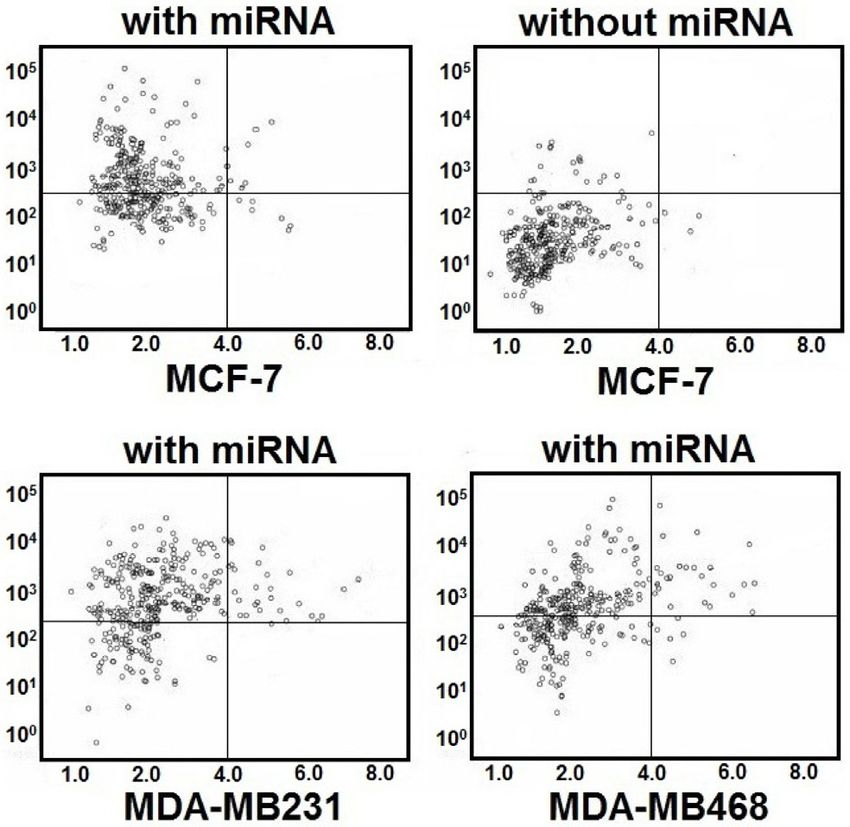
1292 Role of miR-31 and miR-143 in breast cancer
Migration and invasion assays Results
Evaluation of invasion and migration was per-
formed by Matrigel-coated filters with 0.8 micron pore Results of in vitro condition
size. Cells were placed on the filters, incubated for 24 Double-stranded hybridization was confirmed
hrs and removed from the culture medium. The filters’
by electrophoresis. After cloning, an appropriate
bottom were then fixed in paraformaldehyde (PFA) and
stained with crystal violet. In the following step, 10 ran- colony was selected by colony-PCR method and the
dom pictures were taken from each one of the wells. In obtained plasmids were purified. The MDA-MB231,
order to calculate invasion and migration, cell counting MDA-MB468 and MCF-7 cell lines were infected
was performed in two conditions (with Matrigel and FBS with vectors without miRs and with miRs and flow
and without Matrigel). cytometrically analyzed (Figure 1).
Animal studies
The real time PCR was performed with LNA
miR primers to analyze the expression rates. The
The MDA-MB231, MDAMB-468 and MCF-7 cells obtained results before transfection of miR-31
were infected with pLV3 or pLV3-720 and suspended in
construct showed that the miR-31 expression was
PBS. BALB/c nude female mice (20 control and 20 case)
aged 4 to 5 weeks were used in this study. For the experi-
decreased 4, 70 and 100 times in MCF-7, MDA-
ments, mice were implanted with 5×10⁵ cells via intrave- MB468 and MDA-MB231, respectively, as com-
nous tail injection. Six weeks later, mice were sacrificed. pared to normal breast cells. Also, the expression
The breasts of the mice were fixed and stained with he- analysis after treatment of cells with vectors with
matoxylin and eosin. Breast metastasis was quantified miR-31 and without miR-31 revealed that the ex-
by counting the number of tumor foci in 10 randomly pression of miR-31 increased 80 times in MDA-
selected high-power fields. MB231 cell line (Figure 2).
Statistics To determine the potential role of miR-31
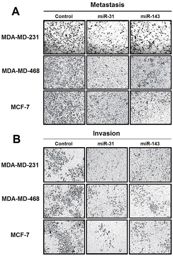
and miR-143 in breast cancer, invasion and mi-
Data from at least three independent experiments
gration assays were performed. The invasion was
are presented as mean ± SD. The t-test was used for com-
parisons between groups unless otherwise noted. Data reduced 15 and 10 times in the MDA-MB231 and
comparisons used paired t-test and group comparisons MDAMB-468 cell lines, respectively. On the other
used one-way analysis of variance (ANOVA). p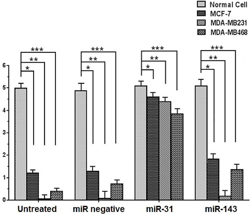
Role of miR-31 and miR-143 in breast cancer 1293
The obtained results before transfection of
miR-143 construct showed that the miR-143 ex-
pression was decreased 2, 10 and 15 times in MCF-
7, MDA-MB468 and MDA-MB231, respectively, as
compared to normal breast cells. Also, expression
analysis after treatment of cells with vectors with
miR-143 and without miR-143 showed that the ex-
pression of miR-143 increased insignificantly in
the cell lines. The invasion was reduced 4, 3 and 4
times in the MDA-MB231, MDAMB-468 and MCF-
7 cell lines, respectively (Figures 3 and 5).
Results of in vivo condition
To confirm that the expression of miRs sup-
presses migration and invasion, nude mice were
injected with MDA-MB231, MDAMB-468 and
MCF-7 cells expressing miR-31 and miR-143 via
intravenous tail injection. Six weeks post-injection,
body weights of the mice did not differ. However,
the obtained results from miR-31 groups were
significantly lighter as compared with the control
group. On the other hand, only 7 out of the 20 mice
(35%) from the miR-31 group showed metastasis.
In the control group, 14 out of the 20 mice (70%)
developed breast metastasis. Also, the obtained re-
sults from miR-143 group were not significantly
different as compared to the control group. In this
group, 13 out of the 20 mice (65%) from the miR-
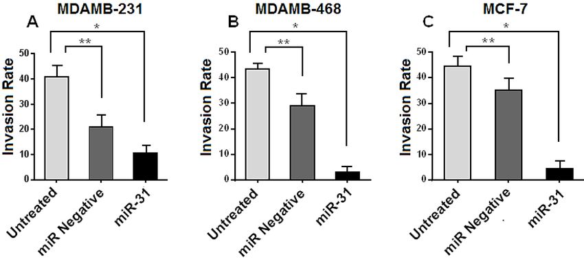
Figure 3. Effects of miR-31 and miR-141 mimics trans- 143 group developed breast metastasis (Figure 6).
fection on invasion and migration of MDA-MB231, MDA-
MB468 and MCF-7 cell lines. The miR-31 and miR-143 de-
creased metastasis (A) and invasion (B) in MDA-MB231,
Discussion
MDA-MB468 and MCF-7 cells. The matrigel assay showed
that the rate of invasion and metastasis was significant- In this study we investigated the expression
ly lower in the miR-31-transfected cells than the control of miR-31 and miR-143 in breast cancer progres-
group (p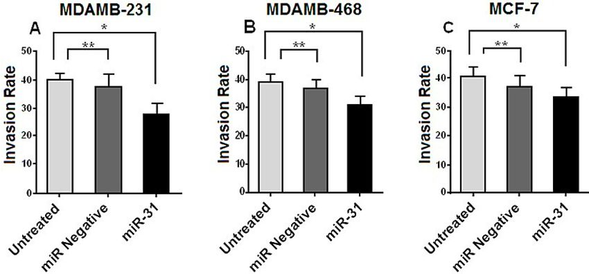
1294                            Role of miR-31 and miR-143 in breast cancer

Figure 5. Cell invasion rate in untreated, treated without miR-143 and treated with miR-143 in MDAMB-231 (A),
MDAMB-468 (B) and MCF-7 (C) cell lines (in vitro). The miR-143 was downregulated in MDAMB-231, MDAMB-468 and
MCF-7 cells. (A) *p=0.099 and **p=0.018; (B) *p=0.699 and **p=0.87; (C) *p=0.79 and **p=0.81.

Figure 6. Five week old female BALB/c nude mice were injected with MDA-MB-231 MDAMB-468 and MCF-7 cells. The
body weights (A) and the breast weights (B) were measured after six weeks. The obtained results showed that the body
and breast weight in miR-31 group was significantly more than in the control group (*p
Role of miR-31 and miR-143 in breast cancer                                         1295

and mimic). Antagonists are used to miR function               results, miR-31-mimic and miR-143-mimic can be
inhibition which has gained function, and mimic                ideal options to consider invasion and metastasis
are used for miR function recycling that have lost             inhibition in breast cancer, but miR-31 is highly
their function [33]. According to previous studies,            efficient than miR-143 in the inhibition of invasion
methods based on miR-mimic are superior com-                   and metastasis in breast cancer.
pared to antagonistic methods [34,35]. Therefore,
the present study was performed using mimic
methods. Studies have shown that miRs, especially              Conflict of interests
those that target metastatic cancers, can be bona
fide tumor suppressors [32]. Thus, according to our                 The authors declare no conflict of interests.

References
1.   Hutchinson L. Breast cancer: challenges, controversies,   16. Yu Z, Creighton C, Fountain MD et al. MiR-31 Is a Tu-
     breakthroughs. Nature Publishing Group; 2010.                 mor Suppressor MicroRNA That Functions in Ovarian
2.   Herranz M, Ruibal A. Optical imaging in breast cancer         Cancer. Biol Reproduction 2010;83:35.
     diagnosis: the next evolution. J Oncol 2012;2012.         17. Edmonds MD, Boyd KL, Moyo T et al. MicroRNA-31
3.   Cuk K, Zucknick M, Heil J et al. Circulating microRNAs        initiates lung tumorigenesis and promotes mutant
     in plasma as early detection markers for breast cancer.       KRAS-driven lung cancer. J Clin Investig 2016;126:349.
     Int J Cancer 2013;132:1602-12.                            18. Yu M, Liang H, Fu Z et al. BAP1 suppresses lung can-
4.   Al-Khanbashi M, Caramuta S, Alajmi AM et al. Tis-             cer progression and is inhibited by miR-31. Oncotarget
     sue and Serum miR Profile in Locally Advanced Breast          2016;7:13742.
     Cancer (LABC) in Response to Neo-Adjuvant Chemo-          19. Li T, Luo W, Liu K, Lv X, Xi T. miR-31 promotes pro-
     therapy (NAC) Treatment. PLoS One 2016;11:e0152032.           liferation of colon cancer cells by targeting E2F2. Bio-
5.   Luo J, Zhao Q, Zhang W et al. A novel panel of micro-         technol Lett 2015;37:523-32.
     RNAs provides a sensitive and specific tool for the di-   20. Kurihara H, Maruyama R, Ishiguro K et al. The rela-
     agnosis of breast cancer. Mol Med Rep 2014;10:785-91.         tionship between EZH2 expression and microRNA-31
                                                                   in colorectal cancer and the role in evolution of the
6.   Chen L, Li Y, Fu Y et al. Role of deregulated microRNAs
                                                                   serrated pathway. Oncotarget 2016;7:12704-17.
     in breast cancer progression using FFPE tissue. PLoS
     One 2013;8:e54213.                                        21. Greenberg E, Hershkovitz L, Itzhaki O et al. Regula-
                                                                   tion of cancer aggressive features in melanoma cells
7.   Zhang H, Li Y, Lai M. The microRNA network and tu-
                                                                   by microRNAs. PLoS One 2011;6:e18936.
     mor metastasis. Oncogene 2010;29:937-48.
                                                               22. Asangani IA, Harms PW, Dodson L et al. Genetic and
8.   Harquail J, Benzina S, Robichaud GA. MicroRNAs and
                                                                   epigenetic loss of microRNA-31 leads to feed-for-
     breast cancer malignancy: an overview of miR-regu-
                                                                   ward expression of EZH2 in melanoma. Oncotarget
     lated cancer processes leading to metastasis. Cancer
                                                                   2012;3:1011-25.
     Biomarkers 2012;11:269-80.
                                                               23. Ma Q, Jiang Q, Pu Q et al. MicroRNA-143 inhib-
9.   van Rooij E. The art of microRNA research. Circulation
                                                                   its migration and invasion of human non-small-cell
     Res 2011;108:219-34.
                                                                   lung cancer and its relative mechanism. Int J Biol Sci
10. Farazi TA, Horlings HM, Jelle J et al. MicroRNA se-            2013;9:680-92.
    quence and expression analysis in breast tumors by
                                                               24. Takagi T, Iio A, Nakagawa Y, Naoe T, Tanigawa N, Akao
    deep sequencing. Cancer Res 2011;71:4443-53.
                                                                   Y. Decreased expression of microRNA-143 and-145 in
11. Wang X, Shi Z, Liu X et al. Upregulation of miR-191            human gastric cancers. Oncology 2009;77:12-21.
    promotes cell growth and invasion via targeting TIMP3      25. Liu Q, Yang W, Luo Y, Hu S, Zhu L. Correlation between
    in prostate cancer. JBUON 2018;23:444-52.                      miR-21 and miR-145 and the incidence and prognosis
12. Bu P, Chen K-Y, Chen JH et al. A microRNA miR-34a-             of colorectal cancer. JBUON 2018;23:29-35.
    regulated bimodal switch targets Notch in colon cancer     26. Pramanik D, Campbell NR, Karikari C et al. Restitution
    stem cells. Stem Cell 2013;12:602-15.                          of tumor suppressor microRNAs using a systemic nan-
13. Mulrane L, Gallagher WM, O’Connor DP. A novel mech-            ovector inhibits pancreatic cancer growth in mice. Mol
    anism of regulation of the anti-metastatic miR-31 by           Cancer Ther 2011;10:1470-80.
    EMSY in breast cancer. Breast Cancer Res 2014;16:467.      27. Wang X, Tang S, Le S-Y et al. Aberrant expression of
14. Viré E, Curtis C, Davalos V et al. The breast cancer on-       oncogenic and tumor-suppressive microRNAs in cervi-
    cogene EMSY represses transcription of antimetastatic          cal cancer is required for cancer cell growth. PLoS One
    microRNA miR-31. Mol Cell 2014;53:806-18.                      2008;3:e2557.
15. Hassan MK, Watari H, Mitamura T et al. P18/Stathmin1       28. Ahmad I, Singh L, Yang Z et al. Mir143 expression in-
    is regulated by miR-31 in ovarian cancer in response           versely correlates with nuclear ERK5 immunoreactivity
    to taxane. Oncoscience 2015;2:294.                             in clinical prostate cancer. Br J Cancer 2013;108:149-54.

                                                                                                 JBUON 2018; 23(5): 1295
1296 Role of miR-31 and miR-143 in breast cancer
29. Osaki M, Takeshita F, Sugimoto Y et al. MicroRNA-143 32. Soriano A, Jubierre L, Almazán-Moga A et al. microR-
regulates human osteosarcoma metastasis by regulat- NAs as pharmacological targets in cancer. Pharmacol
ing matrix metalloprotease-13 expression. Mol Ther Res 2013;75:3-14.
2011;19:1123-30. 33. Bader A, Brown D, Stoudemire J, Lammers P. Devel-
30. Akao Y, Nakagawa Y, Iio A, Naoe T. Role of micro- oping therapeutic microRNAs for cancer. Gene Ther
RNA-143 in Fas-mediated apoptosis in human T-cell 2011;18:1121-6.
leukemia Jurkat cells. Leukemia Res 2009;33:1530- 34. Lu J, Getz G, Miska EA et al. MicroRNA expression pro-
8. files classify human cancers. Nature 2005;435:834-8.
31. Laurila EM, Kallioniemi A. The diverse role of miR-31 35. Kumar MS, Lu J, Mercer KL, Golub TR, Jacks T. Impaired
in regulating cancer associated phenotypes. Genes microRNA processing enhances cellular transforma-
Chromosomes Cancer 2013;52:1103-13. tion and tumorigenesis. Nat Genetics 2007;39:673-7.
JBUON 2018; 23(5): 1296You can also read