NOTES Transduction of Terminally Differentiated Neurons by Avian Sarcoma Virus
←
→
Page content transcription
If your browser does not render page correctly, please read the page content below
JOURNAL OF VIROLOGY, May 2004, p. 4902–4906                                                                                      Vol. 78, No. 9
0022-538X/04/$08.00⫹0 DOI: 10.1128/JVI.78.9.4902–4906.2004
Copyright © 2004, American Society for Microbiology. All Rights Reserved.

                                                            NOTES

          Transduction of Terminally Differentiated Neurons by Avian
                               Sarcoma Virus
                James G. Greger,1,2 Richard A. Katz,1* Konstantin Taganov,1,3 Glenn F. Rall,1
                                         and Anna Marie Skalka1,2
                 Fox Chase Cancer Center, Institute for Cancer Research, Philadelphia, Pennsylvania 19111-24971;
                  Graduate Group in Cell and Molecular Biology, University of Pennsylvania School of Medicine,

                                                                                                                                                  Downloaded from http://jvi.asm.org/ on February 28, 2021 by guest
                   Philadelphia, Pennsylvania 191042; and Department of Molecular Biology and Biotechnology,
                                  Russian State Medical University, Moscow, Russian Federation3
                                          Received 8 October 2003/Accepted 24 December 2003

            Recent studies have demonstrated that avian sarcoma virus (ASV) can transduce cycle-arrested cells. Here,
          we have assessed quantitatively the transduction efficiency of an ASV vector in naturally arrested mouse
          hippocampal neurons. This efficiency was determined by comparing the number of transduced cells after
          infection of differentiated neurons versus dividing progenitor cells. The results indicate that ASV is able to
          transduce these differentiated neurons efficiently and that this activity is not the result of infection of residual
          dividing cells. The transduction efficiency of the ASV vector was found to be intermediate between the relatively
          high and low efficiencies obtained with human immunodeficiency virus type 1 and murine leukemia virus
          vectors, respectively.

   Human immunodeficiency virus type 1 (HIV-1)-based vec-                    for gene delivery into terminally differentiated, postmitotic
tors have been shown to transduce cycle-arrested cells (2, 17,               cells (reviewed in reference 5). To determine if ASV has a
25). This capability has been attributed to (i) an active nuclear            similar ability, we used primary mouse hippocampal neurons
import mechanism mediated by nuclear localization signals                    isolated from embryos between days 14.5 and 16.5 of gestation
(NLSs) on viral proteins and (ii) a DNA flap, which promote                  (Fig. 1) (13). The isolated neural progenitors (Fig. 1A) un-
the entry of lentivirus DNA into the nuclei of nondividing cells             dergo a single division event immediately after isolation and
(reviewed in reference 20). In contrast, it has been reported                differentiate into neurons within approximately 4 days (Fig.
that murine leukemia virus (MLV, a gammaretrovirus) re-                      1B). The resultant cultures contain greater than 90% neurons,
quires passage of the host cell through mitosis for efficient                as indicated by morphology and immunostaining with a neu-
integration and subsequent expression of viral or foreign trans-             ron-specific marker, microtubule-associated protein 2 (MAP2)
duced genes (15, 18). It has been proposed that this require-                (Fig. 1C). This limited cell division allows the comparison of
ment reflects the need for nuclear membrane disassembly to                   retroviral transduction with dividing and nondividing cells in
allow MLV DNA to access and integrate into the host genome                   the same culture system.
(14, 18). Consistent with this notion, MLV appears to lack a                    For these experiments, we used an ASV vector (9) derived
discernible NLS.                                                             from RCASBP(A), which was designed by Barsov and Hughes
   Previous studies in our laboratory identified an NLS in the               (1). This ASV vector is replication competent in avian cells and
integrase of avian sarcoma virus (ASV, an alpharetrovirus) (11,
                                                                             encodes a murine amphotropic envelope gene, which allows
12). This suggested that, like lentiviruses, ASV might possess
                                                                             efficient entry and integration (but not propagation) in mam-
an active import mechanism that would allow transduction of
                                                                             malian cells (1). To identify cells transduced by this ASV vec-
nondividing cells. Furthermore, recent studies in our labora-
                                                                             tor, it has been engineered to express the reporter gene for
tory (9) and others (8) have demonstrated that ASV can trans-
                                                                             enhanced green fluorescent protein (GFP) under control of
duce cells arrested with chemical cell cycle inhibitors. We have
                                                                             the cytomegalovirus (CMV) immediate-early promoter. Exam-
therefore extended our investigations (9) to determine the
                                                                             ination for GFP expression 3 days postinfection showed that
efficiency with which ASV can transduce naturally arrested
neurons.                                                                     this ASV vector could transduce the neuronal progenitors in-
   Many groups have examined the ability of HIV-1-based vec-                 fected within 1 day of explantation, when cell division should
tors to transduce neurons in vivo and ex vivo as model systems               occur (Fig. 2A), as well as differentiated neurons that were
                                                                             infected after either 5 days (Fig. 2A) or 2 weeks in culture
                                                                             (data not shown). The majority of the GFP-positive cells,
                                                                             whether derived from infection at 1 day or 5 days after isola-
  * Corresponding author. Mailing address: Fox Chase Cancer Cen-
                                                                             tion, exhibited neuronal morphology, with dendrites extending
ter, Institute for Cancer Research, 333 Cottman Ave., Philadelphia,
PA 19111-2497. Phone: (215) 728-3668. Fax: (215) 728-2778. E-mail:           from a large cell body, as the cultures have differentiated by the
r_katz@fccc.edu.                                                             time GFP expression is observed.

                                                                      4902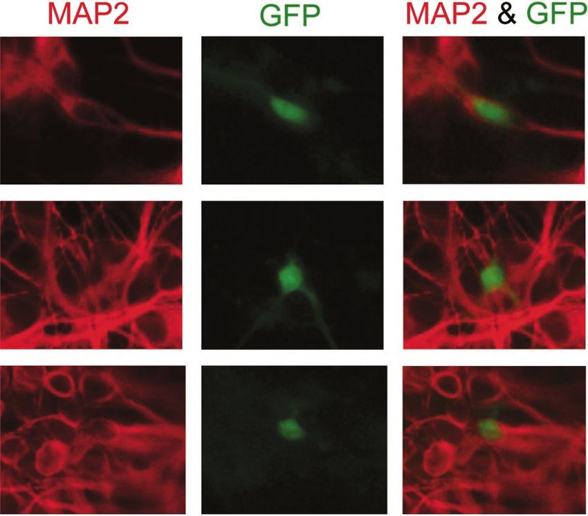
VOL. 78, 2004                                                                                                                  NOTES        4903

                                                                                                                                                    Downloaded from http://jvi.asm.org/ on February 28, 2021 by guest
                                                                            FIG. 2. Transduction of neurons with an ASV-GFP vector.
                                                                         (A) Neuronal cultures were infected with the same virus stock, and
                                                                         GFP reporter expression was examined 3 days postinfection by micros-
                                                                         copy. Representative fluorescent fields (GFP expression, left) and
                                                                         phase-contrast fields (right) are shown. Cultures of neural progenitors
                                                                         infected 1 day after isolation (top) and differentiated neurons infected
                                                                         5 days after isolation (bottom) are shown. (B) ASV transduction cor-
                                                                         relates with integration detected by B2-PCR. Neurons were infected 5
                                                                         days after isolation. Total cellular DNA was isolated 3 days postinfec-
   FIG. 1. Mouse embryonic hippocampal neuron explants. The hip-         tion. B2-PCRs of uninfected neurons, infected neurons, infected mu-
pocampus was isolated from BL/6 mouse embryos between days 14.5          rine fibroblasts, and infected neuron DNA amplified without B2 prim-
and 16.5 of gestation. The hippocampus was stripped of meninges, and     ers are shown.
a single-cell suspension was plated. The neuronal progenitors divide
once and become fully differentiated neurons after approximately 4
days in culture. Representative differential interference contrast mi-
crographs are shown. Panels: A, neuronal progenitors 1 day after
                                                                         after the culture had differentiated were indeed neurons, we
isolation; B, differentiated neurons after 5 days in culture; C, MAP2    asked if the GFP signal colocalized with antibody staining
staining of the same differentiated neurons that were fixed and incu-    against the neuronal marker, MAP2. Examination of MAP2
bated with an anti-MAP2 antibody (red). 4⬘,6⬘-Diamidino-2-phenylin-      expression in the neurons after 5 days in culture verified that
dole (DAPI) (DNA)-stained nuclei are shown in blue.                      this protein was expressed in the dendrites of greater than 90%
                                                                         of the cells (Fig. 1C). In contrast, antibody staining revealed
                                                                         that very few cell bodies (less than 5% of the culture) ex-
   We previously reported that the ASV vector with an inactive           pressed glial fibrillary acidic protein (antibody from DAKO),
integrase does not express detectable GFP (9). To confirm that           an astrocyte-specific marker. We infected these differentiated
GFP expression from the ASV vector corresponded to stable                neuronal cultures with the ASV vector, and 3 days after infec-
transduction, we performed B2-PCR, a modified Alu-PCR                    tion, the cells were fixed in paraformaldehyde and stained with
method (23, 24), to detect the covalent joining of viral DNA to          the MAP2 antibody (Chemicon). Microscopic counting re-
cellular DNA. Chromosomal DNA isolated from differentiated               vealed that 68% of the GFP-positive cells (n ⫽ 113) were also
neurons infected after 5 days in culture was used in this assay          positive for MAP2 (Fig. 3). It was unclear if the remaining
with a primer for mouse B2 genomic repeats (5⬘-TTCACAA                   GFP-positive cells (without detectable MAP2 staining) were
CTCTCGGTGGATGGTGG-3⬘) (7) and a primer for the                           neurons that did not stain with this antibody or were cells that
ASV long terminal repeat (5⬘-GGCTTCGGTTGTACGCGG                          lacked MAP2, such as astrocytes. We note that the milder
TTAGGAGT-3⬘). The samples were subsequently diluted, and                 permeabilization conditions that were used to favor retention
a nested PCR and Southern blotting procedure (4) was per-                of GFP may have resulted in incomplete MAP2 staining. From
formed to generate a unique PCR product for quantitation. A              these results, we conclude that the majority of the cells trans-
strong signal was detected in the infected neuron cultures but           duced with the ASV reporter were differentiated neurons.
not in the uninfected cultures or in a reaction mixture that did            To address our concern that the differentiated neuronal
not include the B2 primer (Fig. 2B). B2-PCR demonstrated                 cultures may contain a small percentage of dividing cells that
that the GFP expression from the ASV reporter virus corre-               would complicate our analyses, we performed a bromode-
lates with integration of the retroviral DNA into the genome of          oxyuridine (BrdU) pulse to label dividing cells as they pro-
the neurons.                                                             ceeded through S phase (22). Both progenitor (1 day after
   To confirm that the GFP-positive cells that were transduced           isolation) and differentiated (5 days after isolation) neuron
4904 NOTES J. VIROL.
Downloaded from http://jvi.asm.org/ on February 28, 2021 by guest
FIG. 3. MAP2-positive neurons are transduced by the ASV re- FIG. 5. Transduction efficiencies of ASV, HIV-1, and MLV. Divid-
porter virus. Neurons were infected 5 days after isolation, and at 3 days ing progenitor cells on the day of isolation and differentiated neurons
postinfection they were fixed and stained for MAP2. MAP2 is localized after 5 days in culture were infected with the same stock of virus. The
to the dendrites and around the cell body, while GFP is localized number of GFP-positive cells was determined by fluorescence-acti-
throughout the cell. GFP-expressing cells were frequently MAP-2 pos- vated cell sorter analysis approximately 3 days postinfection. Trans-
itive. Representative fields of fluorescent staining by MAP2 (left) and duction efficiency was determined by the ratio of GFP-expressing cells
GFP expression (center) and their overlay (right) are shown. in the dividing progenitors to differentiated neurons. The mean trans-
duction efficiency from at least two parallel experiments carried out in
triplicate is shown with the standard deviation between the experi-
ments.
cultures were treated with 50 M BrdU (Sigma) for 30 min at
37°C. Forty-eight hours after the BrdU pulse, we examined
BrdU incorporation by staining the cells with an anti-BrdU
antibody (BD Bioscience) after fixation and treatment with degraded in other phases of the cell cycle (22). Similar to the
DNase I (Promega). As expected, microscopic examination results obtained with BrdU incorporation, a large percentage
showed that a significant number of the progenitor cells (Fig. of the progenitor cells (Fig. 4C) stained with an anti-PCNA
4A) incorporated BrdU, whereas no incorporation (fewer than antibody (DAKO) while cells in the differentiated cultures
1 cell per 10,000) was detected in the differentiated neuronal (Fig. 4D) contained no detectable PCNA (fewer than 1 cell per
culture (Fig. 4B). We also examined the accumulation of pro- 10,000). The results of the BrdU incorporation and PCNA
liferating-cell nuclear antigen (PCNA) in these cells. This pro- staining experiments demonstrate that, unlike the progenitors,
tein accumulates in cells as they enter S phase, but it is rapidly terminally differentiated neuron cultures do not contain cells
that are undergoing significant DNA replication or cell divi-
sion. From these results, we conclude that contamination of
differentiated cells with dividing cells cannot account for the
observed transduction of differentiated neuronal cultures.
The mouse neuron system provided us with a unique oppor-
tunity to compare the relative efficiency with which pseudo-
typed ASV transduces nondividing versus dividing cells. Simi-
lar analyses were carried out with an HIV-1-based vector (3)
pseudotyped with the vesicular stomatitis virus (VSV) G pro-
tein (17) and an MLV vector (pLEGFP-C1; Clontech). The
MLV vector production system yields infectious particles after
transient transfection of a vector plasmid into the AmphoPack-
293 packaging cell line expressing Gag-Pol and the ampho-
tropic Env. As with the ASV vector, expression of enhanced
FIG. 4. Assays for cell cycling. (A) BrdU pulse of dividing progen- GFP encoded in the HIV-1 and MLV vectors is under control
itor cells that were stained for BrdU after 2 days. Representative
differential interference contrast (left) and fluorescence (right) fields of the CMV immediate-early promoter. In these assays, GFP-
are shown. (B) BrdU pulse of differentiated neurons. Differentiated positive cells were quantitated by flow cytometry. Relative
neurons after 5 days in culture were treated as described above. transduction efficiencies were measured by analysis of dividing
(C) PCNA levels in dividing progenitor cells. Dividing progenitor cells neuronal progenitors infected on the day of isolation and dif-
cultured for 1 day were fixed and stained with an anti-PCNA antibody.
(D) PCNA levels in differentiated neurons. Differentiated neurons
ferentiated neurons infected after 5 days in culture with the
were cultured for 5 days before they were fixed and stained with an same virus stock. To compare these vectors, the results are
anti-PCNA antibody. expressed as a transduction ratio, the ratio of the number ofVOL. 78, 2004 NOTES 4905
GFP-positive cells after infection of differentiated neurons enhancing the ability of HIV-1 to transduce nondividing cells
compared to the infection of dividing progenitor cells. but are not required for this function. As ASV shares this
As summarized in Fig. 5, with the ASV vector, there were ability with HIV-1, some determinants may be shared as well.
four times more GFP-positive cells when the cultures were Here, we have confirmed that ASV can transduce naturally
infected prior to differentiation, yielding a transduction ratio of arrested, terminally differentiated neurons. We show that the
ca. 0.25. This value was approximately one-half of the 0.4 ratio terminally differentiated neuronal cultures do not contain de-
observed with the HIV-1 vector. This difference is consistent tectable dividing cells and that therefore such cells cannot
with the titer of the ASV vector being half that of the HIV-1 account for the observed transduction activities of the ASV,
vector on the neuronal cultures (1.57 ⫻ 104 ⫾ 0.9 ⫻ 104 for HIV-1, and MLV vectors. MLV was the most restricted, and
ASV compared to 2.56 ⫻ 104 ⫾ 0.8 ⫻ 104 for HIV-1), even ASV transduction efficiency was reduced compared to that of
though the titers of these vectors were similar on dividing HIV-1. These results demonstrate the ability of a non-lentivi-
progenitors (6.25 ⫻ 104 ⫾ 2.0 ⫻ 104 and 6.6 ⫻ 104 ⫾ 2.4 ⫻ 104, rus vector to transduce postmitotic cells and indicate that the
respectively). Previous studies with differentiated myofibers determinants for this ability are not lentivirus specific but may
(19) and unstimulated G0 hematopoietic stem cells (21) be shared with other retroviruses.
Downloaded from http://jvi.asm.org/ on February 28, 2021 by guest
showed similar HIV transduction ratios. In our studies, the
GFP promoter had little effect on transduction efficiency be- We thank Christoph Seeger and John Taylor for critical comments
cause an HIV-1-based vector with the phosphoglycerol kinase on the manuscript.
promoter (6) gave results similar to those obtained with the This work was supported by National Institutes of Health grants
AI40385, CA71515, CA06927, and GM47903, by the Mathers Chari-
vector containing the CMV promoter (data not shown). The table Foundation, and also by an appropriation from the Commonwealth
MLV vector had the lowest transduction ratio, less than 0.1. of Pennsylvania. The following Fox Chase Cancer Center shared facilities
Previous investigations have reported similar results following were used in the course of this work: Flow Cytometry and Cell Sorting
MLV infection of rat neuronal cultures (16). It is generally Facility, Cell Culture Facility, Biochemistry and Biotechnology Facility
(DNA synthesis), and Research Secretarial Services.
assumed that MLV is unable to infect noncycling cells and that The contents of this report are solely the responsibility of the au-
transduction of such cultures signifies the presence of residual thors and do not necessarily represent the official views of the National
dividing cells. However, the ratio that we obtained with MLV Cancer Institute or any other sponsoring organization.
cannot be attributed to contamination with dividing cells, as REFERENCES
there is no detectable background in these differentiated cul- 1. Barsov, E. V., and S. H. Hughes. 1996. Gene transfer into mammalian cells
tures (Fig. 4). We suspect that the cellular environment pro- by a Rouse sarcoma virus-based retroviral vector with the host range of the
vided in the neuronal cultures, which recently exited the cell amphotropic murine leukemia virus. J. Virol. 70:3922–3939.
2. Bukrinsky, M. I., N. Sharova, M. P. Dempsey, T. L. Stanwick, A. G. Bukrin-
cycle, may provide favorable conditions for completion of early skaya, S. Haggerty, and M. Stevenson. 1992. Active nuclear import of human
events and thereby allow more efficient detection of viral DNA immunodeficiency virus type 1 preintegration complexes. Proc. Natl. Acad.
Sci. USA 89:6580–6584.
nuclear import and integration. 3. Chen, W., X. Wu, D. N. Levasseur, H. Liu, L. Lai, J. C. Kappes, and T. M.
Although the vectors used in this study encode similar re- Townes. 2000. Lentiviral vector transduction of hematopoietic stem cells that
porter cassettes, we note that the vectors were not completely mediate long-term reconstitution of lethally irradiated mice. Stem Cells
18:352–359.
matched. In the MLV and HIV-1 expression vectors, essential 4. Daniel, R., G. Kao, K. Taganov, J. G. Greger, O. Favorova, G. Merkel, T. J.
viral genes are replaced with the reporter cassette, whereas the Yen, R. A. Katz, and A. M. Skalka. 2003. Evidence that the retroviral DNA
ASV vector encodes a full complement of viral genes in addi- integration process triggers an ATR-dependent DNA damage response.
Proc. Natl. Acad. Sci. USA 100:4778–4783.
tion to the reporter cassette and is replication competent in 5. Deglon, N., and P. Aebischer. 2002. Lentiviruses as vectors for CNS diseases.
avian cells. The HIV-1 vector contains the VSV G protein for Curr. Top. Microbiol. Immunol. 261:191–209.
6. Gerolami, R., R. Uch, F. Jordier, S. Chapel, C. Bagnis, C. Brechot, and P.
entry, whereas both the ASV and MLV vectors enter cells via Mannoni. 2000. Gene transfer to hepatocellular carcinoma: transduction
the amphotropic envelope receptor. Nevertheless, any differ- efficacy and transgene expression kinetics by using retroviral and lentiviral
ences in titers caused by vector differences should be irrelevant vectors. Cancer Gene Ther. 7:1286–1292.
7. Ha, H. C., K. Juluri, Y. Zhou, S. Leung, M. Hermankova, and S. H. Snyder.
as our comparison relies on the relative transduction efficiency 2001. Poly(ADP-ribose) polymerase-1 is required for efficient HIV-1 inte-
of each vector on dividing versus nondividing cultures (trans- gration. Proc. Natl. Acad. Sci. USA 98:3364–3368.
duction ratio), rather than on absolute titers. However, it is 8. Hatziioannou, T., and S. P. Goff. 2001. Infection of nondividing cells by Rous
sarcoma virus. J. Virol. 75:9526–9531.
possible that as the cultures differentiate, the surface expres- 9. Katz, R. A., J. G. Greger, K. Darby, P. Boimel, G. F. Rall, and A. M. Skalka.
sion of the amphotropic receptor, Pit-2, could be altered. This 2002. Transduction of interphase cells by avian sarcoma virus. J. Virol.
76:5422–5434.
possibility is less likely for the ubiquitous VSV G receptor. 10. Kim, V. N., K. Mitrophanous, S. M. Kingsman, and A. J. Kingsman. 1998.
Despite these differences between vectors, it is clear that ASV Minimal requirement for a lentivirus vector based on human immunodefi-
can transduce nondividing neuron cultures with reasonable ciency virus type 1. J. Virol. 72:811–816.
11. Kukolj, G., K. S. Jones, and A. M. Skalka. 1997. Subcellular localization of
efficiency. avian sarcoma virus and human immunodeficiency virus type 1 integrases.
We (9) and others (8) have shown that ASV can transduce J. Virol. 71:843–847.
nondividing cells. The results presented here confirm this find- 12. Kukolj, G., R. A. Katz, and A. M. Skalka. 1998. Characterization of the
nuclear localization signal in the avian sarcoma virus integrase. Gene 223:
ing, although the transduction ratio of ASV is less than that of 157–163.
HIV-1. The viral sequences that account for these differences 13. Lawrence, D. M., C. E. Patterson, T. L. Gales, J. L. D’Orazio, M. M. Vaughn,
and G. F. Rall. 2000. Measles virus spread between neurons requires cell
have not been mapped and may include, but not be limited to, contact but not CD46 expression, syncytium formation, or extracellular virus
nuclear import signals. Studies with HIV-1 vectors that lack all production. J. Virol. 74:1908–1918.
accessory proteins, Vpr, Vpu, Vif, and Nef, have shown re- 14. Lewis, P. F., and M. Emerman. 1994. Passage through mitosis is required for
oncoretroviruses but not for the human immunodeficiency virus. J. Virol.
duced transduction efficiencies with cycle-arrested cells (10). 68:510–516.
This suggests that these accessory proteins may play a role in 15. Miller, D. G., M. A. Adam, and A. D. Miller. 1990. Gene transfer by retro-4906 NOTES J. VIROL.
virus vectors occurs only in cells that are actively replicating at the time of of human progenitor hematopoietic stem cells by human immunodeficiency
infection. Mol. Cell. Biol. 10:4239–4242. virus type 1-based vectors is cell cycle dependent. J. Virol. 73:3649–3660.
16. Mitrophanous, K., S. Yoon, J. Rohll, D. Patil, F. Wilkes, V. Kim, S. Kingsman, 22. Takase, K., M. Sawai, K. Yamamoto, J. Yata, Y. Takasaki, H. Teraoka, and K.
A. Kingsman, and N. Mazarakis. 1999. Stable gene transfer to the nervous Tsukada. 1992. Reversible G1 arrest induced by dimethyl sulfoxide in human
system using a non-primate lentiviral vector. Gene Ther. 6:1808–1818. lymphoid cell lines: kinetics of the arrest and expression of the cell cycle marker
17. Naldini, L., U. Blomer, P. Gallay, D. Ory, R. Mulligan, F. H. Gage, I. M. proliferating cell nuclear antigen in Raji cells. Cell Growth Differ. 3:515–521.
Verma, and D. Trono. 1996. In vivo gene delivery and stable transduction of 23. Vandegraaff, N., R. Kumar, C. J. Burrell, and P. Li. 2001. Kinetics of human
immunodeficiency virus type 1 (HIV) DNA integration in acutely infected
nondividing cells by a lentiviral vector. Science 272:263–267.
cells as determined using a novel assay for detection of integrated HIV
18. Roe, T., T. C. Reynolds, G. Yu, and P. O. Brown. 1993. Integration of murine DNA. J. Virol. 75:11253–11260.
leukemia virus DNA depends on mitosis. EMBO J. 12:2099–2108. 24. Vandegraaff, N., R. Kumar, H. Hocking, T. R. Burke, Jr., J. Mills, D. Rhodes,
19. Sakoda, T., N. Kasahara, Y. Hamamori, and L. Kedes. 1999. A high-titer C. J. Burrell, and P. Li. 2001. Specific inhibition of human immunodeficiency
lentiviral production system mediates efficient transduction of differentiated virus type 1 (HIV-1) integration in cell culture: putative inhibitors of HIV-1
cells including beating cardiac myocytes. J. Mol. Cell. Cardiol. 31:2037–2047. integrase. Antimicrob. Agents Chemother. 45:2510–2516.
20. Sherman, M. P., and W. D. Greene. 2002. Slipping through the door: HIV 25. Weinberg, J. B., T. J. Matthews, B. R. Cullen, and M. H. Malim. 1991.
entry into the nucleus. Microbes Infect. 4:67–73. Productive human immunodeficiency virus type 1 (HIV-1) infection of non-
21. Sutton, R. E., M. J. Reitsma, N. Uchida, and P. O. Brown. 1999. Transduction proliferating human monocytes. J. Exp. Med. 174:1477–1482.
Downloaded from http://jvi.asm.org/ on February 28, 2021 by guestYou can also read