Comparison of initial high-resolution computed tomography (HRCT) features of coronavirus disease 2019 (COVID-19) pneumonia and other viral pneumonias
←
→
Page content transcription
If your browser does not render page correctly, please read the page content below
Original Article

Comparison of initial high-resolution computed tomography
(HRCT) features of coronavirus disease 2019 (COVID-19)
pneumonia and other viral pneumonias
Yilong Huang1#, Yuanming Jiang1#, Zhenhui Li2#, Dan Han1, Li Wu1, Jiyao Ma1, Peng Wang3, Ying Xie4,
Zhipeng Li5, Xiang Li6, Minchang Hong7, Jialong Zhou8, Chuwei Duan9, Yunhui Yang10, Wei Zhao1,
Feng Yuan1, Kunhua Wang11, Wenfang Yi1, Bo He1,11
1
Medical Imaging Department, First Affiliated Hospital of Kunming Medical University, Kunming, China; 2Medical Imaging Department, Yunnan
Cancer Hospital & The Third Affiliated Hospital of Kunming Medical University, Kunming, China; 3Radiology Department, the Second People’s
Hospital of Yunnan Province, Kunming, China; 4Department of Radiology, Calmette Hospital & The First Hospital of Kunming, Kunming, China;
5
    Medical Imaging Department, Yunnan Provincial Infectious Disease Hospital (Yunnan AIDS Care Center), Kunming, China; 6Department of
Radiology, The 3rd People’s Hospital of Kunming, Kunming, China; 7Department of Radiology, the First People’s Hospital of Zhaotong, Zhaotong,
China; 8MRI Department, The First People’s Hospital of Yunnan Province, Kunming, China; 9Radiology Department, Yan’an Hospital of Kunming,
Kunming, China; 10Department of Medical Imaging in People’s Hospital of Xishuangbanna Dai Autonomous Prefecture, Jinghong, China; 11NHC
Key Laboratory of Drug Addiction Medicine, First Affiliated Hospital of Kunming Medical University, Kunming Medical University, Kunming,
China
Contributions: (I) Conception and design: Y Huang, W Yi, B He; (II) Administrative support: D Han, K Wang, B He; (III) Provision of study
materials or patients: P Wang, Y Xie, Z Li, X Li, M Hong, J Zhou, C Duan, Y Yang, W Zhao; (IV) Collection and assembly of data: Y Jiang, Z Li, L
Wu, J Ma, F Yuan; (V) Data analysis and interpretation: Y Huang, Y Jiang, Z Li, L Wu, J Ma, B He; (VI) Manuscript writing: All authors; (VII) Final
approval of manuscript: All authors.
#
These authors contributed equally to this work.
Correspondence to: Bo He. NHC Key Laboratory of Drug Addiction Medicine, Medical Imaging Department, First Affiliated Hospital of Kunming
Medical University, Kunming Medical University, Kunming, China. Email: hebo_ydyy@qq.com; Kunhua Wang. NHC Key Laboratory
of Drug Addiction Medicine, First Affiliated Hospital of Kunming Medical University, Kunming Medical University, Kunming, China.
Email: wangkunhua_ydyy@163.com; Wenfang Yi. Medical Imaging Department, First Affiliated Hospital of Kunming Medical University, Kunming,
China. Email: 471991019@qq.com.

                  Background: Multicenter retrospective comparison of the first high-resolution computed tomography
                  (HRCT) findings of coronavirus disease 2019 (COVID-19) and other viral pneumonias.
                  Methods: We retrospectively collected clinical and imaging data from 262 cases of confirmed viral pneumonia
                  in 20 hospitals in Yunnan Province, China, from March 1, 2015 to March 15, 2020. According to the virus
                  responsible for the pneumonia, the pneumonias were divided into non-COVID-19 (141 cases) and COVID-19
                  (121 cases). The non-COVID-19 pneumonias comprised cytomegalovirus (CMV) (31 cases), influenza A
                  virus (82 cases), and influenza B virus (20 cases). The differences in the basic clinical characteristics, lesion
                  distribution, location and imaging signs among the four viral pneumonias were analyzed and compared.
                  Results: Fever and cough were the most common clinical symptoms of the four viral pneumonias.
                  Compared with the COVID-19 patients, the non-COVID-19 patients had higher proportions of fatigue,
                  sore throat, expectorant and chest tightness (all P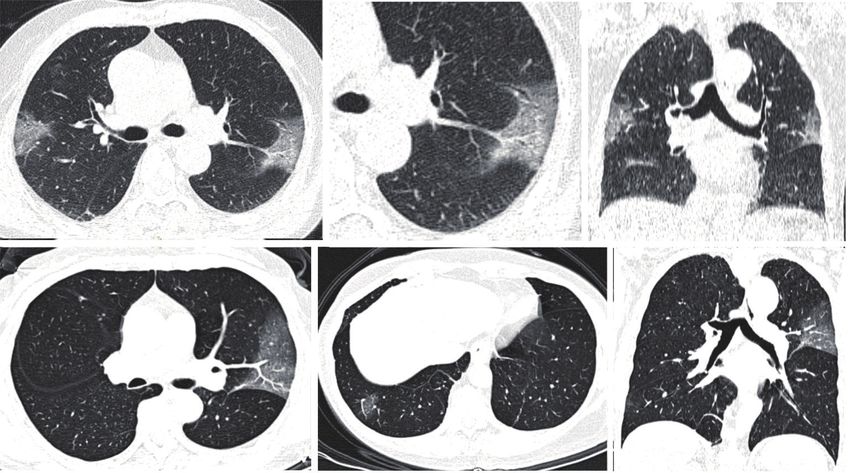
Annals of Palliative Medicine, Vol 10, No 1 January 2021                                                                                   561

                 thickening and pleural effusion were rare (all P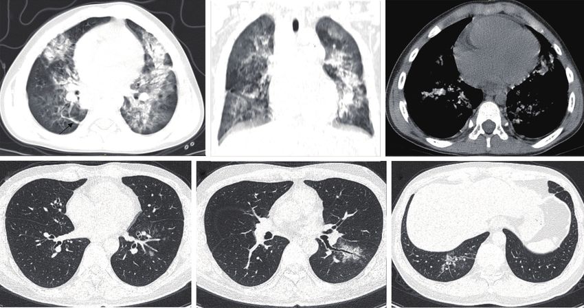
562                                                                                     Huang et al. HRCT of COVID-19 and other viral pneumonias

                                    Patients with viral pneumonia in Yunnan (n=353)

                                                             Exclusion step 1, n=22        No clinical and imaging data (n=22)

                               Confirmed patients with clinical and imaging data (n=331)

                                                                                           1. Initial chest examination were DR (n=8)
                                                             Exclusion step 2, n=51        2. Not enough CT images (n=12)
                                                                                           3. Image format were not DICOM (n=9)
                                                                                           4. Thickness> 2 mm (n=22)

                                        Complete clinical and CT data (n=280)
                                                                                           1. Obvious image artifacts (n=8)
                                                             Exclusion step 3, n=18        2. Combining severe bacterial infections,
                                                                                           fungal infections and tuberculosis (n=10)

                                  Patients with standard clinical and CT data (n=262)

                            Non-COVID-19 (n=141)
                                                                     COVID-19 (n=121)
                                  CMV (n=31)
                            Influenza A virus (n=82)               No CT findings (n=33)
                            Influenza B virus (n=20)             Positive CT findings (n=88)
                         Other viruses with less than 2
                                  cases (n=8)

Figure 1 Flowchart of the study design and the participants included in the analysis. COVID-19, coronavirus disease 2019; CMV,
cytomegalovirus; CT, computed tomography. DR, digital radiography; DICOM, digital imaging and communications in medicine.

diarrhea or no obvious symptoms), underlying diseases                            1.5 mm. CT machines dedicated to COVID-19 patients are
[hypertension, diabetes, coronary heart disease, liver disease,                  used for chest examinations, using disposable sheets to avoid
tumor, acquired immunodeficiency syndrome (AIDS) and                             cross-infection. After the patient’s CT examination, disinfect
leukopenia], hospital admission time and CT examination                          the examination bed and floor, and disinfect the computer
time were recorded. Patients completed the chest CT                              room and air with ultraviolet rays.
examination within 48 h of admission. According to the
virus found in the lungs, the patients were divided into four
                                                                                 Chest CT analysis
groups: cytomegalovirus (CMV), influenza A virus, influenza
B virus and COVID-19. The number of cases from each                              Three radiologists were blinded to the qRT-PCR results, all
hospital is shown in Table 1. All patients were from Yunnan                      patient information, and the type of viral pneumonia. First,
Province, China.                                                                 two experienced radiologists (Yilong Huang and Yuanming
                                                                                 Jiang) in the cardiothoracic group independently read
                                                                                 the radiographs. When their opinions were inconsistent,
CT protocol
                                                                                 they discussed them and reached a consensus, which was
HRCT examination: CT scanners with ≥16 detector rows                             reviewed and confirmed by the third senior radiologist in
(Siemens, Germany; Philips, The Netherlands; and GE,                             the cardiothoracic group (Bo He). The morphological signs
USA) were used. The patient was scanned in the supine                            of the first CT examination after admission were analyzed.
position while breath holding after inhalation. The scanning                     The CT imaging evaluation included lesion distribution
range was from the thoracic inlet to the costophrenic angles.                    (peripheral, central), location (left upper lobe, left lower
Scanning parameters: detector collimation width 64 mm ×                          lobe, right upper lobe, right middle lobe and right lower
0.6 mm or 128 mm × 0.6 mm, tube voltage 120 kV, adaptive                         lobe) and signs (GGO, partial consolidation, multifocal
tube current, high-resolution algorithm reconstruction,                          consolidation, focal consolidation, fibrous stripes, septal
reconstruction layer thickness 1 or 1.5 mm and layer spacing                     thickening, intralobular interstitial thickening, subpleural

© Annals of Palliative Medicine. All rights reserved.                   Ann Palliat Med 2021;10(1):560-571 | http://dx.doi.org/10.21037/apm-20-2479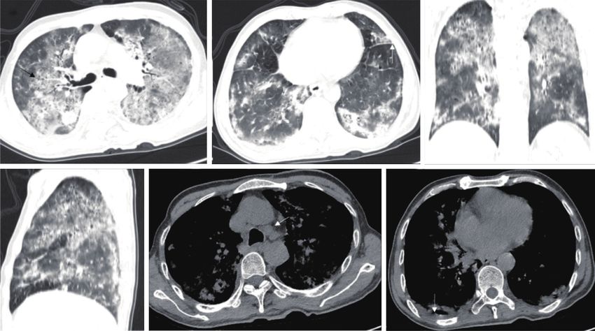
Annals of Palliative Medicine, Vol 10, No 1 January 2021                                                                                      563

 Table 1 Institution and number of cases in this multicenter study
 Institution                                            Total No. of cases   CMV       Influenza A virus     Influenza B virus     COVID-19

 Kunming Third People’s Hospital                               71             2               31                     5                 30

 Yunnan Provincial Infectious Disease Hospital                 50             27               8                     0                 12

 Kunming First People’s Hospital                               30             0               18                    11                 0

 The First People’s Hospital of Zhaotong                       22             0                4                     1                 17

 Third People’s Hospital of Yunnan Province                    15             1               12                     2                 0

 Xishuangbanna People’s Hospital                               13             0                3                     1                 9

 Qujing First People’s Hospital                                12             0                0                     0                 12

 Dali People’s Hospital                                        11             0                0                     0                 11

 Yuxi People’s Hospital                                        10             0                0                     0                 10

 Lijiang People’s Hospital                                      6             0                0                     0                 6

 Second People’s Hospital of Yunnan Province                    5             1                4                     0                 0

 Chuxiong People’s Hospital                                     4             0                0                     0                 4

 Lancang First People’s Hospital                                3             0                0                     0                 3

 First People’s Hospital of Yunnan Province                     2             0                2                     0                 0

 Baoshan People’s Hospital                                      2             0                0                     0                 2

 Honghe People’s Hospital                                       2             0                0                     0                 2

 Pu’er People’s Hospital                                        1             0                0                     0                 1

 Qiaojia People’s Hospital                                      1             0                0                     0                 1

 Ruili People’s Hospital                                        1             0                0                     0                 1

 Total                                                         262            31              82                    20                121
 Data are number of patients. CMV, cytomegalovirus; COVID-19, coronavirus disease 2019.

lines, crazy-paving pattern, tree-in-bud, bronchial wall                      The Z-test (Bonferroni method) was used for pairwise
thickening, bronchiectasis, vascular thickening, air                          comparisons. A P value
564                                                                       Huang et al. HRCT of COVID-19 and other viral pneumonias

other COVID-19 patients. Five patients with influenza                 signs (59.09% vs. 14.89%, P
Annals of Palliative Medicine, Vol 10, No 1 January 2021                                                                                        565

 Table 2 Basic clinical characteristics of 262 patients with viral pneumonia
                                                        Influenza A virus        Influenza B virus    Non-COVID-19         COVID-19
 Clinical characteristics                CMV (n=31)                                                                                       P value
                                                              (n=82)                   (n=20)            (n=141)            (n=121)

 Age (av.), years                        41.55±10.69      55.50±18.46*             40.85±18.75         48.50±19.47*       42.88±17.67      0.000

 Age range                                  28–76             6–93                     6–72                0.5–93             3–79

 Sex, n (%)                                                                                                                                0.000

   Male                                   26 (83.87)*      56 (68.29)*              11 (55.00)           97 (68.79)*        54 (44.63)

   Female                                 5 (16.13)*       26 (31.71)*               9 (45.00)           44 (31.21)*        67 (55.37)

 Epidemiological history, n (%)

   Travel/residence history in Hubei       0 (0.00)          0 (0.00)                0 (0.00)             0 (0.00)          98 (80.99)

   Close contact with patients             0 (0.00)         16 (19.51)               2 (10.00)           19 (13.48)         23 (19.01)

   Exposure to infected poultry            0 (0.00)          5 (6.10)                0 (0.00)             5 (3.55)           0 (0.00)

 Symptoms, n (%)

   Fever                                  22 (70.97)       72 (87.80)*              16 (80.00)          118 (83.69)*        60 (49.59)     0.000

   Cough                                  20 (64.52)       75 (91.46)*               8 (40.00)          103 (73.05)*        53 (43.80)     0.000

   Fatigue                                12 (38.71)*      32 (39.02)*               5 (25.00)           49 (34.75)*        17 (14.05)     0.000

   Difficulty breathing                    3 (9.68)          6 (7.32)                1 (5.00)             11 (7.80)          3 (2.48)      0.349

   Sore throat                             0 (0.00)        23 (28.05)*              2 (10.00)*           25 (17.73)         12 (9.92)      0.001

   Runny nose                              4 (12.90)        11 (13.41)               1 (5.00)            16 (11.35)          8 (6.61)      0.450

   Expectorant                            10 (32.26)*       32 (39.02)              8 (40.00)*           50 (35.46)*        18 (14.88)     0.001

   Headache                                1 (3.23)          4 (4.88)                1 (5.00)             6 (4.26)           7 (5.79)      0.972

   Muscle ache                             0 (0.00)          6 (7.32)                1 (5.00)             7 (4.96)           9 (7.44)      0.551

   Chest tightness                        21 (67.74)*        4 (4.88)                7 (35.00)           32 (22.70)*         9 (7.44)      0.000

   Chest pain                              0 (0.00)          2 (2.44)                1 (5.00)             3 (2.13)           3 (2.48)      0.838

   Nausea and vomiting                     0 (0.00)          0 (0.00)                0 (0.00)             0 (0.00)           2 (1.65)      0.336

   Diarrhea                                0 (0.00)          0 (0.00)                0 (0.00)             0 (0.00)           1 (0.83)      0.686

   No obvious symptoms                     0 (0.00)          0 (0.00)*               0 (0.00)             0 (0.00)*         15 (12.40)     0.000

 Underlying disease, n (%)

   Hypertension                            3 (9.68)        28 (34.15)*              10 (50.00)*          42 (29.79)*         9 (7.44)      0.000

   Diabetes                                2 (6.45)        19 (23.17)*               2 (10.00)           23 (16.31)*         7 (5.79)      0.004

   Coronary heart disease                  1 (3.23)          2 (2.44)                1 (5.00)             4 (2.84)           3 (2.48)      0.977

   Liver disease                           6 (19.35)         3 (3.66)                2 (10.00)            11 (7.80)          2 (1.65)      0.003

   Malignant tumor                         1 (3.23)          0 (0.00)                0 (0.00)             1 (0.71)           0 (0.00)      0.215

   AIDS                                   22 (70.97)*        0 (0.00)                0 (0.00)            22 (15.60)*         0 (0.00)      0.000

   Leukopenia                             6 (19.35)*         0 (0.00)                0 (0.00)             6 (4.26)           0 (0.00)      0.000
 Continuous data are expressed as mean ± SD, and categorical data are presented as n (%). *, compared with COVID-19, significance was
 considered when P
566 Huang et al. HRCT of COVID-19 and other viral pneumonias
Distribution Involved lobes Upper left lobe
Peripheral + central Left lower lobe
Right upper lobe
Peripheral
Right lung middle lobe
Percentage of involved lobes (%)
100 Right lower lobe
Percentage of cases (%)
100
75
75
50
25 50
0 25
19
19
19
19
V
V
s
s
s
s
ru
ru
M
ru
M
ru
D-
D-
D-
D-
vi
vi
C
C
vi
vi
VI
VI
VI
VI
B
B
A
A
O
O
O
O
za
za
za
za
C
C
C
C
-
-
en
en
en
en
on
on
flu
flu
flu
flu
N
N
In
In
In
In
Figure 2 Distribution and location of COVID-19 pneumonia and other viral pneumonias: (A) lesion distribution and (B) lesion location.
Data are reported as percentages. *, PAnnals of Palliative Medicine, Vol 10, No 1 January 2021 567
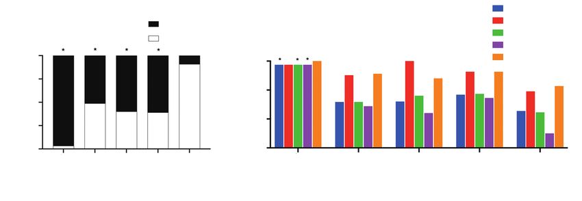
Table 4 Diagnosis performance of CT findings in distinguishing COVID-19 from Non-COVID-19
CT findings Sensitivity (%) Specificity (%) Accuracy (%)
GGO 96.59 53.19 69.87
Multifocal consolidation 7.95 60.28 40.17
Fibrous stripes 15.91 65.25 46.29
Intralobular interstitial thickening 63.64 80.85 74.24
Subpleural lines 7.95 75.18 49.34
Crazy-paving pattern 0.00 85.82 52.84
Tree-in-bud 1.14 72.34 44.98
Vascular thickening 67.05 95.04 84.28
Halo sign 59.09 85.11 75.11
Mediastinal lymphadenectasis 1.14 85.82 53.28
Pleural thickening 38.64 43.26 41.48
Pleural effusion 0.00 74.47 45.85
COVID-19, coronavirus disease 2019; GGO, ground-glass opacities.
A B C
D E F
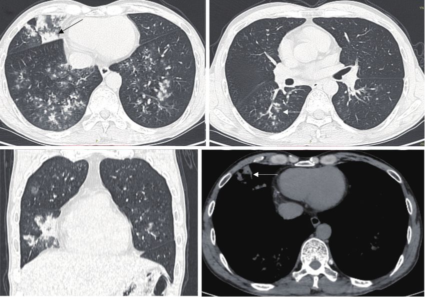
Figure 3 Chest CT findings in 54-year-old male with CMV pneumonia combined with AIDS and leukopenia. (A,B,C,D,E,F) Diffuse
GGO and multifocal consolidation in both lungs. (A) Bronchiectasis (black arrow), (B) left intralobular interstitial thickening (white arrow),
(E) mediastinal lymphadenectasis (white arrow), and (F) left pleural effusion (white arrow). (A,B,E,F): axial view; (C): coronal view; (D):
sagittal view. AIDS, acquired immunodeficiency syndrome; COVID-19, coronavirus disease 2019; CMV, cytomegalovirus; CT, computed
tomography; GGO, ground-glass opacity.
other viral pneumonias, and multifocal consolidation was between COVID-19 and other viral pneumonias are
more common in other viral pneumonias, a result that was still rare. The GGO, intralobular interstitial thickening,
consistent with previous studies (17,19). To the best of our vascular thickening and halo sign were more likely to occur
knowledge, studies comparing the microscopic differences in COVID-19 than in other viral pneumonias, and fibrous
© Annals of Palliative Medicine. All rights reserved. Ann Palliat Med 2021;10(1):560-571 | http://dx.doi.org/10.21037/apm-20-2479568 Huang et al. HRCT of COVID-19 and other viral pneumonias
A B
C D
Figure 4 Chest CT findings of influenza A pneumonia in a 44-year-old male (H1N1 infection). (A,B,C,D) Multifocal GGO, partial
consolidation and consolidation in both lungs (arrow in A, D shows consolidation in right middle lobe); (B) tree-in-bud (white arrow). (C)
Coronal view. CT, computed tomography; GGO, ground-glass opacity.
A B C
D E F
Figure 5 Chest CT findings of influenza B pneumonia in (A,B,C) 6-year-old male and (D,E,F) 39-year-old male. (A,B,C) Multifocal
GGO with partial consolidation in both lungs with a parenchymal band (A, black arrow), air bronchogram, pleural thickening, and pleural
effusion. (D) Multifocal GGO and tree-in-bud in lower lobes of both lungs, with bronchial wall thickening (E) and partial consolidation and
parenchymal band (F). (A,B,C,D,E,F) Axial view.
stripes, subpleural lines, crazy-paving pattern, tree-in- characteristic imaging changes are still unclear. However,
bud, pleural effusion and mediastinal lymphadenectasis the difference in CT findings in COVID-19 and other viral
occurred less frequently in COVID-19 than in other viral pneumonia may be closely related to the difference in virus
pneumonias. The pathophysiological mechanisms of these types. After autopsy of patients who died of COVID-19 and
© Annals of Palliative Medicine. All rights reserved. Ann Palliat Med 2021;10(1):560-571 | http://dx.doi.org/10.21037/apm-20-2479Annals of Palliative Medicine, Vol 10, No 1 January 2021 569
A B C
D E F
Figure 6 Chest CT findings of COVID-19 pneumonia in (A,B,C) 63-year-old female and (D,E,F) 66-year-old female. (A,B,C) Multifocal
GGO in both lungs. (B) Vascular thickening and intralobular interstitial thickening, and (C) pleural thickening. (D,E,F) Multifocal GGO
in both lungs. (D) Intralobular interstitial thickening, (D,E) vascular thickening and (F) pleural thickening. (A,B,D,F) Axial view; (C,F)
coronal view.
influenza (20), histological results revealed diffuse alveolar elderly, lung lesions are more likely to progress. Study has
injury with perivascular T cell infiltration in the peripheral shown that age is an independent wind direction factor for
lung tissue of COVID-19. The pulmonary vessels of COVID-19 (26). In-hospital mortality increases linearly
Covid-19 patients have severe endothelial damage, and with age, with the highest mortality in patients over
extensive thrombosis is accompanied by microvascular 80 years of age.
disease. The incidence of alveolar capillary thrombosis This study has some limitations. First, although we tried
in Covid-19 patients is 9 times that of influenza patients. our best to collect the clinical and imaging data of patients
And this also may be related to the slower development with viral pneumonia in Yunnan Province, the numbers of
of COVID-19 than other viral pneumonias (19). confirmed cases of adenovirus, measles virus, herpes virus
It is worth noting that tree-in-bud was observed in 27.66% and other viruses were relatively small, but the differences
of influenza pneumonia cases, and similar results were in imaging signs among them are very interesting and will
observed by Shiley et al. (21), but almost no tree-in- be studied in the future. Second, most of the included CMV
bud was observed in COVID-19. Compared with other pneumonia patients had AIDS, which is likely to cause a
viral pneumonias, CMV pneumonia often presents as a selection bias regarding other CMV pneumonia patients.
characteristic bronchiectasis, and immunocompromise is Third, different populations, such as infants, children
an important risk factor. In previous studies, it has been and elderly adults, may be susceptible to different viruses,
called “AIDS associated bronchiectasis” (22). With this and their signs of pulmonary lesions on imaging might be
information combined with the clinical data, radiologists different. Because of the sample size, we did not perform
can better identify the type of viral pneumonia (23). a subgroup analysis for age. In future studies, more effort
Currently, Duan et al. and Gu et al. observe the CT findings should be made to determine the differences in the imaging
of COVID-19 patients in different populations, such as characteristics of different populations.
children, adults, and the elderly (24,25). They found that In summary, the analysis and comparison of the chest CT
compared with adults, pediatric COVID-19 patients are findings of COVID-19 and other viral pneumonias showed
characterized by lower incidence, milder clinical symptoms, that the chest CT findings partially overlapped, but many
shorter course of disease, and fewer severe cases. In the significant imaging features could still be observed, which
© Annals of Palliative Medicine. All rights reserved. Ann Palliat Med 2021;10(1):560-571 | http://dx.doi.org/10.21037/apm-20-2479570 Huang et al. HRCT of COVID-19 and other viral pneumonias
is helpful for the early differential diagnosis of COVID-19 from Patients with Pneumonia in China, 2019. N Engl J
and the development of more accurate clinical diagnosis and Med 2020;382:727-33.
treatment strategies. 2. Guan WJ, Ni ZY, Hu Y, et al. Clinical Characteristics
of Coronavirus Disease 2019 in China. N Engl J Med
2020;382:1708-20.
Acknowledgments
3. Qiu H, Wu J, Hong L, et al. Clinical and epidemiological
The authors express their appreciation for all the hospital features of 36 children with coronavirus disease 2019
staff for their efforts to combat the COVID-19 outbreak. (COVID-19) in Zhejiang, China: an observational cohort
Thank you to all the patients who participated in this study. study. Lancet Infect Dis 2020;20:689-96.
Funding: None. 4. Chen N, Zhou M, Dong X, et al. Epidemiological
and clinical characteristics of 99 cases of 2019 novel
coronavirus pneumonia in Wuhan, China: a descriptive
Footnote
study. Lancet 2020;395:507-13.
Reporting Checklist: The authors have completed the 5. Fowlkes A, Steffens A, Temte J, et al. Incidence of
STROBE reporting checklist. Available at http://dx.doi. medically attended influenza during pandemic and
org/10.21037/apm-20-2479 postpandemic seasons through the Influenza Incidence
Surveillance Project, 2009-13. Lancet Respir Med
Data Sharing Statement: Available at http://dx.doi. 2015;3:709-18.
org/10.21037/apm-20-2479 6. Moriyama M, Hugentobler WJ, Iwasaki A. Seasonality of
Respiratory Viral Infections. Annu Rev Virol 2020;7:83-101.
Conflicts of Interest: All authors have completed the ICMJE 7. Li Y, Reeves RM, Wang X, et al. Global patterns in
uniform disclosure form (available at http://dx.doi. monthly activity of influenza virus, respiratory syncytial
org/10.21037/apm-20-2479). The authors have no conflicts virus, parainfluenza virus, and metapneumovirus:
of interest to declare. a systematic analysis. Lancet Glob Health
2019;7:e1031-e1045.
Ethical Statement: The authors are accountable for all 8. Fang Y, Zhang H, Xie J, et al. Sensitivity of Chest CT
aspects of the work in ensuring that questions related for COVID-19: Comparison to RT-PCR. Radiology
to the accuracy or integrity of any part of the work are 2020;296:E115-E117.
appropriately investigated and resolved. The study was 9. Koo HJ, Lim S, Choe J, et al. Radiographic and CT Features
conducted in accordance with the Declaration of Helsinki (as of Viral Pneumonia. Radiographics 2018;38:719-39.
revised in 2013). The retrospective study was approved by 10. Miller WT Jr, Mickus TJ, Barbosa E Jr, et al. CT of viral
the institutional committee of the First Affiliated Hospital lower respiratory tract infections in adults: comparison
of Kunming Medical University. Informed consent was among viral organisms and between viral and bacterial
waived because the study was retrospective in design. infections. AJR Am J Roentgenol 2011;197:1088-95.
11. Ai T, Yang Z, Hou H, et al. Correlation of Chest CT
Open Access Statement: This is an Open Access article and RT-PCR Testing in Coronavirus Disease 2019
distributed in accordance with the Creative Commons (COVID-19) in China: A Report of 1014 Cases. Radiology
Attribution-NonCommercial-NoDerivs 4.0 International 2020;296:E32-E40.
License (CC BY-NC-ND 4.0), which permits the non- 12. Chung M, Bernheim A, Mei X, et al. CT Imaging Features
commercial replication and distribution of the article with of 2019 Novel Coronavirus (2019-nCoV). Radiology
the strict proviso that no changes or edits are made and the 2020;295:202-7.
original work is properly cited (including links to both the 13. Caruso D, Zerunian M, Polici M, et al. Chest CT
formal publication through the relevant DOI and the license). Features of COVID-19 in Rome, Italy. Radiology
See: https://creativecommons.org/licenses/by-nc-nd/4.0/. 2020;296:E79-E85.
14. Pan F, Ye T, Sun P, et al. Time Course of Lung Changes
On Chest CT During Recovery From 2019 Novel
References
Coronavirus (COVID-19) Pneumonia. Radiology
1. Zhu N, Zhang D, Wang W, et al. A Novel Coronavirus 2020;295:715-21.
© Annals of Palliative Medicine. All rights reserved. Ann Palliat Med 2021;10(1):560-571 | http://dx.doi.org/10.21037/apm-20-2479Annals of Palliative Medicine, Vol 10, No 1 January 2021 571
15. Pan Y, Guan H, Zhou S, et al. Initial CT findings and features of community-acquired respiratory viral infections
temporal changes in patients with the novel coronavirus in adult inpatients with lower respiratory tract infections. J
pneumonia (2019-nCoV): a study of 63 patients in Wuhan, Thorac Imaging 2010;25:68-75.
China. Eur Radiol 2020;30:3306-9. 22. McGuinness G, Naidich DP, Garay S, et al. AIDS
16. China National Health Commission. Diagnosis and associated bronchiectasis: CT features. J Comput Assist
treatment of pneumonitis caused by new coronavirus (trial Tomogr 1993;17:260-6.
version 6). Available online: http://www.nhe.gov.en/yzygj/ 23. Bai HX, Hsieh B, Xiong Z, et al. Performance of
s7653p/202002/8334a8326dd94d329df351d7da8aefe2.s radiologists in differentiating COVID-19 from viral
html. Accessed February 19, 2020. pneumonia on chest CT. Radiology 2020;296:E46-E54.
17. Shi H, Han X, Jiang N, et al. Radiological findings from 24. Duan YN, Zhu YQ, Tang LL, et al. CT features of novel
81 patients with COVID-19 pneumonia in Wuhan, China: coronavirus pneumonia (COVID-19) in children. Eur
a descriptive study. Lancet Infect Dis 2020;20:425-34. Radiol 2020;30:4427-33.
18. Zhao D, Yao F, Wang L, et al. A comparative study on 25. Gu Q, Ouyang X, Xie A, et al. A retrospective study of
the clinical features of COVID-19 pneumonia to other the initial chest CT imaging findings in 50 COVID-19
pneumonias. Clin Infect Dis 2020;71:756-61. patients stratified by gender and age. J Xray Sci Technol
19. Tang X, Du R, Wang R, et al. Comparison of hospitalized 2020;28:875-84.
patients with ARDS caused by COVID-19 and H1N1. 26. Rosenthal N, Cao Z, Gundrum J, et al. Risk Factors
Chest 2020;158:195-205. Associated With In-Hospital Mortality in a US National
20. Ackermann M, Verleden SE, Kuehnel M, et al. Pulmonary Sample of Patients With COVID-19. JAMA Netw Open
Vascular Endothelialitis, Thrombosis, and Angiogenesis in 2020;3:e2029058.
Covid-19. N Engl J Med 2020;383:120-8.
21. Shiley KT, Van Deerlin VM, Miller WT Jr. Chest CT (English Language Editor: A. Kassem)
Cite this article as: Huang Y, Jiang Y, Li Z, Han D, Wu L,
Ma J, Wang P, Xie Y, Li Z, Li X, Hong M, Zhou J, Duan C,
Yang Y, Zhao W, Yuan F, Wang K, Yi W, He B. Comparison of
initial high-resolution computed tomography (HRCT) features
of coronavirus disease 2019 (COVID-19) pneumonia and other
viral pneumonias. Ann Palliat Med 2021;10(1):560-571. doi:
10.21037/apm-20-2479
© Annals of Palliative Medicine. All rights reserved. Ann Palliat Med 2021;10(1):560-571 | http://dx.doi.org/10.21037/apm-20-2479You can also read