Ultrasonographic Diagnosis of Urachal Anomalies in Cats and Dogs: Retrospective Study of 98 Cases (2009-2019)
←
→
Page content transcription
If your browser does not render page correctly, please read the page content below
veterinary
           sciences
Article
Ultrasonographic Diagnosis of Urachal Anomalies in
Cats and Dogs: Retrospective Study of 98 Cases
(2009–2019)
Francesca Perondi 1 , Caterina Puccinelli 1 , Ilaria Lippi 1, *, Daniele Della Santa 2 ,
Michelangelo Benvenuti 2 , Tommaso Mannucci 1 and Simonetta Citi 1
 1    Department of Veterinary Sciences, University of Pisa, Via Livornese Lato Monte, 56121 Pisa, Italy;
      f.perondi87@gmail.com (F.P.); caterina.puccinelli@phd.unipi.it (C.P.); tommy.mannucci@gmail.com (T.M.);
      simonetta.citi@unipi.it (S.C.)
 2    Vet Hospital H24, 50142 Firenze, Italy; danieledellasanta@yahoo.it (D.D.S.);
      miche-sportster.hd@libero.it (M.B.)
 *    Correspondence: ilaria.lippi@unipi.it; Tel.: +39-0502210100

 Received: 8 May 2020; Accepted: 25 June 2020; Published: 2 July 2020

 Abstract: This retrospective study investigated the prevalence of different urachal anomalies (UA) in
 cats (n = 60) and dogs (n = 38) and their association with clinical symptoms and urinalysis alterations.
 Among UA, the vesicourachal diverticulum was the most prevalent UA diagnosed in both cats (96.7%)
 and dogs (89.5%): the intramural vesicourachal diverticulum was diagnosed in 76.7% of cats and
 71.1% of dogs, followed by extramural vesicourachal diverticulum (20.0% and 18.4% respectively).
 In both cats and dogs, bladder wall diffuse or regional thickening was the most prevalent alteration.
 The most common alterations of the urinary bladder content were urolithiasis sediment in cats (33.3%)
 and in dogs (31.6%). Dogs with UA were more often asymptomatic (p = 0.01). No difference was
 found in cats. Stranguria, hematuria, and urethral obstruction were the most frequently reported
 clinical signs, while hematuria and leukocyturia were the most prevalent abnormalities at urinalysis.
 In conclusion, our study confirmed UA as uncommon, and often incidental findings, with a high
 prevalence of animals without clinical signs.

 Keywords: dog; cat; urachal anomalies; vesicourachal diverticula; ultrasound

1. Introduction
      The urachus is a fetal connection allowing urine to pass between the developing urinary bladder
and the placenta [1–3]. Urachal anomalies (UA) result from failure of the urachus to undergo
complete atrophy by the time of birth: at this time it should be nonfunctional and typically look like a
fibrous connective tissue remnant connecting the bladder vertex with the umbilicus [4,5]. The factors
responsible for incomplete closure and atrophy of the urachus have not yet been defined [5]. UA are
relatively uncommon congenital diseases of the lower urinary tract of dogs and cats [3,4,6].
      Four different UA have been described in dogs and cats: patent urachus, urachal cysts,
vesicourachal diverticula, and urachal sinus (Figure 1) [3,4,6].

Vet. Sci. 2020, 7, 84; doi:10.3390/vetsci7030084                                   www.mdpi.com/journal/vetsci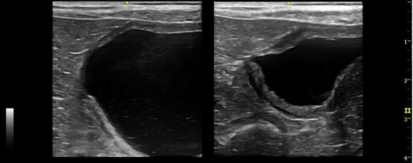
Vet. Sci. 2020, 7, x 2 of 12
from the bladder apex [3,4]. Vesicourachal diverticula may be classified as intramural or extramural
based on their anatomical extent [4,8]. A urachal sinus occurs when the distal part of the urachus
remains patent and communicates with the umbilicus. This is often asymptomatic and rarely
Vet. Sci. 2020, 7, 84 2 of 12
recognized [4].
Figure 1. Illustration of the different types of urachal anomalies. (a) Normal bladder with a completely
atrophied urachus; (b) urachal sinus: the distal part of the urachus remains patent and communicates
with the umbilicus; (c) patent urachus: the urachal canal remains patent and connects the bladder to the
Figure 1. Illustration
umbilicus; (d) urachalofcyst:
the different types ofofurachal
a focal segment anomalies.
the patent urachus(a) Normalpersistent;
remains bladder with a completely
(e) vesicourachal
atrophied
diverticulumurachus; (b) urachal
(extramural): thesinus: the distal
portion of thepart of thelocated
urachus urachusatremains patentvertex
the bladder and communicates
fails to close,
with the umbilicus;
resulting (c) patent urachus:
in a blind diverticulum the urachal
protruding beyond canal remains
the serosal patent
surface andbladder;
of the connects (f)the bladder to
vesicourachal
the umbilicus; (d) urachal cyst: a focal segment of the patent urachus remains
diverticulum (intramural): in this case the blind diverticulum is limited to the thickness of the persistent; (e)
vesicourachal
bladder wall. diverticulum (extramural): the portion of the urachus located at the bladder vertex fails
to close, resulting in a blind diverticulum protruding beyond the serosal surface of the bladder; (f)
A patent (ordiverticulum
vesicourachal persistent) (intramural):
urachus occurs in thiswhen theblind
case the urachal canal remains
diverticulum is limitedfunctionally
to the thicknesspatent
between
of thethe bladder
bladder wall.and the umbilicus. It is characterized by inappropriate urine loss through the
umbilicus and can also be associated with omphalitis and urinary tract infection [3,4]. Urachal cysts
UA when
develop are often
the under-diagnosed
umbilical and bladder conditions
ends of that
themay be found
urachus incidentallybut
are obliterated, at imaging. Associated
a focal segment of a
clinicalurachus
patent signs may be attributed
remains persistentto [4,7].
urinary tract infectiondiverticulum
A vesicourachal (UTI) and may be indistinguishable
occurs when a portion of from
the
other
urachusacquired
locatedcauses of lowervertex
at the bladder urinaryfailstract diseases
to close, (LUTD)
resulting in a or affected
blind patientsthat
diverticulum canprotrudes
be completely
from
asymptomatic
the bladder apex [5,7,9].
[3,4]. Vesicourachal diverticula may be classified as intramural or extramural based
In human
on their medicine,
anatomical extentthe reported
[4,8]. incidence
A urachal sinusofoccurs
urachal anomalies
when is relatively
the distal low,
part of the approximately
urachus remains
one in 5000
patent for adults (lesswith
and communicates among the infants).
umbilicus. TheThis
mostis common type of urachal
often asymptomatic anomaly
and rarely reported[4].
recognized is a
patentUAurachus
are often (47%), followed by urachal
under-diagnosed conditions cystthat(30%),
may sinus (18%),incidentally
be found and, least at commonly,
imaging.
vesicourachal
Associated diverticulum
clinical signs may (3%) [7]. In veterinary
be attributed medicine,
to urinary tract the(UTI)
infection prevalence
and may is be
unknown, because
indistinguishable
there are only
from other few studies
acquired causes and reviews
of lower [3,8,10]
urinary tractor case reports
diseases (LUTD) [11–13].
or affected patients can be completely
Although [5,7,9].
asymptomatic UA can be diagnosed by different imaging techniques (ultrasonography, contrast
cystography,
In humanormedicine,
cystoscopy)the ultrasonography
reported incidence is of
theurachal
most commonly
anomaliesused, because
is relatively of its
low, widespread
approximately
availability
one in 5000 and absence
for adults of among
(less ionizinginfants).
radiation Theexposure [3,6,7,14–16].
most common type of urachal anomaly reported is a
patentThe purposes
urachus (47%),offollowed
the present retrospective
by urachal study
cyst (30%), are(18%),
sinus to determine
and, least(1) the prevalence
commonly, of the
vesicourachal
different types
diverticulum of urachal
(3%) anomaliesmedicine,
[7]. In veterinary in cats and thedogs (2) the association
prevalence is unknown,ofbecauseUA with urinary
there clinical
are only few
signs
studiesand
andurinalysis abnormalities.
reviews [3,8,10] or case reports [11–13].
Although UA can be diagnosed by different imaging techniques (ultrasonography, contrast
cystography, or cystoscopy) ultrasonography is the most commonly used, because of its widespread
availability and absence of ionizing radiation exposure [3,6,7,14–16].
The purposes of the present retrospective study are to determine (1) the prevalence of the different
types of urachal anomalies in cats and dogs (2) the association of UA with urinary clinical signs and
urinalysis abnormalities.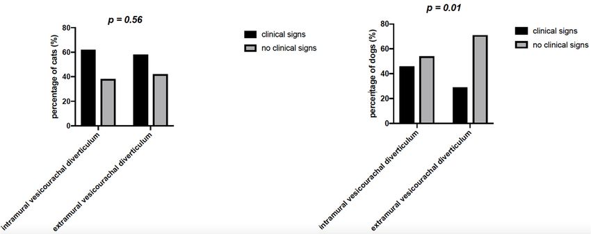
Vet. Sci. 2020, 7, 84                                                                              3 of 12

2. Materials and Methods
     Medical records (n = 28,174) of the Veterinary Teaching Hospital of the Department of Veterinary
Science (University of 61 Pisa, Italy) and Veterinary Clinical Practice “Vet Hospital H24” (Florence,
Italy) between July 2009 and July 2019 were retrospectively reviewed for client-owned dogs and cats
with an ultrasonographic diagnosis of urachal anomalies.
     For each case with diagnosis of UA, data regarding signalment, history, physical examination and
urological clinical findings (hematuria, pollakiuria, stranguria or urinary incontinence), abdominal
ultrasound, and urinalysis findings (bacteriuria, leukocyturia, hematuria and crystalluria) were
collected from the medical record when available. Patients were considered symptomatic in case at
least one of the following signs of lower urinary tract disease was present: hematuria, pollakiuria,
stranguria, urinary incontinence. In case none of the symptoms was present, patients were defined
as asymptomatic. All cases with uncertain ultrasonographic diagnosis have been excluded.
     Ultrasonography was performed using a Toshiba Aplio 400 (Canon Medical Systems Europe B.V.,
Zoetermeer, The Netherlands), and General Electrics Logiq E, Logiq E R6 and logiq E R7 equipped
with 3,5-10 MHz microconvex (8C-RS), 4,5-13 MHz (12L) and 6,7-18 (L8-18i-RS) linear probes; patients
were scanned in lateral recumbency.
     Ultrasonographic records, images, and videos were reviewed for each patient with urachal
anomalies. Data regarding ultrasound appearance of the wall and the content of the urinary bladder
were also recorded for each patient. Urachal anomalies were then classified as follows: patent
urachus, defined as a fluid-filled tubular structure, extending from the cranioventral bladder wall
to the umbilicus; urachal cyst, defined as a thin-walled, fluid-filled structure, cranial to the bladder;
vesicourachal diverticulum defined as a fluid filled structure extending from the bladder cranioventrally
as a convex out-pouching of the lumen, subclassified as intramural (limited to the thickness of the
bladder wall) or extramural (protruding beyond the serosal surface of the bladder) [3,17–19].
     Moreover, the presence of other urinary bladder anomalies was also recorded: bladder anomalies
were then divided into content anomalies and wall anomalies. Content anomalies were classified as
the presence of calculi (intraluminal mobile structures, of variable size, number and shape, with a
hyperechoic interface and forming distal shadow); sediment (intraluminal material of variable
echogenicity, which layers in the dependent portion of the bladder and suspends with agitation of the
urinary bladder); hyperechoic bands (multiple intraluminal suspended hyperechoic strips, compatible
with necrotic, fibrinous and haemorrhagic material); and blood clot (free intraluminal nonshadowing
structure, of variable size, shape, and echogenicity) [17,18,20]. Wall anomalies were defined as the
presence of wall thickening and were classified as diffuse or regional thickening and focal thickening
(wall mass extending into the bladder lumen) [17,18].
     Urine samples were collected and analyzed within 12 h from collection (IDEXX VetLab UA
Analyzer and Idexx UA Strips, Idexx, Milan, Italy). Urinary sediment reports were reviewed to check
for the presence of bacteriuria, leukocyturia, hematuria, and crystalluria.
     The D’Agostino and Pearson normality test was used to test data for normality using Graphpad
Prism 4 (Graph Pad, San Diego, CA, USA). Chi-squared test was used to compare the prevalence of
asymptomatic and symptomatic dogs and cats in relation to different urachal anomalies.

3. Results
     During the study period, 28,174 abdominal ultrasounds (6450 cats and 21,724 dogs) were performed
for different clinical indications, resulting in an ultrasonographic diagnosis of UA in 98 cases (60 cats
and 38 dogs). The prevalence of UA was 0.93% in cats and 0.18% in dogs.
     Most of the cats were of mixed lineage (n = 56, 93.3%), four cats were reported as pure breed
represented breed include Persian Cats (2 cats, 3.3%), Ragdoll and Norwegian Forest Cat (one cat
each, 1.6%). Forty-six cats (76.7%) were male (6/46 intact male and 40/46 were castrated male) and
14 cats (23.3%) were female (3/14 were intact female and 11/14 spayed female). The median age was
5 years (ranging from 4 months to 15 years), 95% of cats were >1 years. Dogs were represented by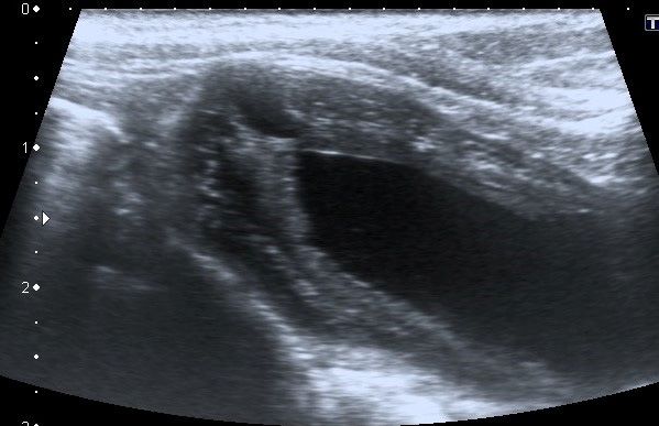
Vet. Sci. 2020, 7, x                                                                                                     4 of 12

following
     Vet. Sci. breeds:
               2020, 7, 84 mixed breed (12 dogs, 31.5%), Boxer (6 dogs, 15.8%), Golden Retriever4 (4            dogs,
                                                                                                            of 12
10.5%), Newfoundlander, English Cocker Spaniel, Labrador Retriever (2 dogs each, 5.3%) and Beagle,
German Shepherd dog, Dalmatian, Papillon, American Staffordshire Terrier, Dogue de Bordeaux,
     the following breeds: mixed breed (12 dogs, 31.5%), Boxer (6 dogs, 15.8%), Golden Retriever (4 dogs,
ShihTzu, Jack Russell Terrier, Rottweiler, and Poodle (one dog each, 2.6%). Thirty-one dogs (81.6%)
     10.5%), Newfoundlander, English Cocker Spaniel, Labrador Retriever (2 dogs each, 5.3%) and Beagle,
wereGerman
       male (25/31Shepherd were  castrated
                              dog,           male
                                     Dalmatian,    and 6/31
                                                 Papillon,    were intact
                                                           American       male) and
                                                                    Staffordshire       seven
                                                                                   Terrier,     (18.4%)
                                                                                            Dogue        were intact
                                                                                                   de Bordeaux,
female.   The    median      age  was  8.5  years (ranging  from 2 months    to 15  years)  and  18%  were
     ShihTzu, Jack Russell Terrier, Rottweiler, and Poodle (one dog each, 2.6%). Thirty-one dogs (81.6%)
Vet. Sci. 2020, 7, x
Vet. Sci. 2020, 7, 84
5 of 12
5 of 12
Vet. Sci. 2020, 7, x 5 of 12
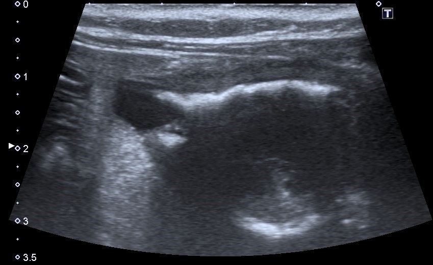
Figure 3. Longitudinal ultrasound image of the urinary bladder in a cat, showing the presence of a
small (Vet.
Vet.Sci. 2020,7,7,84
Sci.2020, x 6 of 12
6 of 12
(a) (b) (c)
Figure5.5.Longitudinal
Figure Longitudinal ultrasound
ultrasound images
imagesofofaapatent
patenturachus
urachus inin
a dog.
a dog.AAtubular structure
tubular structure(≤5 (≤5
mmmm
diameter)with
diameter) withaasmall
smallamount
amountof ofanechoic
anechoiccontent
content extends
extends from
from the
the cranio-ventral
cranio-ventral bladder wall to the
the umbilical
umbilical region.
region. MildMild reactivity
reactivity ofperitoneal
of the the peritoneal fat and
fat and a small
a small quantity
quantity of peritoneal
of peritoneal fluidfluid are
are present
inpresent in proximity
proximity of the (a)
of the urachus. urachus. (a) urachus
Terminal Terminalpart
urachus part the
reaching reaching the umbilical
umbilical region; (b)region; (b)
intermediate
intermediate urachus part; (c) urachus part in continuity with
urachus part; (c) urachus part in continuity with the bladder apex. the bladder apex.
Nourachal
No urachalsinus
sinus was
was diagnosed
diagnosed during
duringthe
theperiod
periodofofthe
theretrospective
retrospective study.
study.
Twenty-three out of 60 (38.3%) cats did not present bladder content
Twenty-three out of 60 (38.3%) cats did not present bladder content anomalies anomalies at ultrasound
at ultrasound
evaluation, and 28/60 (46.6%) cats did not present any bladder wall
evaluation, and 28/60 (46.6%) cats did not present any bladder wall anomaly; 20/60anomaly; 20/60 (33.3%) hadhad
(33.3%)
sediment and 30/60 (50%) reported diffuse wall thickening (Table
sediment and 30/60 (50%) reported diffuse wall thickening (Table 2). 2).
Eighteen out of 38 dogs (47.4%) did not have any other bladder anomalies at ultrasound
evaluation. Twenty out
Table 2. Prevalence of 38 dogs
of urinary (52.6%)
bladder had
content andother alterations:
wall anomalies calculi
in all were
patients the most
included withprevalent
different
content anomaly (13/38, 31.6%),
type of urachal anomalies. and diffuse thickening was the most prevalent wall anomaly (16/38,
42.1%) (Table 2).
Urinary Bladder Urinary Bladder
Urachal Anomalies Species n n
Table 2. Prevalence of urinary bladder Content
content Anomalies Wall Anomalies
and wall anomalies in all patients included with
different type of urachal anomalies. Calculi 8/45
Diffuse or regional
Sediment 15/45 25/45
Urinary Blood
Bladder Content
thickening
Urinary Bladder Wall
Intramural vesicourachal Cats clot 1/45 1/45
Urachal Anomalies Species n Focal thickening n
diverticulum Anomaliesbands
Hyperechoic 3/45 Anomalies 19/45
No anomalies
NoCalculi
anomalies 18/45
8/45
Diffuse or regional
Sediment 15/45 Diffuse or regional 25/45
Calculi 10/28 thickening 14/28
Cats Blood clot 1/45 thickening 1/45
Dogs Sediment 8/28 Focal 4/28
Hyperechoic bands 3/45 Focal thickening
thickening 19/45
Intramural vesicourachal No anomalies 10/28 No 10/28
No anomalies 18/45 No anomalies
anomalies
diverticulum Calculi 5/12
Diffuseor
orregional
regional
Diffuse 4/12
Extramural vesicourachal Cats Calculi
Sediment 10/28
3/12
thickening 14/28
thickening 8/12
diverticulum Dogs Sediment
No anomalies 8/28
4/12
No anomalies 4/28
Focal thickening
No anomalies 10/28
Diffuse or regional 10/28
Calculi 1/7 No anomalies 1/7
Dogs thickening
NoCalculi
anomalies 5/12
6/7 Diffuse or regional 6/7
4/12
No anomalies
Cats Sediment 3/12 thickening
Diffuse or regional 8/12
Extramural vesicourachal No anomalies 4/12 No anomalies 1/2
Cats Sediment 2/2 thickening
Urachal cysts
diverticulum Diffuse or regional 1/2
Calculi 1/7 Focal thickening 1/7
Dogs thickening
No anomalies 6/7 Diffuse or regional 6/7
Calculi 1/3 No anomalies 1/3
Dogs thickening
No anomalies 2/3 Diffuse or regional 2/3
No anomalies 1/2
Patent urachus Cats
Dogs Sediment
No anomalies 2/2
1/1 thickening
No anomalies 1/1
1/2
Focal thickening
Urachal cysts
Diffuse or regional
Calculi 1/3 1/3
Eighteen out of 38 dogs (47.4%)Dogs did not have any other bladder anomalies thickening
at ultrasound evaluation.
No anomalies 2/3 2/3
No anomalies
Twenty out of 38 dogs
Patent urachus (52.6%) had
Dogs other alterations:
No anomaliescalculi were the
1/1 most prevalent
No anomalies content anomaly
1/1
(13/38, 31.6%), and diffuse thickening was the most prevalent wall anomaly (16/38, 42.1%) (Table 2).
OnOnhistory,
history, 23/60 cats (38.3%)
23/60 cats (38.3%)andand22/38
22/38 dogs
dogs (57.9%)
(57.9%) hadhad no urinary
no urinary clinical
clinical signs (Table
signs (Table 3). In 3).
Inparticular,
particular, in cats,
in cats, no statistically
no statistically significant
significant difference
difference in the percentage
in the percentage of patientsofwith
patients with or
or without
clinical signs
without wassigns
clinical foundwas
(p =found
0.56) for = 0.56)
(p both intramural
for both andintramural
extramuraland
vesicourachal
extramural diverticulum,
vesicourachal
although a non-significant
diverticulum, trend of highertrend
although a non-significant percentage of patients
of higher withof
percentage clinical signs
patients withwasclinical
noticed.signs
was noticed. While in dogs the percentage of animals with no clinical signs was significantly higherVet. Sci. 2020, 7, 84 7 of 12
(p = 0.01) than the percentage of animals with clinical signs, for both intramural and extramural
vesicourachal diverticulum.
Table 3. Prevalence of clinical urinary findings in all patients included with different type of
urachal anomalies.
Urachal Anomalies Urinary Symptoms Cats Dogs
Hematuria 5/45 3/28
Pollakiuria 8/45 5/28
Intramural vesicourachal diverticulum Stranguria 13/45 6/28
Urinary incontinence 0 3/28
Asymptomatic 17/45 15/28
Hematuria 4/12 1/7
Pollakiuria 2/12 1/7
Extramural vesicourachal diverticulum Stranguria 4/12 0
Urinary incontinence 0 0
Asymptomatic 5/12 5/7
Hematuria 0 0
Pollakiuria 1/2 1/3
Urachal cysts Stranguria 0 0
Urinary incontinence 0 0
Asymptomatic 1/2 2/3
Hematuria 0 0
Pollakiuria 1/1 0
Patent urachus Stranguria 0 0
Urinary incontinence 0 0
Asymptomatic 0 0
For intramural vesicourachal diverticulum, stranguria was the most frequent alteration (28.8%
and 21.4% for cats and dogs, respectively), followed by pollakiuria (17.7% for cats and 17.8% for dogs),
while 37.7% of cats and 53.6% of dogs were without clinical signs.
Thirteen out of 37 cats (35.0%) and 6/16 dogs (37.5%) with urinary clinical signs were presented in
emergency for urethral obstruction.
Fifty-two out of 98 (36 cats and 16 dogs) had urinalysis available for review. Leukocyturia and
hematuria were the most prevalent urinalysis anomalies in both cats (24/36, 66.6% and 30/36, 83.3%,
respectively) and dogs (12/16, 75.0% and 10/16, 62.5%, respectively). Crystalluria was present in 31.2%
of dogs and in 52.7% of cats, while bacteriuria was present, respectively, in 25.0% and 27.8% of dogs
and cats. In particular for intramural vesicourachal diverticulum, leukocyturia and hematuria were
present, respectively, in 83.3% and 58.3% of dogs, and in 63.3% and 86.6% of cats.
In cats, crystalluria was present in 53.3% of cats with intramural vesicourachal diverticula, 25.0%
of cats with extramural vesicourachal diverticula and in one cat with urachal cyst and patent urachus.
Crystalluria was present in 41.6% of dogs with intramural vesicourachal diverticulum, while it was
absent in the remaining UA. In cats, crystalluria was due to struvite in 13/16 cats (81.3%) and to calcium
oxalate in 3/16 cats (18.7%). In dogs, crystalluria was due to struvite in 3/5 dogs (60.0%), to calcium
oxalate in 1/5 dogs (20.0%), and to cystine in 1/5 dogs (20.0%).
4. Discussion
Urachal anomalies are uncommon disorders of the urinary bladder, and often incidental findings
in veterinary medicine [3–5,8]. The real incidence is yet unknown because there are only a few
case reports [11–13,21] or reviews [3–5,10] concerning this topic. Similar to veterinary medicine,
the incidence of urachal anomalies in human medicine is relatively low [7,22], with the majority of
cases showing no clinical signs, and being diagnosed incidentally [7]. In particular, urachal anomalies
are more frequently diagnosed in children, where an incidence of 1 case on 7.610 for patent urachus
and 1 case on 5.000 of urachal cyst was reported [22].Vet. Sci. 2020, 7, 84 8 of 12
In our study, the prevalence of UA was 0.93% in cats and 0.18% in dogs, similar to what is reported
Vet. Sci. 2020, 7, x 8 of 12
in humans. Interestingly, only 5% of cats and 18% of dogs diagnosed with UA were < 1 year old.
In our
old. Instudy, a median
our study, age ofage
a median 5 years (4 months–15
of 5 years (4 months–15years)years)
was reported for cats,
was reported forand a median
cats, and a medianage of
8.5 years (2 months–15 years) for dogs. Median age at diagnosis seems
age of 8.5 years (2 months–15 years) for dogs. Median age at diagnosis seems higher in veterinary higher in veterinary patients
compared to humans.
patients compared This finding
to humans. Thisisfinding
in agreement with previous
is in agreement with studies
previous in studies
veterinary medicine,
in veterinary
in which mean
medicine, in whichage mean
at diagnosis was far above
age at diagnosis was far 1 year
above old1 [4,8]. In a[4,8].
year old study In of 24 clinically
a study normal
of 24 clinically
cats with radiographic evidence of an intramural vesicourachal
normal cats with radiographic evidence of an intramural vesicourachal diverticulum, a mean age diverticulum, a mean age of 2.5of
years
2.5 yearswaswasreported, while
reported, whilein another
in another study of 149
study symptomatic
of 149 symptomatic catscats
with diverticula,
with diverticula, a mean
a mean ageageof
3.7 ± 2.7
of 3.7 years
± 2.7 years was
wasreported
reported[4].[4].Mean
Meanage ageatatdiagnosis
diagnosiswas waseven
evenhigher
higherin in dogs,
dogs, where
where Groesslinger
Groesslinger
and colleagues reported a mean age of 10.4 ± 4.4 years, with only 8% of dogs < 1 <
and colleagues reported a mean age of 10.4 ± 4.4 years, with only 8% of dogs 1 years
years oldThe
old [8]. [8].
The authors
authors hypothesize
hypothesize thatincreased
that the the increased median median
age atage at diagnosis
diagnosis of UAofinUA ourincohort
our cohort
is relatedis related
to the
to the high prevalence of asymptomatic patients. UA was often an
high prevalence of asymptomatic patients. UA was often an incidental finding in patients requiring incidental finding in patients
requiring ultrasonography
ultrasonography for reasons forother
reasons thanother than urinary
urinary tract disease.
tract disease. In our In our study
study the majority
the majority of dogs of
dogs
(57.9%) (57.9%)
were were asymptomatic.
asymptomatic. This finding
This finding was in was in agreement
agreement withwith a previous
a previous study study
[8], [8], in which
in which the
the diagnosis of UA was not associated with clinical signs of UTI in 34%
diagnosis of UA was not associated with clinical signs of UTI in 34% of dogs [8]. In the cats of our of dogs [8]. In the cats of
our
cohortcohort
the the prevalence
prevalence of asymptomatic
of asymptomatic subjects
subjects waswas lowerthan
lower thanthat
thatfound
found in in dogs
dogs (38.3%).
(38.3%). In In
particular, while the number of dogs with no clinical signs was significantly
particular, while the number of dogs with no clinical signs was significantly higher than the number higher than the number
of
of dogs
dogs withwith clinical
clinical signs,
signs, for
for both
both intramural
intramural and and extramural
extramural diverticulum,
diverticulum, this this difference
difference was was notnot
statistically
statistically evident
evident in in cats
cats (Figure
(Figure 6). 6). Unpublished
Unpublished clinical clinical experience
experience showed
showed that that clinical
clinical signs
signs of of
LUTD
LUTD are a more frequent reason for ultrasonography in feline, rather than in canine patients. As aa
are a more frequent reason for ultrasonography in feline, rather than in canine patients. As
consequence,
consequence, the the number
number of of cats
cats requiring
requiring ultrasonography,
ultrasonography, with with signs
signs of of LUTD
LUTD may may be be relatively
relatively
higher
higher thanthan inin dogs. Although it
dogs. Although it is
is possible
possible that that the
the diagnosis
diagnosis of of UA
UA is is more
more frequently
frequently associated
associated
with
with clinical
clinicalsigns
signsofofLUTD
LUTDinincats catsthan
than inindogs,
dogs, this finding
this findingis difficult to interpret
is difficult to interpretdue due
to the tofact
the that
fact
previous
that previousdata data
in cats
in are
catslimited to few
are limited tocase
few reports [5,11].[5,11].
case reports
6. Chi squared test of the percentage of cats
Figure 6. cats and
and dogs
dogs with
with clinical
clinical signs
signs and
and without
without clinical
clinical
signs, for both intramural and
and extramural
extramural vesicourachal
vesicourachal diverticulum.
diverticulum.
In symptomatic
symptomaticanimals,
animals,hematuria,
hematuria, dysuria,
dysuria, pollakiuria,
pollakiuria, andand urethral
urethral obstruction
obstruction werewere the
the most
most commonly
commonly reported
reported clinical
clinical signs associated
signs associated with UAwithin UA
the in the current
current literature
literature [4,5,12].
[4,5,12]. In our
In our cohort
cohort of patients,
of patients, the most
the most prevalent
prevalent clinical
clinical symptoms
symptoms associated
associated withUA
with UAwere were stranguria
stranguria andand
pollakiuria, particularly in dogs and cats cats with
with intramural
intramural vesicourachal
vesicourachal diverticula.
diverticula. Interestingly,
urethral obstruction was present in 35% of of cats
cats and
and in
in 37.5%
37.5% ofof dogs
dogs with
with urinary
urinary symptoms.
symptoms. It is
plausible that in patients with urethral obstruction the diagnosis of UA may be
plausible that in patients with urethral obstruction the diagnosis of UA may be facilitated facilitated by a condition
by a
of bladderof
condition overfill.
bladderThis finding
overfill. seems
This in agreement
finding with the results
seems in agreement with theof results
a previous
of a study of Osborne
previous study of
and colleagues,
Osborne in which urethral
and colleagues, in which obstruction was reported
urethral obstruction wasin the 54.5% in
reported of cats with vesicourachal
the 54.5% of cats with
diverticula [4]. diverticula
vesicourachal The largest [4].
diverticula were found
The largest in catswere
diverticula withfound
complete urethral
in cats withoutflow
completeobstruction,
urethral
supposedly due to the
outflow obstruction, increased intraluminal
supposedly pressureintraluminal
due to the increased in the bladder lumenin
pressure [4].
the bladder lumen [4].
Unfortunately, urinalysis was only available in a limited number of dogs and cats. Interestingly,
the majority of dogs and cats for which urinalysis was available had signs of inflammation, such as
hematuria, leukocyturia and crystals. However, it is to be noted that dogs and cats with urinalysisVet. Sci. 2020, 7, 84 9 of 12
Unfortunately, urinalysis was only available in a limited number of dogs and cats. Interestingly,
the majority of dogs and cats for which urinalysis was available had signs of inflammation, such as
Vet. Sci. 2020, 7, x 9 of 12
hematuria, leukocyturia and crystals. However, it is to be noted that dogs and cats with urinalysis were
thosethose
were patients for which
patients abdominal
for which ultrasonography
abdominal was performed
ultrasonography to investigate
was performed urologic
to investigate clinical
urologic
signs. Therefore, the prevalence of urinary signs of inflammation may be
clinical signs. Therefore, the prevalence of urinary signs of inflammation may be overestimated.overestimated.
Historically,the
Historically, thediagnosis
diagnosisof ofUA
UAin inveterinary
veterinarymedicine
medicinewas wasperformed
performedby bythe
theuse
useofofdifferent
different
imaging techniques, such as ultrasonography, contrast cystography,
imaging techniques, such as ultrasonography, contrast cystography, and cystoscopy [3,14,23]. and cystoscopy [3,14,23].
In
In previous
previous studies,
studies, contrast
contrast cystography
cystography waswas used
used toto diagnoseUA
diagnose UA[8,12].
[8,12].According
AccordingtotoOsborne
Osborneand and
colleagues, positive
colleagues, antegrade
positive cystourethrography
antegrade and retrograde
cystourethrography and positive contrastpositive
retrograde urethrocystography
contrast
were considered the procedures of choice for the diagnosis of vesicourachal
urethrocystography were considered the procedures of choice for the diagnosis of diverticula [4].vesicourachal
In the present
study the diagnosis
diverticula [4]. In theofpresent
UA wasstudy
basedthe on diagnosis
abdominalofultrasonography
UA was based on only. Abdominal
abdominal ultrasound has
ultrasonography
only. Abdominal ultrasound has the advantages to be a routinely available and non-invasivein
the advantages to be a routinely available and non-invasive imaging technique. Similarly, human
imaging
medicine, abdominal ultrasound is considered a fast and readily available
technique. Similarly, in human medicine, abdominal ultrasound is considered a fast and readily technique to diagnose UA
in absence
available of the risks
technique connectedUA
to diagnose to radiation
in absence exposure [7]. However,
of the risks connectedwe to should consider
radiation exposurethat[7].
the
diagnostic power of abdominal ultrasonography in identifying UA may be
However, we should consider that the diagnostic power of abdominal ultrasonography in identifying reduced by a condition of
inadequate
UA bladder filling.
may be reduced In one ofof
by a condition theinadequate
patients ofbladder
our study, the diagnosis
filling. In one ofoftheintramural
patients ofvesicourachal
our study,
diverticulum was made at the second ultrasonography, when the bladder appeared
the diagnosis of intramural vesicourachal diverticulum was made at the second ultrasonography, less filled (Figure 7).
Therefore, we cannot exclude that small diverticula may be undiagnosed by the
when the bladder appeared less filled (Figure 7). Therefore, we cannot exclude that small diverticula use of ultrasonography,
in case
may of inadequateby
be undiagnosed bladder
the usefilling.
of ultrasonography, in case of inadequate bladder filling.
(a) (b)
Figure
Figure7.7. Two
Twolongitudinal
longitudinalultrasound
ultrasoundimages
imagesofofthe
theurinary
urinarybladder
bladderofofaadog
dogwith
withanan intramural
intramural
vesicourachal
vesicourachaldiverticulum.
diverticulum.TheTheimages
imagesshow
showtwo
twodifferent
differentvolumes
volumesofofurine
urineinto
intothe
thebladder
bladderlumen.
lumen.
(a)Overly
(a) Overly distended:
distended: the
thediverticulum
diverticulumappears
appearsasas
a not very
a not visible
very focalfocal
visible depression in thein
depression cranial-ventral
the cranial-
aspect of
ventral the bladder,
aspect of the with thinning
bladder, withofthinning
the correspondent bladder wall;bladder
of the correspondent (b) ddequately
wall; (b) distended:
ddequatelythe
diverticulum
distended: theappears as an evident,
diverticulum appears focal,
as an evagination in the
evident, focal, cranio-ventral
evagination in theaspect of the bladder.
cranio-ventral aspect of
the bladder.
In our cohort, the vesicourachal diverticulum was the most frequently diagnosed UA, with a
prevalence
In our of 96.7%the
cohort, in cats and 89.5% in
vesicourachal dogs. In particular,
diverticulum was thethemostprevalence of intramural
frequently diagnosedvesicourachal
UA, with a
diverticulum was 76.7% in cats
prevalence of 96.7% in cats and 89.5% and 71.1% in dogs. This was followed by extramural vesicourachal
dogs. In particular, the prevalence of intramural
diverticulum diverticulum
vesicourachal (18.4% and 20% was in76.7%
dogs and cats,
in cats andrespectively).
71.1% in dogs. In veterinary medicine,by
This was followed vesicourachal
extramural
diverticulum diverticulum
vesicourachal (both microscopic(18.4%andandmacroscopic)
20% in dogs and seemed
cats, to be the most
respectively). In frequent
veterinaryUA in dogs
medicine,
and cats [4,8]. For feline patients, one study [24] reported a prevalence of
vesicourachal diverticulum (both microscopic and macroscopic) seemed to be the most frequent UA 40% of microscopic
diverticula,
in while
dogs and cats another
[4,8]. study
For feline [4] reported
patients, a prevalence
one study of vesicourachal
[24] reported a prevalencediverticulum of 22% in
of 40% of microscopic
cats with urinary symptoms. For canine patients, one study reported a prevalence
diverticula, while another study [4] reported a prevalence of vesicourachal diverticulum of 22% in of vesicourachal
diverticulum
cats of 34%
with urinary [8]. In veterinary
symptoms. For caninemedicine, there
patients, oneisstudy
only one study reporting
reported a different
a prevalence prevalence
of vesicourachal
between intramural
diverticulum of 34%(19/33,
[8]. In57.6%) and extramural
veterinary medicine, diverticula
there is only (14/33,
one42.4%)
study in 33 cats with
reporting bladder
a different
prevalence between intramural (19/33, 57.6%) and extramural diverticula (14/33, 42.4%) in 33 cats
with bladder diverticula [4]. In another study of 50 dogs, only 1/17 vesicourachal diverticula was
classified as extramural, confirming also in the dog, the lower prevalence of extramural diverticula
compared to intramural [8]. Veterinary patients seem to differ from human patients, for whom patent
urachus was the most prevalent UA (47%), followed by urachal cyst (30%), and vesicourachalVet. Sci. 2020, 7, 84 10 of 12
diverticula [4]. In another study of 50 dogs, only 1/17 vesicourachal diverticula was classified as
extramural, confirming also in the dog, the lower prevalence of extramural diverticula compared to
intramural [8]. Veterinary patients seem to differ from human patients, for whom patent urachus was
the most prevalent UA (47%), followed by urachal cyst (30%), and vesicourachal diverticulum (3%) [7].
In our study the prevalence of urachal cysts and patent urachus was very low in both cats and dogs.
This finding was in agreement with what was previously described in both case reports [11,21] and
reviews [4,5]. The urachal cysts may be under-diagnosed because they are located along the course of
the urachus, but they are not necessarily associated and/or adjacent to the bladder, and for example
they could be misinterpreted as mesenteric cysts.
No urachal sinus was diagnoses during the period of the retrospective study. One possible
explanation could be that the urachal sinus is often associated with non-specific symptoms and is
largely asymptomatic unless it develops a complication (most commonly infection). [4,25,26] Another
possible explanation could be its anatomical location; indeed, it is not located near the urinary bladder
and if asymptomatic it may not be observed during an abdominal ultrasound examination.
In our cohort of patients, UA was frequently associated with other ultrasound alterations of the
bladder in terms of content and/or wall anomalies. In both dogs and cats, diffuse or regional thickening
was the most prevalent bladder wall alteration. The most common alterations of the urinary bladder
content were urolithiasis in dogs and sediment in cats. Similar findings were reported by the study
of Osborne and Colleagues [4], in which the most prevalent abnormalities in patients with UA were
thickened bladder wall (75%), irregular mucosa (44%), calculi and sediment (33%). These findings,
together with unpublished clinical experiences may suggest a potential role of UA in promoting
urolithiasis, and/or inflammatory changes of the bladder mucosa. UA may host bacteria, or protein
and mineral debris, which may be difficult to clear by the physiologic urine flow. Some studies also
reported that macroscopic diverticula may develop from microscopic diverticula, with the concomitant
presence of disorders of the lower urinary tract, such as bacterial infections, urolithiasis, urethral plugs,
sediment, or idiopathic cystitis. However, the exact relationship between vesicourachal diverticula
and other disorders of the lower urinary tract is still unknown [4,27–30].
This study has some limitations. Due to the retrospective nature of the study, cases with incomplete
or uncertain ultrasound diagnosis were excluded from the study. Therefore, it is possible that the exact
prevalence of UA may be slightly affected. Data regarding urinalysis were available only for a limited
number of subjects. In some cases, urinalysis was not performed as the patients were referred to the
hospital only for the ultrasonographic examination, in other cases urinalysis was not included in the
initial diagnostic plan. In both cases, the lack of urinalysis might affect our results, in terms of prevalence
of signs of urinary inflammation. Moreover, as in the majority of our cases UA were incidental findings,
no urine culture was available. The lack of urine culture did not allow us to make any speculation on
the prevalence of UTI in these patients. Finally, as abdominal ultrasonography was the only imaging
technique used to identify UA, it is possible that some patients with UA were undiagnosed, especially
if urinary bladder was inadequately replete, and/or in the case of small diverticula.
5. Conclusions
In conclusion, our study confirmed urachal anomalies as uncommon and often incidental findings,
with a high number/proportion of patients without urinary clinical signs. When present, clinical
signs were mostly characterized by hematuria, stranguria, and/or urethral obstruction. Different from
human medicine, UA were diagnosed in dogs and cats of any age, with a lower prevalence in patients
< 1 year old. Among different UA, intramural and extramural vesicourachal diverticula were the
most prevalent in both canine and feline populations, often associated with other ultrasound signs
of bladder inflammation. Abdominal ultrasound is a routinely available and non-invasive imaging
technique to investigate UA. However, additional imaging techniques (such as contrast cystography)
or repeated abdominal ultrasounds should be considered in the case of inadequate bladder filling
and/or in cases of patients with clinical signs where a specific management could be important.Vet. Sci. 2020, 7, 84 11 of 12
Author Contributions: Conception, S.C, T.M., D.D.S., M.B.; writing—original draft preparation, F.P.;
writing—review and editing, F.P., S.C., I.L., C.P., D.D.S.; statistical analysis, F.P., I.L.; supervision, S.C.; project
administration, S.C. All authors have read and agreed to the published version of the manuscript.
Funding: This research received no external funding.
Acknowledgments: The authors thank Alessandro Antonelli for his contribution to realize Figure 1.
Conflicts of Interest: The authors declare no conflict of interest.
References
1. Buddha, S.; Menias, C.O.; Katabathina, V.S. Imaging of urachal anomalies. Abdom. Radiol. (N. Y.)
2019, 44, 3978–3989. [CrossRef]
2. Wilson, A.L.; Gandhi, J.; Seyam, O.; Rahmani, B.; Patel, S.; Joshi, G.; Smith, N.L.; Khan, S.A. Urachal anomalies:
A review of pathological conditions, diagnosis, and management. Transl. Res. Anat. 2019, 16, 100041.
[CrossRef]
3. Bartges, J.W.; Callens, A.J. Congenital Diseases of the Lower Urinary Tract. Vet. Clin. N. Am. Small
Anim. Pract. 2015, 45, 703–719. [CrossRef] [PubMed]
4. Osborne, C.A.; Johnston, G.R.; Kruger, J.M.; O’Brien, T.D.; Lulich, J.P. Etiopathogenesis and biological
behavior of feline vesicourachal diverticula. Don’t just do something stand there. Vet. Clin. N. Am. Small
Anim. Pract. 1987, 17, 697–733. [CrossRef]
5. Kruger, J.M.; Osborne, C.A.; Lulich, J.P.; Oakley, R.E. Inherited and congenital diseases of the feline lower
urinary tract. Vet. Clin. N. Am. Small Anim. Pract. 1996, 26, 265–279. [CrossRef]
6. Bartges, J.W.; Kruger, J.M. Congenital disease of the lower urinary tract. In Nephrology and Urology of
Small Animals; Bartges, J., Polzin, D.J., Eds.; John Wiley & Sons, Ltd.: West Sussex, UK, 2011; pp. 809–817.
7. Parada Villavicencio, C.; Adam, S.Z.; Nikolaidis, P.; Yaghmai, V.; Miller, F.H. Imaging of the Urachus:
Anomalies, Complications, and Mimics. Radiographics 2016, 36, 2049–2063. [CrossRef]
8. Groesslinger, K.; Tham, T.; Egerbacher, M.; Lorinson, D. Prevalence and radiologic and histologic appearance
of vesicourachal diverticula in dogs without clinical signs of urinary tract disease. J. Am. Vet. Med. Assoc.
2005, 226, 383–386. [CrossRef] [PubMed]
9. Tazi, F.; Ahsaini, M.; Khalouk, A.; Mellas, S.; Stuurman-Wieringa, E.R.; Elfassi, J.M.; Farih, H.M. Abscess of
urachal remnants presenting with acute abdomen: A case series. J. Med. Case Rep. 2012, 6, 226. [CrossRef]
10. Hansen, J.S. Urachal remnant in the cat: Occurrence and relationship to the feline urological syndrome.
Vet. Med. Small Anim. Clin. 1977, 72, 1735–1746.
11. Hansen, J.S. Parent urachus in a cat (a case history). Vet. Med. Small Anim. Clin. 1972, 67, 379–381.
12. Wilson, J.W.; Kluasner, J.S.; Stevens, J.B.; Osborne, C.A. Canine Vesicourachal Diverticula. Vet. Surg.
1979, 8, 63–67. [CrossRef]
13. Łojszczyk-Szczepaniak, A.; Smiech, A.; Wojnowski, T. Congenital urachal diverticulum in dogs: A case report.
Medycyna Wet. 2010, 66, 412–424.
14. Brovida, C. Urogenital defects in dogs. Vet. Focus. 2014, 24, 2–9. [CrossRef]
15. Boscia, D.; Baracchini, L.; Rossi, F.; Vignoli, M. Radiologia Del Cane e Del Gatto, 1st ed.; Poletti editore: Gudo
Visconti, Milano, Italy, 2005.
16. Yu, J.S.; Kim, K.W.; Lee, H.J.; Lee, Y.J.; Yoon, C.S.; Kim, M.J. Urachal remnant diseases: Spectrum of CT and
US findings. Radiographics. 2001, 21, 451–461. [CrossRef]
17. Sutherland, J.; Penninck, D. Bladder and Urethra. In Atlas of Small Animal Ultrasonography, 2nd ed.; Pennick, D.,
D’Anjou, M.A., Eds.; John Wiley & Sons, Inc.: Oxford, UK, 2015; pp. 363–401.
18. Nyland, T.G.; Widmer, W.R.; Mattoon, J.S. Urinary Tract. In Small Animal Diagnostic Ultrasound, 3rd ed.;
Mattoon, J.S., Nyland, T.G., Eds.; Saunders: St. Louis, MO, USA, 2015; pp. 557–607.
19. Rahal, S.C.; Mamprim, M.J.; Torelli, S.R. What is your diagnosis? Patent urachus. J. Am. Vet. Med. Assoc.
2004, 225, 1041–1042. [CrossRef]
20. Le Boedec, K.; Pastor, M.L.; Lavoué, R.; Reynolds, B.S. Pseudomembranous cystitis, an unusual condition
associated with feline urine outflow obstruction: Four cases. J. Feline Med. Surg. 2011, 13, 588–593. [CrossRef]
21. Laverty, P.H.; Salisbury, S.K. Surgical management of true patent urachus in a cat. J. Small Anim. Pract.
2002, 43, 227–229. [CrossRef]Vet. Sci. 2020, 7, 84 12 of 12
22. Choi, Y.J.; Kim, J.M.; Ahn, S.Y.; Oh, J.T.; Han, S.W.; Lee, J.S. Urachal anomalies in children: A single
center experience. Yonsei Med. J. 2006, 47, 782–786. [CrossRef]
23. Hecht, S. Diagnostic imaging of lower urinary tract disease. Vet. Clin. N. Am. Small Anim. Pract.
2015, 45, 639–663. [CrossRef]
24. Wilson, G.P.; Dill, L.S.; Goodman, R.Z. The relationship of urachal defects in the feline urinary bladder to
feline urological syndrome. In Proceedings of the 7th Kal Kan Symposium, Columbus, OH, USA, 24–25
September 1983; p. 125.
25. Risher, W.H.; Sardi, A.; Bolton, J. Urachal abnormalities in adults: The ochsner experience. South. Med. J.
1990, 83, 1036–1039. [CrossRef]
26. El Ammari, J.E.; Ahallal, Y.; El Yazami Adli, O.; El Fassi, M.J.; Farih, M.H. Urachal sinus presenting with
abscess formation. ISRN Urol. 2011, 2011, 1–3. [CrossRef] [PubMed]
27. Osborne, C.A.; Kroll, R.A.; Lulich, J.P. Medical management of vesicourachal diverticula in 15 cats with
lower urinary tract disease. J. Small Anim. Pract. 1989, 30, 608–612. [CrossRef]
28. Osborne, C.A.; Kalkstein, T.S.; Kruger, J.M. Le affezioni idiopatiche delle basse vie urinarie del gatto.
Veterinaria 2001, 15, 53–61.
29. Visser, J.; Kummeling, A.; Van Nugteren, M.A.; Grinwis, G.C.M.; Brocks, B.A.W. Resection of urachal
anomalies in dogs with recurrent lower urinary tract disease. Vet. Surg. 2019, 49, 214–221. [CrossRef]
30. Gotthelf, L.N. Persistent urinary tract infection and urolithiasis in a cat with a urachal diverticulum.
Vet. Med. Small Anim. Clin. 1981, 76, 1745–1747. [PubMed]
© 2020 by the authors. Licensee MDPI, Basel, Switzerland. This article is an open access
article distributed under the terms and conditions of the Creative Commons Attribution
(CC BY) license (http://creativecommons.org/licenses/by/4.0/).You can also read