Right heart function during simulated altitude in patients with pulmonary arterial hypertension - Open Heart
←
→
Page content transcription
If your browser does not render page correctly, please read the page content below
Pulmonary vascular disease

                                                                                                                                                           Open Heart: first published as 10.1136/openhrt-2016-000532 on 20 January 2017. Downloaded from http://openheart.bmj.com/ on March 21, 2021 by guest. Protected by copyright.
                                 Right heart function during simulated
                                 altitude in patients with pulmonary
                                 arterial hypertension
                                 Leigh M Seccombe,1,2 Vincent Chow,2,3 Wei Zhao,3 Edmund M T Lau,2
                                 Peter G Rogers,1 Austin C C Ng,2,3 Elizabeth M Veitch,1 Matthew J Peters,1,2
                                 Leonard Kritharides2,3

To cite: Seccombe LM,            ABSTRACT
Chow V, Zhao W, et al. Right                                                                   KEY QUESTIONS
                                 Objective: Patients with pulmonary arterial
heart function during
                                 hypertension (PAH) are often recommended                      What is already known about this subject?
simulated altitude in patients
                                 supplemental oxygen for altitude travel due to the            ▸ Patients with pulmonary arterial hypertension
with pulmonary arterial
hypertension. Open Heart         possible deleterious effects of hypoxia on pulmonary            may experience deleterious effects of exposure
2017;4:e000532.                  haemodynamics and right heart function. This includes           to altitude-related hypoxia due to further ele-
doi:10.1136/openhrt-2016-        commercial aircraft travel; however, the direct effects         vated pulmonary artery pressure from increased
000532                           and potential risks are unknown.                                sympathetic activity and hypoxic pulmonary
                                 Methods: Doppler echocardiography and gas                       vasoconstriction. The effects of altitude, com-
                                 exchange measures were investigated in group 1                  parable to commercial aircraft flight, on right
Received 7 September 2016        patients with PAH and healthy patients at rest breathing        ventricle function in these patients are unknown.
Revised 21 November 2016         room air and while breathing 15.1% oxygen, at rest for
Accepted 22 November 2016        20 min and during mild exertion.                              What does this study add?
                                 Results: The 14 patients with PAH studied were                ▸ This novel study investigated the effects of mild
                                 clinically stable on PAH-specific therapy, with                 altitude simulation on right ventricular function
                                 functional class II (n=11) and III (n=3) symptoms               in patients with pulmonary arterial hypertension.
                                 when tested. Measures of right ventricular size and             Pulmonary artery pressure increased signifi-
                                 function were significantly different in the PAH group at       cantly during hypoxia, with no evidence of pro-
                                 baseline as compared to 7 healthy patients (p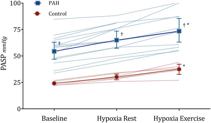
Open Heart

                                                                                                                                              Open Heart: first published as 10.1136/openhrt-2016-000532 on 20 January 2017. Downloaded from http://openheart.bmj.com/ on March 21, 2021 by guest. Protected by copyright.
minimal symptoms and arterial desaturation of 5–10% in             Respiratory Society criteria (MasterScreen Body, Jaeger,
flight and during simulation studies in patients with FC            Hoechberg, Germany).14 15 Predicted values were
I–IV PAH,6 7 there is no information regarding right ven-          derived from the recommendations of the Global Lung
tricular (RV) function at altitude that may provide more           Initiative for spirometry and European Community for
appropriate insight into potential hypoxic stress and risk         Coal and Steel for diffusing capacity.16 17
of a medically adverse event.
   Arterial desaturation from hypoxic exposure is miti-            Echocardiography
gated by increased sympathetic activity that elevates              All images were acquired from standard echocardio-
cardiac output.8 Increased pulmonary vascular resistance           graphic views in accordance with the American Society
has been shown to occur in the presence of hypoxic pul-            of Echocardiography recommendations.2 18 All images
monary vasoconstriction that can occur within minutes              were stored digitally and analysed offline using commer-
of exposure to altitudes from 1500 to 2400 m.9 10 These            cially available software (GE EchoPac 7.0.1, Horten,
combined effects result in higher pulmonary artery pres-           Norway). All prospectively acquired images were per-
sures at altitude than observed at the comparative                 formed by an experienced operator (WZ) based on pre-
cardiac output at sea level11 with increased afterload             determined protocols. All analyses were performed in
that may induce changes in RV function.12 For those                batches on anonymised images by a single analyst (VC)
with PAH, any further elevation in pulmonary artery                blinded to clinical details of patients. Resting echocardi-
pressures may further impair RV function that subse-               ography was performed to assess RV function and esti-
quently may compromise the ability to adequately                   mate the pulmonary artery systolic pressure (PASP). RV
elevate cardiac output to combat hypoxia, particularly             dimensions were measured at end-diastole from an
during exertion. However, no previous studies have eval-           RV-focused apical four-chamber view with care to avoid
uated the effects of hypoxia (at a level comparable to             foreshortening and to ensure that the crux and apex of
commercial air travel) on RV function in patients with             the heart were in view. The basal diameter is generally
PAH.                                                               defined as the maximal short-axis dimension in the
   We sought to describe the effects of simulated 2400 m           basal one-third of the RV seen on the four-chamber
altitude at rest and during a mild exercise task on mea-           view.19
sures of RV function and gas exchange in patients with                The PASP was derived as the sum of the tricuspid
PAH and in a comparison group of healthy normal                    regurgitant pressure gradient obtained from continuous-
patients.                                                          wave Doppler and the right atrial pressure as estimated
                                                                   from the inferior vena cava assessment.2 RV longitudinal
                                                                   systolic function was measured by determining the tricus-
METHODS                                                            pid annular plane systolic excursion (TAPSE)2 and a
Subjects                                                           cut-off of 0.6.18 All mea-
abnormalities of the chest wall, recent upper respiratory          surements were averaged over three consecutive cardiac
tract infection or claustrophobia. Healthy normal                  cycles. Intraobserver variability for individual echocar-
patients without known heart or lung disease were                  diographic parameters in our laboratory has been previ-
recruited as a comparison group. All testing was per-              ously published23 and was derived from 20 patients,
formed with the patient taking their usual medications,            repeating all analyses on three separate occasions with
including their pulmonary hypertension therapy.                    the operator (VC) blinded to previous results.
Medical history and current medications were sourced
and documented from the patient’s treating physician.              Hypoxic challenge
                                                                   While lying supine in a semirecumbent position,
Lung function                                                      breath-by-breath gas analysis was monitored continuously
Spirometry and diffusing capacity were performed                   (Oxycon Pro, Jaeger-Toennies, Hochberg, Germany)
according to the American Thoracic Society/European                with the patient breathing through a large two-way, non-

2                                            Seccombe LM, Chow V, Zhao W, et al. Open Heart 2017;4:e000532. doi:10.1136/openhrt-2016-000532
Pulmonary vascular disease
Open Heart: first published as 10.1136/openhrt-2016-000532 on 20 January 2017. Downloaded from http://openheart.bmj.com/ on March 21, 2021 by guest. Protected by copyright.
rebreathing valve (Y-shape 2730, Hans Rudolph, Kansas minute in ‘hypoxia during exercise’. The change in
City, Missouri, USA) from a sealed face mask. Leaks echocardiograph and ventilatory parameters between
were monitored and addressed when required to ensure each phase were assessed using a two-way analysis of vari-
that the fraction of inspired carbon dioxide was near 0% ance (ANOVA) rather than repeated measures as three
and fraction of inspired oxygen was stable. The system patients with PAH did not complete all three test phases.
was calibrated for flow, gas concentration and response Group comparisons were via an unpaired two-tailed
time prior to each test incorporating the current t-test. Associations between measured variables were
ambient conditions. assessed using Pearson’s correlation analysis. A p value
Measurements were initially collected at rest, while theOpen Heart
Open Heart: first published as 10.1136/openhrt-2016-000532 on 20 January 2017. Downloaded from http://openheart.bmj.com/ on March 21, 2021 by guest. Protected by copyright.
of significant LV dysfunction in any of the study patients. did not complete the exercise task and were not
Forced vital capacity and alveolar volume were lower in included in the ‘hypoxia during exercise’ analysis, two
the patients with PAH compared to controls ( pPulmonary vascular disease
Open Heart: first published as 10.1136/openhrt-2016-000532 on 20 January 2017. Downloaded from http://openheart.bmj.com/ on March 21, 2021 by guest. Protected by copyright.
Table 3 Gas exchange parameters
Baseline sea level Hypoxia at rest Hypoxia during exercise* p Value
SpO2,%
PAH 97 (1) 90 (4)† 91 (2)† 0.01
Control 98 (1) 94 (2) 94 (2) 0.01
PaO2, mm Hg
PAH 86 (10) 55 (6)* – 0.01
PaCO2, mm Hg
PAH 33 (4) 33 (5)* – 0.87
VE, L/min
PAH 13.5 (3.8)† 13.6 (4.8)† 21.9 (6.6) 0.01
Control 9.5 (2.9) 8.4 (2.2) 18.4 (3.5) 0.01
VT, L
PAH 0.73 (0.25) 0.71 (0.24) 1.11 (0.45) 0.01
Control 0.67 (0.10) 0.66 (0.13) 0.97 (0.23) 0.01
RR, bpm
PAH 19 (4)† 19 (5)† 22 (7) 0.55
Control 14 (4) 13 (5) 20 (3) 0.02
VO2, mL.min.kg
PAH 4.68 (0.75) 4.54 (1.33) 6.92 (1.97) 0.01
Control 4.26 (0.74) 3.57 (0.51) 8.54 (2.11) 0.01
Borg 1–10
PAH 0.4 (0.5)† 1.0 (1.1)† 1.7 (0.8)† 0.01
Control 0.0 (0.0) 0.0 (0.0) 0.1 (0.4) 0.39
Data presented as mean (SD). N=14 PAH, N=7 Control.
*N=11 PAH.
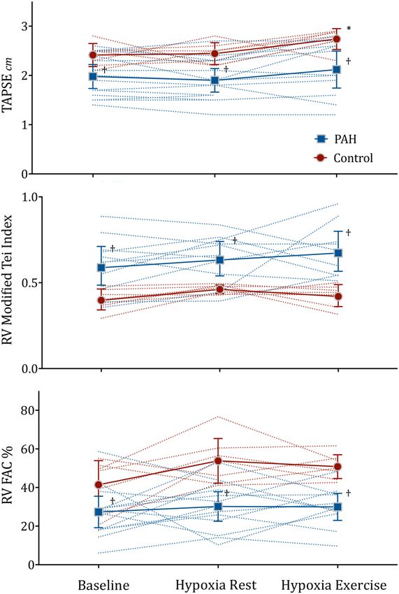
†versus Control unpaired t-test p0.6, (PAH) and 7 healthy patients (control). *pOpen Heart
Open Heart: first published as 10.1136/openhrt-2016-000532 on 20 January 2017. Downloaded from http://openheart.bmj.com/ on March 21, 2021 by guest. Protected by copyright.
following 1 hour at simulated 2400 m altitude at rest.26
The authors attributed this rise to a mildly elevated PVR
that was also seen in the control group. In comparison
to our study participants who have PAH with normal LV
systolic function, their patients had predominantly
impaired LV systolic dysfunction, with a large proportion
of patients (90%) on β-blockers. The latter would have
limited a patient’s ability to increase cardiac output due
to chronotropic and inotropic blunting.
The patients with PAH in this study had moderately
elevated PASP and the majority had evidence of RV dila-
tion and RV dysfunction at baseline sea level. The
observed group mean increase in PASP from baseline
after mild exercise during hypoxia was ≈20 mm Hg.
Despite this increase in PASP, we did not document any
deterioration or change in measures of RV size or func-
tion on echocardiography, and no adverse events were
reported despite a mild increase in the perception of
breathlessness. This includes the single patient who did
not complete the exercise stress due to significant desat-
uration. The postulated deleterious effects on RV func-
tion from hypoxic pulmonary vasoconstriction and/or
RV ischaemia in patients with PAH were not apparent in
this study. Hypoxia equivalent to commercial air travel
(2400 m) may not have been sufficient to elicit detect-
able adverse effect on the RV. However, it should be
noted that controls displayed augmentation of TAPSE
during hypoxia with exertion, a phenomenon that was
Figure 3 Mean with 95% CI error bars and individual data not observed in the PAH group. It has been demon-
points of right ventricular function parameters at rest breathing strated that an increase in TAPSE occurs during exercise
room air (baseline), during 20 min breathing 15.1% O2 at rest in healthy controls.27 The lack of increase in TAPSE in
(hypoxia rest) and following mild exercise (hypoxia exercise) the PAH group may indicate reduced RV reserve or pos-
in 14 patients with pulmonary arterial hypertension (PAH) and
sibly reflect early signs of hypoxic decompensation.
7 healthy patients (control). *pPulmonary vascular disease
Open Heart: first published as 10.1136/openhrt-2016-000532 on 20 January 2017. Downloaded from http://openheart.bmj.com/ on March 21, 2021 by guest. Protected by copyright.
Patients with PAH desaturated to a greater extent from European Respiratory Society (ERS) Endorsed by: Association for
European Paediatric and Congenital Cardiology (AEPC),
baseline during ‘hypoxia at rest’ than healthy patients International Society for Heart and Lung Transplantation (ISHLT).
and the pressure of arterial oxygen strongly correlated Eur Heart J 2016;37:67–119.
with PASP; three patients with a PaO2 that fell below 3. Ahmedzai S, Balfour-Lynn IM, Bewick T, et al. Managing
passengers with stable respiratory disease planning air travel:
50 mm Hg also recorded significantly elevated PASP and British Thoracic Society recommendations. Thorax 2011;66
higher perceptions of breathlessness. Absolute minute (Suppl 1):i1–30.
4. Code of Federal Regulations. 14, part 25841. Washington: US
ventilation and respiratory rate were higher in PAH in Goverment Printing Office, 1986.
resting conditions compared to normal patients, possibly 5. Kelly PT, Seccombe LM, Rogers PG, et al. Directly measured cabin
pressure conditions during Boeing 747–400 commercial aircraft
attributed to their reduced diffusing capacity that may flights. Respirology 2007;12:511–15.
have contributed to their higher perception of 6. Roubinian N, Elliott CG, Barnett CF, et al. Effects of
breathlessness. commercial air travel on patients with pulmonary hypertension
air travel and pulmonary hypertension. Chest 2012;142:
It is acknowledged that our study population included 885–92.
mainly WHO FC II patients who were stabilised on 7. Burns RM, Peacock AJ, Johnson MK, et al. Hypoxaemia in patients
with pulmonary arterial hypertension during simulated air travel.
PAH-specific therapies including ERA and/or PDE5-I. Respir Med 2013;107:298–304.
Thus, our study findings pertain strictly to this popula- 8. Vogel JA, Harris CW. Cardiopulmonary responses of resting man
tion which had well compensated disease. It is unknown during early exposure to high altitude. J Appl Physiol
1967;22:1124–8.
whether a more prolonged period of hypoxia (beyond 9. Talbot NP, Balanos GM, Dorrington KL, et al. Two temporal
our protocol duration) will lead to deleterious effects on components within the human pulmonary vascular response to
approximately 2 h of isocapnic hypoxia. J Appl Physiol
RV function, particularly considering the observed non- 2005;98:1125–39.
changing TAPSE. Thus, our study findings do not contra- 10. Swenson ER. Hypoxic pulmonary vasoconstriction. High Alt Med
dict current recommended guidelines regarding the use Biol 2013;14:101–10.
11. Groves BM, Reeves JT, Sutton JR, et al. Operation Everest II:
of supplemental oxygen in flight. Similarly, further elevated high-altitude pulmonary resistance unresponsive to oxygen.
studies are needed to determine the effect of simulated J Appl Physiol 1987;63:521–30.
12. Huez S, Retailleau K, Unger P, et al. Right and left ventricular
altitude in patients with PAH with more advanced FC adaptation to hypoxia: a tissue Doppler imaging study. Am J Physiol
status. Heart Circ Physiol 2005;289:H1391–8.
13. McLaughlin VV, Archer SL, Badesch DB, et al. ACCF/AHA 2009
expert consensus document on pulmonary hypertension a report
of the American College of Cardiology Foundation Task Force on
CONCLUSION Expert Consensus Documents and the American Heart
Stable group 1 patients with PAH on vasodilator treat- Association developed in collaboration with the American College
of Chest Physicians; American Thoracic Society, Inc.; and the
ment did not experience any acute deterioration in RV Pulmonary Hypertension Association. J Am Coll Cardiol
function during simulated mild altitude, at rest or fol- 2009;53:1573–619.
lowing mild exertion. PASP increased both at rest and 14. Miller MR, Hankinson J, Brusasco V, et al. Standardisation of
spirometry. Eur Respir J 2005;26:319–38.
during mild exercise that was associated with arterial 15. Macintyre N, Crapo RO, Viegi G, et al. Standardisation of the
desaturation. Despite a mild increase in symptoms, the single-breath determination of carbon monoxide uptake in the lung.
Eur Respir J 2005;26:720–35.
level and duration of hypoxia in this study protocol was 16. Quanjer PH, Stanojevic S, Cole TJ, et al. Multi-ethnic reference
tolerated well. values for spirometry for the 3–95-yr age range: the global
lung function 2012 equations. Eur Respir J 2012;40:
Acknowledgements The authors thank Ms Sharon Quirk and Dr Tommy 1324–43.
Chung of the Department of Cardiology, Concord Hospital, for their assistance 17. Quanjer PH. Standardized lung function testing. Report
with patient recruitment. working party. Bull Eur Physiopathol Respir 1983;19(Suppl
5):1–95.
Competing interests None declared. 18. Lang RM, Badano LP, Mor-Avi V, et al. Recommendations for
cardiac chamber quantification by echocardiography in adults: an
Ethics approval Sydney Local Area Health District, New South Wales, update from the American Society of Echocardiography and the
Australia, HREC. European Association of Cardiovascular Imaging. Eur Heart
J Cardiovasc Imaging 2015;16:233–70.
Provenance and peer review Not commissioned; internally peer reviewed. 19. Wright LM, Dwyer N, Celermajer D, et al. Follow-up of pulmonary
hypertension with echocardiography. JACC Cardiovasc Imaging
Open Access This is an Open Access article distributed in accordance with
2016;9:733–46.
the Creative Commons Attribution Non Commercial (CC BY-NC 4.0) license, 20. Forfia PR, Fisher MR, Mathai SC, et al. Tricuspid annular
which permits others to distribute, remix, adapt, build upon this work non- displacement predicts survival in pulmonary hypertension. Am
commercially, and license their derivative works on different terms, provided J Respir Crit Care Med 2006;174:1034–41.
the original work is properly cited and the use is non-commercial. See: http:// 21. Harada K, Tamura M, Toyono M, et al. Comparison of the right
creativecommons.org/licenses/by-nc/4.0/ ventricular Tei index by tissue Doppler imaging to that obtained by
pulsed Doppler in children without heart disease. Am J Cardiol
2002;90:566–9.
22. Rudski LG, Lai WW, Afilalo J, et al. Guidelines for the
REFERENCES echocardiographic assessment of the right heart in adults: a report
1. Lau EM, Tamura Y, McGoon MD, et al. The 2015 ESC/ERS from the American Society of Echocardiography endorsed by the
Guidelines for the diagnosis and treatment of pulmonary European Association of Echocardiography, a registered branch of
hypertension: a practical chronicle of progress. Eur Respir J the European Society of Cardiology, and the Canadian Society of
2015;46:879–82. Echocardiography. J Am Soc Echocardiogr 2010;23:685–713; quiz
2. Galie N, Humbert M, Vachiery JL, et al. 2015 ESC/ERS Guidelines 86–8.
for the diagnosis and treatment of pulmonary hypertension: the Joint 23. Chow V, Ng ACC, Seccombe L, et al. Impaired 6-min walk test,
Task Force for the Diagnosis and Treatment of Pulmonary heart rate recovery and cardiac function post pulmonary embolism in
Hypertension of the European Society of Cardiology (ESC) and the long-term survivors. Respir Med 2014;108:1556–65.
Seccombe LM, Chow V, Zhao W, et al. Open Heart 2017;4:e000532. doi:10.1136/openhrt-2016-000532 7Open Heart
Open Heart: first published as 10.1136/openhrt-2016-000532 on 20 January 2017. Downloaded from http://openheart.bmj.com/ on March 21, 2021 by guest. Protected by copyright.
24. Jette M, Sidney K, Blumchen G. Metabolic equivalents (METS) in 28. Gong H Jr, Tashkin DP, Lee EY, et al. Hypoxia-altitude simulation
exercise testing, exercise prescription, and evaluation of functional test. Evaluation of patients with chronic airway obstruction. Am Rev
capacity. Clin Cardiol 1990;13:555–65. Respir Dis 1984;130:980–6.
25. Borg GA. Psychophysical bases of perceived exertion. Med Sci 29. Allemann Y, Sartori C, Lepori M, et al. Echocardiographic and
Sports Exerc 1982;14:377–81. invasive measurements of pulmonary artery pressure correlate
26. Hobkirk JP, Damy T, Walters M, et al. Effects of reducing inspired closely at high altitude. Am J Physiol Heart Circ Physiol 2000;279:
oxygen concentration for one hour in patients with chronic heart H2013–16.
failure: implications for air travel. Eur J Heart Fail 2013;15:505–10. 30. D’Alto M, Romeo E, Argiento P, et al. Accuracy and precision of
27. Chia EM, Lau EM, Xuan W, et al. Exercise testing can unmask right echocardiography versus right heart catheterization for the
ventricular dysfunction in systemic sclerosis patients with normal assessment of pulmonary hypertension. Int J Cardiol
resting pulmonary artery pressure. Int J Cardiol 2016;204:179–86. 2013;168:4058–62.
8 Seccombe LM, Chow V, Zhao W, et al. Open Heart 2017;4:e000532. doi:10.1136/openhrt-2016-000532You can also read