Happy Hypoxia in COVID-19 Patients at Kinshasa University Hospital (Democratic Republic of the Congo): Frequency and Vital Outcome - ResearchGate
←
→
Page content transcription
If your browser does not render page correctly, please read the page content below
Journal of Biosciences and Medicines, 2021, 9, 12-20
                                                                                               https://www.scirp.org/journal/jbm
                                                                                                         ISSN Online: 2327-509X
                                                                                                           ISSN Print: 2327-5081

Happy Hypoxia in COVID-19 Patients at
Kinshasa University Hospital (Democratic
Republic of the Congo): Frequency and Vital
Outcome

Ben Bepouka1, Hippolyte Situakibanza1, Ossam Odio1, Jean Robert Makulo2, Madone Mandina1,
Murielle Longokolo1, Nadine Mayasi1, Kazadi Mutombo1, Tresor Pata1, Godelive Nsangana1,
Felly Tshikangu1, Donatien Mangala1, Dupont Maheshe1, Christine Namasale1,
Serge Nkarnkwin1, Jonathan Muamba1, Gorby Ndaie1, Rodrigue Ngwizani3, Hervé Mole3,
Gabriel Makeya3, Tharcisse Mabiala3, Patrick Mukuna3, Roger Kabango3, Patricia Kabuni3,
Yves Yanga3, Aliocha Nkodila4, Hervé Keke5, Nice Musangu5, Papy Tshimanga5, Yamin Kokusa1,
Bertin Nsitwayizatadi6, Eric Mukenge7, Guyguy Kamwiziku7, Gabriel Mbunsu7,
Jean Claude Makangara7, Marcel Mbula1, Jean Marie Kayembe8
1
 Unit of Infectious Diseases, Kinshasa University Hospital, Kinshasa, Democratic Republic of the Congo
2
 Unit of Nephrology, Kinshasa University Hospital, Kinshasa, Democratic Republic of the Congo
3
 Unit of Reanimation, Kinshasa University Hospital, Kinshasa, Democratic Republic of the Congo
4
 Unit of Vaccinology, World Health Organization, Kinshasa, Democratic Republic of the Congo
5
 Department of Epidemiology, Ministry of Health, Kinshasa, Democratic Republic of the Congo
6
 Unit of Physiotherapy, Kinshasa Hospital University, Kinshasa, Democratic Republic of the Congo
7
 Unit of Microbiology, Kinshasa Hospital University, Kinshasa, Democratic Republic of the Congo
8
 Unit of Pneumology, Kinshasa University Hospital, Kinshasa, Democratic Republic of the Congo

How to cite this paper: Bepouka, B., Situ-     Abstract
akibanza, H., Odio, O., Makulo, J.R., Man-
dina, M., Longokolo, M., Mayasi, N., Mu-       Background: Happy hypoxia is a new feature found in COVID-19 patients. It
tombo, K., Pata, T., Nsangana, G., Tshi-       consists of the presence of severe hypoxemia but normal breathing rate. Fail-
kangu, F., Mangala, D., Maheshe, D., Na-
                                               ure to identify this hypoxia may have negative consequences on the survival
masale, C., Nkarnkwin, S., Muamba, J.,
Ndaie, G., Ngwizani, R., Mole, H., Makeya,
                                               of the patient. The objective of the present study was to measure the frequen-
G., Mabiala, T., Mukuna, P., Kabango, R, .     cy of patients with happy hypoxia and to evaluate their survival at the Kin-
Kabuni, P., Yanga, Y., Nkodila, A., Keke,      shasa University Hospital (KUH). Methods: This was a historical cohort of
H., Musangu, N., Tshimanga, P., Kokusa, Y.,    141 hospitalized patients with COVID-19 at KUH from March 23 to June 15,
Nsitwayizatadi, B., Mukenge, E., Kamwiziku,
G., Mbunsu, G., Makangara, J.C., Mbula, M.
                                               2020. Happy hypoxia was defined as oxygen saturation below 90% without
and Kayembe, J.M. (2021) Happy Hypoxia in      dyspnea. Socio-demographic data, co-morbidities, follow up time of hospita-
COVID-19 Patients at Kinshasa University       lization and outcomes were studied. Survival was assessed using the Kaplan
Hospital (Democratic Republic of the Congo):   Meier curve. Results: Out of 141 hospitalized patients with COVID-19, 79
Frequency and Vital Outcome. Journal of
Biosciences and Medicines, 9, 12-20.           (56%) patients were at the severe or critical stage and 9 (6.4%) had a happy
https://doi.org/10.4236/jbm.2021.92002         hypoxia on admission. Patients who had happy hypoxia on admission were

DOI: 10.4236/jbm.2021.92002 Feb. 9, 2021                         12                         Journal of Biosciences and Medicines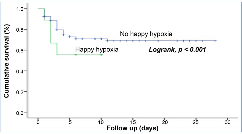
B. Bepouka et al.

Received: January 8, 2021
Accepted: February 6, 2021                      generally older than 60 years of age (55.6%) (p = 0.023). Comparison of sur-
Published: February 9, 2021                     vival curves, based on the presence or absence of happy hypoxia, shows a sta-
                                                tistically significant difference (p = 0.001). The presence of happy hypoxia
Copyright © 2021 by author(s) and
Scientific Research Publishing Inc.             reduces survival. Conclusion: The frequency of happy hypoxia among
This work is licensed under the Creative        COVID-19 patients was low. Survival was reduced in patients with happy
Commons Attribution International               hypoxia. Prehospital pulse oximetry could serve as an early warning signal for
License (CC BY 4.0).
                                                the detection of happy hypoxemia in COVID-19 patients.
http://creativecommons.org/licenses/by/4.0/
               Open Access
                                                Keywords
                                                COVID-19, Happy Hypoxia, Frequency, Outcome, DR Congo

                                              1. Introduction
                                              The world is facing a severe pandemic called Coronavirus 2019 Disease
                                              (COVID-19) caused by the new Severe Acute Respiratory Syndrome Coronavi-
                                              rus-2 (SARS-COV-2), which broke out in China in December 2019. COVID-19
                                              can be particularly severe and fatal in certain groups such as the elderly or
                                              people with co-morbidities. In symptomatic patients, the clinical presentation is
                                              mild in about 80% of cases, moderate to severe in 15% of cases and critical in 5%
                                              of cases. Patients with severe disease may develop acute respiratory distress syn-
                                              drome (ARDS), shock, thromboembolic manifestations, multiple organ failure
                                              and are at risk of death [1]. Patients with SARS-COV-2 pneumonia may de-
                                              compensate due to hypoxemic respiratory failure. Autopsy data show diffuse al-
                                              veolar injury, occasional inflammation, hyaline membranes and accumulation of
                                              alveolar fluid, consistent with acute respiratory distress syndrome (ARDS) [2].
                                                One aspect of COVID-19 that puzzles clinicians who must manage pneumo-
                                              nia is that one of the complications of the disease is the presentation of patients
                                              with very low blood saturation but no sensation of dyspnea. SARS-COV-2
                                              pneumonia can present clinically in a very heterogeneous fashion. For the same
                                              degree of hypoxemia, some patients have a faster breathing rate than others.
                                              COVID-19 patients with a very low degree of blood oxygenation (deep hypox-
                                              emia) may present very different symptoms. Some may have normal breathing.
                                              This phenomenon is called “happy hypoxia” [3]. In the Wuhan cohort of
                                              SARS-COV-2 infected patients, only 19% complained of shortness of breath;
                                              62% of patients with severe illness and 46% of those who were intubated, venti-
                                              lated or died did not have shortness of breath [4]. In cases of happy hypoxia, a
                                              persistent normal respiratory rate was inconsistent with the severity of the hy-
                                              poxia. Roca et al. described the discrepancy between pre-hospital initial respira-
                                              tory rate (IRR) and initial oxygen saturation (SpO2) (i.e., before oxygen supple-
                                              mentation) in COVID-19 patients. They found that some patients had a normal
                                              respiratory rate with very low saturation. Measurement of saturation on admis-
                                              sion identified non-clinically evident acute respiratory failure [5]. In addition,
                                              awake patients who are very ill with COVID-19 therefore require in some re-

DOI: 10.4236/jbm.2021.92002                                        13                         Journal of Biosciences and Medicines
B. Bepouka et al.
spects as high a level of care as those on mechanical ventilation. Maneuvers that
can safely improve oxygenation without requiring additional resources are there-
fore of immense value during an exacerbation of COVID-19 [6]. COVID-19 pa-
tients can rapidly and dangerously desaturate when disconnected from their
oxygen source, especially if apparently not in dyspnea. Patients with high oxygen
requirements who are managed with alternatives to mechanical ventilation re-
quire vigilant monitoring and frequent and careful reassessment [7].
To our knowledge, the socio-demographic and clinical profile and outcome of
severe patients with happy hypoxia in Africa subjects are not known. The objec-
tive of the study was to measure the frequency of happy hypoxia on admission,
to describe the co-morbidities as well as the outcome of these patients who have
been hospitalized at the Kinshasa University Hospital.
2. Materials & Methods
This was a historical cohort. It covered the period from March 23 to June 15,
2020. This study was conducted at the Kinshasa University Hospital, a large re-
gional hospital in Kinshasa, DRC. Kinshasa University Hospital is the tertiary
level hospital in the city of Kinshasa. This study included all hospitalized pa-
tients with COVID-19 confirmed by RT-PCR.
2.1. Data Collection
The variables of interest in this study were socio-demographic characteristics
(age, sex, education level), co-morbidities (hypertension, diabetes mellitus, asthma,
cardiovascular diseases), pregnancy, follow up time of hospitalization and out-
come.
2.2. Operational Definition
According to some previous study about COVID-19, we define Happy Hypoxia
has been defined by an oxygen saturation lower than 90% on room air without
dyspnea with a normal initial respiratory rate at the admission of the patient [7]
[8].
2.3. Laboratory Procedures
Confirmation of diagnosis
Pharyngeal swabs were taken from each patient for the detection of SARS-COV-2
viral nucleic acids by the RT-PCR assay described above [9].
2.4. Statistical Analyses
The analysis was performed with SPSS version 21 software. The descriptive ana-
lyses performed were mean, standard deviation for quantitative data with Gaus-
sian distribution and median (IQE = interquartile standard deviation) for data
with non-Gaussian distribution. The Pearson Chi-square or Fisher’s exact test,
as appropriate, was performed to compare proportions. The Kaplan Meier curve
DOI: 10.4236/jbm.2021.92002 14 Journal of Biosciences and MedicinesB. Bepouka et al.
was used to assess survival. A value of p < 0.05 was considered the threshold of
significance.
2.5. Ethical Considerations
The study was approved by the University of Kinshasa School of Public Health’s
Ethics Committee (ESP/CE/179/2020). The principle of anonymity and confi-
dentiality was maintained in the study. No conflict of interest was declared in the
conduct of this study.
3. Results
3.1. General Characteristics
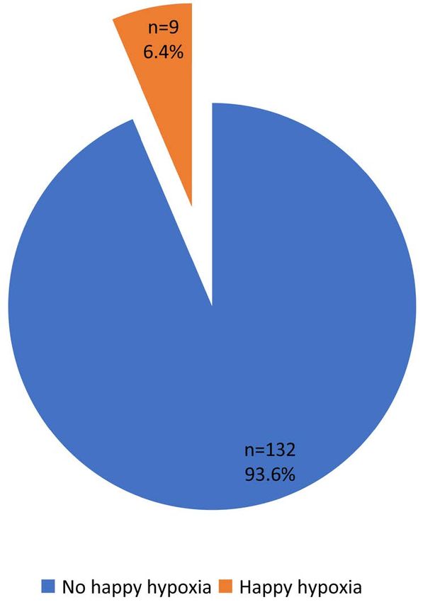
Out of 141 hospitalized patients with COVID-19, 79 (56%) patients were at the
severe or critical stage and 9 (6.4%) had a happy hypoxia on admission (Figure
1). Six patients with happy hypoxia on admission were male (p = 0.963). Five
patients (55.6%) older than 60 years of age (p = 0.023) and seven married pa-
tients (77.8%) (p = 0.275) had happy hypoxia. No patients with happy hypoxia
had comorbidity (Table 1).
3.2. Outcome of Patients with Happy Hypoxia
Of the 9 patients with happy hypoxia, 4 were deceased and 5 survived. All 4 died
within 4 days of hospitalization. Comparison of the survival curves, depending
on the presence or absence of happy hypoxia shows a statistically significant dif-
ference (p = 0.001). The presence of happy hypoxia reduces survival (Figure 2).
Figure 1. Frequency of happy hypoxia at the COVID-19 treatment center of the Kinshasa
University Hospital, from March 23rd to June 15th, 2020.
DOI: 10.4236/jbm.2021.92002 15 Journal of Biosciences and MedicinesB. Bepouka et al.
Table 1. General characteristics of COVID-19 patients at the COVID-19 treatment center
of the Kinshasa University Hospital, from March 23rd to June 15th, 2020.
No happy hypoxia Happy hypoxia Over all
Variables p
n = 132 n=9 n = 141
Sex 0.963
Male 89 (67.4) 6 (66.7) 95 (67.4)
Female 43 (32.6) 3 (33.3) 46 (32.6)
Age 0.023B. Bepouka et al.
saturation lower than 90% which corresponds to a PO2 of 60 mmHg, whereas
Brouqui et al. considered 80 mmHg as threshold because the gasometry was
performed in all patients.
Happy hypoxia has some explanations in the literature. In situations of happy
hypoxia, we found that some patients are tachycardic and have respiratory alka-
losis. These signs suppose that part of the sensory information reaches the brain
stem to induce a partial compensatory reflex respiratory response and lowers the
level of CO2, which diffuses more rapidly in the alveoli than oxygen. The related
homeostatic information emanating from the body is part of our interception
system, which detects the physiological state of the body, creates consciousness
and leads to conscious feelings or symptoms [11]. While in half of ARDS cases
the lung loses some of its elastic properties, it remains distensible in the initial
stage of COVID-19 pneumonia. This is because the lung retains its ability to
vary its volume normally in response to changes in intra-pulmonary pressure.
Experts consider this to be “compliance” preserved. This means that the lung
retains its elasticity and remains aerated with an amount of intrapulmonary air
close to normal while in ARDS outside of COVID-19 the lung becomes stiff. The
severe hypoxemia observed in patients with COVID-19 is accompanied by
near-normal compliance, especially in the near stage. SARS-COV-2 could also
interfere with mitochondrial O2-sensing and causes mitochondrial-induced in-
jury, which impairs carotid body function resulting in impaired respiratory drive
and reduced dyspnea [12] [13].
This maintenance of ventilation is associated with the disruption of vascular
perfusion due to the prothrombotic phase of COVID-19. Autopsy results for
COVID-19 showed the presence of microthrombi in the pulmonary microvas-
culature [14] [15] [16]. This suggests that happy or silent hypoxemia may be due
to conserved ventilation and disrupted perfusion due to capillary obstruction.
The anatomical distribution on this peripheral vascular bed undermines the
predominantly distal and uneven distribution of radiological infiltrates [17].
This process occurs through projections from the brain stem to the cortex that
allows the brain to process homeostatic signals. When hypoxia occurs, the brain
receives the internal hypoxic signal, it gives the sensation of “hunger for air” and
a need to breathe but in severe COVID-19 patients this sensation is curiously
absent. In the presence of hypoxia, respiratory responses occur due to the pres-
ence of sensory nerves in the chemoreceptive zones. Changes in the internal en-
vironment are recognized by these nerves, they transmit information to the
brain stem and these nerves stimulate an increase in the ventilatory drive. Res-
piratory pathologies provoke auto-nomic reflexes, such as coughing, secretions
and bronchospasm. Several clinical conditions can trigger dyspnea. In cardi-
opulmonary diseases, dyspnea is caused by the entry of multiple homeostatic af-
ferents. The interceptive processing of these signals creates a feeling of breath-
lessness and a desire to breathe. This primitive brain stem reflex is essential for
survival because it can respond to many conditions including hypoxia, acidosis,
airway collapse, hypercapnia, irritants and pulmonary vascular congestion [18].
DOI: 10.4236/jbm.2021.92002 17 Journal of Biosciences and MedicinesB. Bepouka et al.
Happy hypoxia characterized by the dissociation between the absence of
dyspnea and the profound hypoxemia does not have clear pathophysiological
mechanisms. Among the reasons evoked, there are also lesions of the vagus and
glossopharyngeal nerves after neck cancer, a congenital neuropathy. In the con-
text of COVID-19, the possibility that SARS-COV-2 has neuroinvasive action is
controversial. On the one hand, there is the presence of neurological signs such
as convulsions, delirium, altered mental status and anosmia. The possible dam-
age to neurons could be due to the direct action of SARS-COV-2 on nerve fibres
and the intense cytokine storm after the inflammatory phase. On the other hand,
SARS-COV-2 could enter the brain through pathways connected to synapses
and is found in the cerebral fluid of the spinal column [19]. On the other hand,
the results of brain magnetic resonance imaging (MRI) studies and pathology
reports in fatal cases of COVID-19 are inconsistent and do not provide a patho-
physiological correlation to explain the absence of dyspnea. The common brain
pathology findings in fatal COVID-19 cases are multiple areas of ischemic stroke
and hemorrhagic microbleeding with only small areas of inflammation; howev-
er, it should be noted that at least 40% of the brain imaging studies were normal
and there were no signals of brain stem abnormalities on MRI scans [20].
Regardless of the uncertain underlying pathology, reduced perception of
dyspnea is a disorder of blood gas interception. It can mask the severity of the
condition and ultimately delay the time at which the patient can seek urgent
medical attention. Patients admitted with COVID-19 may suffer sudden death
after voluntary “cuts” in oxygen supplementation. No patient with happy hy-
poxia had comorbidity. The small number of patients with happy hypoxia in this
study does not allow us to confirm this assertion. The data in the literature to
date do not provide a correlation between happy hypoxia and comorbidities.
Happy hypoxia reduced survival. This result is similar to the study by Brouqui et
al. who found that happy hypoxia was associated with poor outcomes (33.3%
were transferred to the intensive care unit and 25.9% died) [10]. These patients
who do not have warning signs of poor outcome could be falsely considered to
be clinically well, and caregivers may be distracted leading to a negative outcome
if monitoring is not rigorous.
5. Limitations
The study was retrospective with the possibility of missing data. We did not
measure hypoxia by gasometry by looking for the partial pressure of oxygen but
by pulse oximetry. The small sample size of patients with happy hypoxia limit
did us to better establish the characteristics of this new entity.
6. Conclusion
Happy hypoxia was present in our cohort. The frequency of happy hypoxia
among COVID-19 patients was low. Elderly people were predominant. Survival
was reduced in patients with happy hypoxia. Prehospital pulse oximetry could
DOI: 10.4236/jbm.2021.92002 18 Journal of Biosciences and MedicinesB. Bepouka et al.
serve as an early warning signal for the detection of happy hypoxemia in patients
with COVID-19. Continued research is important to establish the link between
happy hypoxia and advanced age.
Acknowledgements
The authors acknowledge ALIMA NGO for support in the management of
COVID-19 patients, especially Drs Armand, Christian MASUDI and Junior
MUWALAWALA. I acknowledge also ANDEMIA project for epistat and scien-
tific writing capacity building.
Conflicts of Interest
All authors declare no conflicts of interest.
References
[1] Wu, Z. and McGoogan, J.M. (2020) Characteristics of and Important Lessons from
the Coronavirus Disease 2019 (COVID-19) Outbreak in China: Summary of a Re-
port of 72314 Cases from the Chinese Center for Disease Control and Prevention.
JAMA, 323, 1239-1242.
[2] Archer, S.L., Sharp, W.W. and Weir, E.K. (2020) Differentiating COVID-19 Pneu-
monia from Acute Respiratory Distress Syndrome (ARDS) and High-Altitude Pul-
monary Edema (HAPE): Therapeutic Implications. Circulation, 142, 101-104.
https://doi.org/10.1161/CIRCULATIONAHA.120.047915
[3] Couzin-Frankel, J. (2020) The Mystery of the Pandemic’s “Happy Hypoxia”.
Science, 368, 455-456.
[4] Guan, W.-J., Ni, Z.-Y., Hu, Y., et al. (2020) Clinical Characteristics of Coronavirus
Disease 2019 in China. The New England Journal of Medicine, 382, 1708-1720.
https://doi.org/10.1056/NEJMoa2002032
[5] Roca, O., Messika, J., Caralt, B., et al. (2016) Predicting Success of High-Flow Nasal
Cannula in Pneumonia Patients with Hypoxemic Respiratory Failure: The Utility of
the ROX Index. Journal of Critical Care, 35, 200-205.
https://doi.org/10.1016/j.jcrc.2016.05.022
[6] World Health Organization (2020) Clinical Management of Severe Acute Respira-
tory Infection (SARI) When COVID-19 Disease Is Suspected.
https://www.who.int/docs/default-source/coronaviruse/clinical-management-of-no
vel-cov.pdf
[7] Caputo, N.D., Strayer, R.J. and Levitan, R. (2020) Early Self-Proning in Awake,
Non-Intubated Patients in the Emergency Department: A Single ED’s Experience
during the COVID-19 Pandemic. Academic Emergency Medicine, 27, 375-378.
https://doi.org/10.1111/acem.13994
[8] Widysanto, A., Wahyuni, T., Simanjuntak, L., et al. (2020) Happy Hypoxia in Criti-
cal COVID-19 Patient: A Case Report in Tangerang, Indonesia. Physiological Re-
ports, 8, e14619. https://doi.org/10.14814/phy2.14619
[9] World Health Organization (2020) Coronavirus Disease (COVID-19) Technical
Guidance: Laboratory Testing for 2019-nCoV in Humans.
https://www.who.int/emergencies/diseases/novel-coronavirus-2019/technical-guida
nce/laboratory-guidance
[10] Brouqui, P., Amrane, S., Million, M., et al. (2020) Asymptomatic Hypoxia in
DOI: 10.4236/jbm.2021.92002 19 Journal of Biosciences and MedicinesB. Bepouka et al.
COVID-19 Is Associated with Poor Outcome. International Journal of Infectious
Diseases, 102, 233-238. https://doi.org/10.1016/j.ijid.2020.10.067
[11] Robinson, D. and Gebhart, F. (2008) Inside Information: The Unique Features of
Visceral Sensation. Molecular Interventions, 8, 242-253.
https://doi.org/10.1124/mi.8.5.9
[12] Gattinoni, L., Coppola, S., Cressoni, M., et al. (2020) COVID-19 Does Not Lead to a
“Typical” Acute Respiratory Distress Syndrome. American Journal of Respiratory
and Critical Care Medicine, 201, 1299-1300.
[13] Negri, E.M., et al. (2020) Heparin Therapy Improving Hypoxia in COVID-19 Pa-
tients—A Case Series.
https://www.medrxiv.org/content/10.1101/2020.04.15.20067017v3
[14] Tian, S., Hu, N. and Niu, L. (2020) Pulmonary Pathology of Early-Phase 2019 Novel
Coronavirus. Pneumonia in Two Patients with Lung Cancer. Journal of Thoracic
Oncology, 15, 700-704. https://doi.org/10.20944/preprints202002.0220.v2
[15] Yao, X.H., Li, T.Y., He, Z.C., et al. (2020) A Pathological Report of Three
COVID-19 Cases by Minimally Invasive Autopsies. Chinese Journal of Pathology,
49, 411-417.
[16] Dolhnikoff, M., Duarte-Neto, A.N., de Almeida Monteiro, R.A., et al. (2020) Patho-
logical Evidence of Pulmonary Thrombotic Phenomena in Severe COVID-19.
Journal of Thrombosis and Haemostasis, 18, 1517-1519.
https://doi.org/10.1111/jth.14844
[17] Ye, Z., Zhang, Y., Wang, Y., et al. (2019) Chest CT Manifestations of New Corona-
virus Disease.
[18] Burki, N. and Lee, L. (2010) Mechanisms of Dyspnea. Chest, 138, 1196-1201.
https://doi.org/10.1378/chest.10-0534
[19] Duarte, A.G. and Kauffmann, L.N. (2020) Is “Happy Hypoxia” in COVID-19 a Dis-
order of Autonomic Interoception? A Hypothesis. Clinical Autonomic Research, 30,
331-333.
[20] Coolen, T., Lolli, V. and Sadeghi, N. (2020) Early Postmortem Brain MRI Findings
in COVID-19 Non-Survivors. Neurology, 95, e2016-e2027.
https://doi.org/10.1101/2020.05.04.20090316
DOI: 10.4236/jbm.2021.92002 20 Journal of Biosciences and MedicinesYou can also read