Grey matter changes in Meige syndrome: a voxel based morphology analysis - Nature
←
→
Page content transcription
If your browser does not render page correctly, please read the page content below
www.nature.com/scientificreports
OPEN Grey matter changes in Meige
syndrome: a voxel‑based
morphology analysis
Jiayu Liu1,5, Lei Li2,5, Lei Chen3, Ruen Liu1,4*, Yongan Jiang4, Jixia Fang1, Dongliang Wang1,
Zhi Liu1 & Jia Ouyang1
To investigate the changes and clinical significance of brain structural abnormalities in patients with
Meige syndrome and related depressive symptoms. We retrospectively analysed clinical data, imaging
examinations, and Hamilton Depression Rating scale scores in 46 patients with Meige syndrome
from January 2017 to January 2019. We compared the Meige syndrome group with the healthy
control group, and the definite depression group with the non-definite depression group. Voxel-based
morphometry (VBM) was used to compare grey matter (GM) volumes. We conducted two-sample
t-tests corrected for subject age and gender. We tested at a level of significance of p < 0.001 with a
false discovery rate (FDR) correction. VBM demonstrated decreased GM volume (p < 0.001 and cluster
size > 50 voxels) in the left hemisphere in the middle frontal orbital gyrus, temporal pole (superior
temporal gyrus) and insula and in the right hemisphere in the temporal pole (middle temporal
gyrus), precuneus, inferior parietal, inferior temporal and olfactory cortices in the Meige syndrome
group. Comparing VBM-MRI measures in Meige syndrome patients with and without depression,
decreased GM volume was found in the left hemisphere in the cuneus and hippocampus and in the
right hemisphere in the angular gyrus, middle frontal gyrus and middle occipital gyrus in the definite
depression group. Unlike other dystonia studies that have suggested an involvement of the basal
ganglia and motor cortex in the pathophysiology of the disorder , we believe that the precuneus
is involved in the development of Meige syndrome. Additionally, our findings suggest that the
hippocampus plays a role in the pathogenesis of depression in patients with Meige syndrome.
Meige syndrome, also known as idiopathic blepharospasm-oromandibular dystonia syndrome, is a rare type of
segmental dystonia disorder with features of adult hyperactivity disorder and characterized by blepharospasm
and oromandibular dystonia d isorder1–3. Meige syndrome usually occurs between 30 and 60 years of age, with
rare cases appearing in teenage y ears4. This disease is more common in females, and the ratio of males to females
is approximately 1:3. Meige syndrome can be divided into three types according to clinical manifestations: blepha-
rospasm, blepharospasm-oromandibular dystonia, and oromandibular dystonia, among which blepharospasm
with oromandibular dystonia is regarded as the complete type of Meige s yndrome3. Meige syndrome is classi-
fied into primary and secondary Meige syndromes. Although plastic changes and reduced cortical inhibition
have been documented by neurophysiological and neuroimaging t echniques5, the aetiology of primary Meige
syndrome is still unknown.
In recent years, the application of the voxel-based morphometry method of magnetic resonance imaging
(VBM-MRI) for structural imaging research has provided a new and important method for structural research
of neurologic movement disorders6. Some VBM-MRI studies have shown significant reductions in grey matter
(GM) volume mainly in the cerebellar vermis and bilateral superior frontal gyri7,8. However, they did not study
Meige patients, and the number of included patients was small. Therefore, a VBM study on Meige syndrome is
necessary to clarify the pathogenesis of this disease. In addition to blepharospasm and oromandibular dystonia,
1
Department of Neurosurgery, Peking University People’s Hospital, 11th Xizhimen South St., Beijing 100044,
China. 2Department of Nuclear Medicine, Zhuhai People’s Hospital (Zhuhai hospital affiliated with Jinan
University), No. 79 Kangning Road, Xiangzhou District, Zhuhai 519000, Guangdong, China. 3Department of
Radiology, Peking University People’s Hospital, 11th Xizhimen South St., Beijing 100044, China. 4Department of
Neurosurgery Jiangxi Provincial People’s Hospital Affiliated to Nanchang University, Nanchang 330006, Jiangxi,
China. 5These authors contributed equally: Jiayu Liu and Lei Li. *email: liuruen@pku.edu.cn
Scientific Reports | (2020) 10:14533 | https://doi.org/10.1038/s41598-020-71479-9 1
Vol.:(0123456789)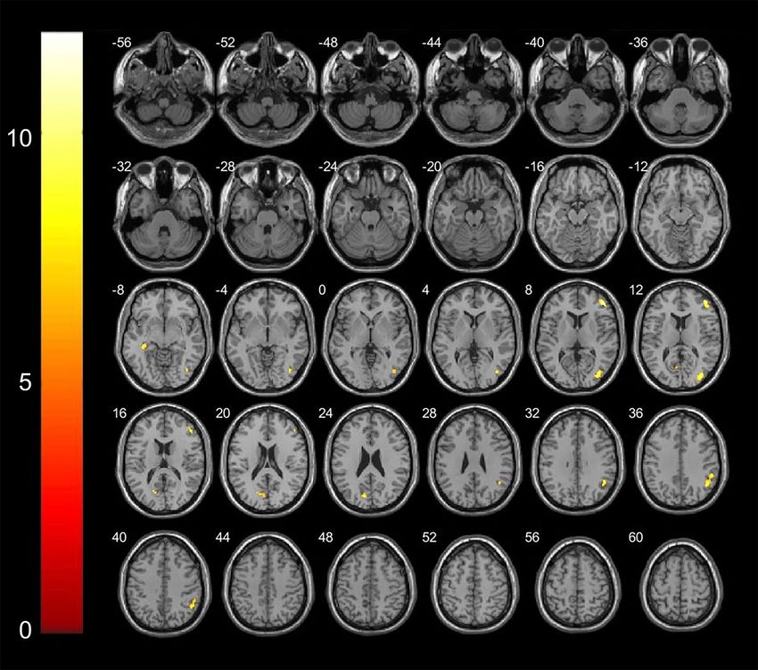
www.nature.com/scientificreports/
Patients Controls P value
Gender (female) 35 39 0.104
Age (years) 57.00 ± 8.86 52.71 ± 6.26 0.083
BFMDRS movement scores 7.87 ± 2.45 0.43 ± 0.49 0.000
HAMD scores 16.96 ± 4.84 5.51 ± 2.95 0.000
Disease duration (years) 4.57 ± 2.23 – –
Distribution (n) – –
Blepharospasm 18 – –
Oromandibular dystonia 5 – –
Both 23 – –
Total 46 64 –
Table 1. Clinical data for Meige syndrome patients and control subjects.
Depression group Non-depression group P value
Gender (female) 18 17 0.514
Age (years) 56.96 ± 9.91 57.67 ± 7.33 0.111
BFMDRS movement scores 9.56 ± 1.62 6.17 ± 1.38 0.726
Table 2. Clinical data for Meige syndrome patients with and without depression.
Meige syndrome is more commonly associated with mood disorders, with a higher incidence of d epression3. In
this study, VBM analysis was used to investigate the changes and clinical significance of brain structural abnor-
malities in patients with Meige syndrome and related depressive symptoms.
Results
Baseline characteristics. A total of 46 cases (11 males; 35 females) were included, and the age of the par-
ticipants ranged from 37 to 73 years (57.00 ± 8.86). The duration of Meige syndrome ranged from 3 to 8 years
(4.57 ± 2.23). Blepharospasm was present in 18 patients, and 5 had oromandibular dystonia; 23 patients had
both blepharospasm and oromandibular dystonia. The BFMDRS movement scores of participants ranged from
3.25 to 13.5 (7.87 ± 2.45). The HAMD scores of the participants ranged from 9 to 29 (16.96 ± 4.84). In the control
group, a total of 64 individuals (25 males; 39 females) were included, and the average age was 52.71 ± 6.26. The
BFMDRS and HAMD scores were significantly higher in the Meige syndrome group than in the healthy control
group (P < 0.05) (Table 1).
We divided the patients into a definite depression group (< 18) and a non-definitive depression group (> 18)
according to HAMD scores. Finally, there were 25 patients in the definitive depression group and 21 in the non-
definitive depression group. There were no significant differences between the two groups in terms of age, gender
or BFMDRS movement scores (P > 0.05) (Table 2).
Comparison of VBM‑MRI in patients with Meige syndrome and controls. Conventional MRI
showed normal results in all patients with Meige syndrome and controls. The VBM analysis in the current study
revealed multiple significant differences between the patients with Meige syndrome and controls. We found
decreased GM volume (p < 0.001 and cluster size > 50 voxels) in the left hemisphere in the middle frontal orbital
gyrus, temporal pole (superior temporal gyrus) and insula and in the right hemisphere in the temporal pole
(middle temporal gyrus), precuneus, inferior parietal, inferior temporal and olfactory cortices (Table 3;
Fig. 1).
Comparison of VBM‑MRI in patients with Meige syndrome with and without depression. We
found decreased GM volume (p < 0.001 and cluster size > 50 voxels) in the left hemisphere in the cuneus and hip-
pocampus and in the right hemisphere in the angular gyrus, middle frontal gyrus and middle occipital gyrus
(Table 4; Fig. 2).
Discussion
Meige syndrome was named after French neurologist Henry Meige. In 1910, Henry Meige described approxi-
mately 10 patients with blepharospasm, one of whom also had mandibular dystonia9. Currently, most researchers
and clinicians prefer the term “Meige syndrome” to describe blepharospasm-oromandibular dystonia syndrome10.
Meige syndrome usually occurs between 30 and 60 years of age, with rare cases presenting during teenage y ears4.
This disease is more common in females, and the ratio of males to females is approximately 1:311. In our study,
a total of 46 patients (11 males; 35 females) were included, and the age of the participants ranged from 37 to
73 years (57.00 ± 8.86), which was consistent with previous studies. Meige syndrome can be divided into three
Scientific Reports | (2020) 10:14533 | https://doi.org/10.1038/s41598-020-71479-9 2
Vol:.(1234567890)www.nature.com/scientificreports/
Peak MNI coordinate
Structure Number of voxels x y z P value T value
Frontal mid Orb_L 424 − 19.5 63 − 15 < 0.001 − 8.83
Temporal pole Sup_L 1,231 − 54 15 − 22.5 < 0.001 − 8.20
Insula_L 107 − 40.5 15 −6 < 0.001 − 7.98
Temporal pole Mid_R 260 42 10.5 − 31.5 < 0.001 − 7.32
Precuneus_R 243 12 − 55.5 67.5 < 0.001 − 5.99
Parietal Inf_R 86 37.5 − 55.5 45 < 0.001 − 5.84
Temporal_Inf_R 196 58.5 − 12 − 36 < 0.001 − 5.03
Olfactory_R 71 6 22.5 − 4.5 < 0.001 − 4.95
Table 3. Areas of decreased GM volume (p < 0.001) in Meige syndrome patients and control subjects.
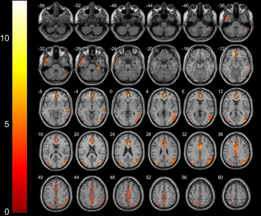
Figure 1. Areas showing decreased GM volume in axial slices (p < 0.001) in Meige syndrome patients and
control subjects. In the left hemisphere in the middle frontal orbital gyrus (t = − 8.83, p < 0.001), temporal pole
superior temporal gyrus (t = − 8.20, p < 0.001) and insula (t = − 7.98, p < 0.001); in the right hemisphere in the
temporal pole middle temporal gyrus (t = − 7.32, p < 0.001), precuneus (t = − 5.99, p < 0.001), inferior parietal
(t = − 5.84, p < 0.001), inferior temporal (t = − 5.03, p < 0.001)and olfactory cortices (t = − 4.95, p < 0.001).
Peak MNI coordinate
Structure Number of voxels x y z P value T value
Angular_R 203 46.5 − 54 34.5 < 0.001 − 4.84
Cuneus_L 159 − 10.5 − 76 22.5 < 0.001 − 4.82
Frontal mid_R 284 45 45 9 < 0.001 − 6.29
Hippocampu_L 52 − 34.5 − 30 − 7.5 < 0.001 − 4.23
Occipital mid_R 51 36 − 82.6 10.5 < 0.001 − 5.80
Table 4. Areas of decreased GM volume (p < 0.001) in Meige syndrome patients with and without depression.
Scientific Reports | (2020) 10:14533 | https://doi.org/10.1038/s41598-020-71479-9 3
Vol.:(0123456789)www.nature.com/scientificreports/
Figure 2. Areas showing decreased GM volume in axial slices in Meige syndrome patients with and without
depression. In the left hemisphere in the cuneus (t = − 4.82, p < 0.001) and hippocampus (t = − 4.23, p < 0.001); in
the right hemisphere in the angular gyrus(t = − 4.84, p < 0.001), middle frontal gyrus(t = − 6.29, p < 0.001) and
middle occipital gyrus(t = − 5.80, p < 0.001).
types according to clinical manifestations: blepharospasm, blepharospasm-oromandibular dystonia, and oro-
mandibular dystonia, among which blepharospasm with oromandibular dystonia is regarded as the complete
type of Meige syndrome10. In our study, blepharospasm was present in 18 patients, and 5 had oromandibular
dystonia; 23 patients had both blepharospasm and oromandibular dystonia. The BFMDRS movement scores of
participants ranged from 3.25 to 13.5 (7.87 ± 2.45).
Discussion
The aetiology of primary Meige syndrome is still unknown, but changes in plasticity and reduced cortical
inhibition due to environmental triggers and genetic susceptibility may be associated with the onset of Meige
syndrome5. Many movement disorders are not limited to a particular region but involve multiple brain regions12.
Therefore, morphological studies of the whole brain are helpful to understand the pathogenesis of Meige syn-
drome. VBM techniques quantitatively analyses the GM and WM volumes in each voxel in MRI images, thereby
allowing an evaluation of the anatomical structural differences across the whole b rain13. Recently, researchers
using VBM observed conflicting results of increased, decreased, or no change in GM volume in patients with pri-
mary blepharospasm and cervical dystonia8. A VBM study by Piccinin7 showed that patients with craniocervical
dystonia had reduced GM volume in the cerebellar vermis, bilateral superior frontal gyri, precuneus, cingulate,
insular cortex, and corpus callosum fissure. However, all of these studies examined patients with craniocervical
dystonia, and to the best of our knowledge, there is no research on Meige syndrome.
In this study, VBM analysis was used to investigate the changes and clinical significance of brain structural
abnormalities in patients with Meige syndrome. We found decreased GM volume (p < 0.001 and cluster size > 50
voxels) in the left hemisphere in the middle frontal orbital gyrus, temporal pole (superior temporal gyrus) and
insula and in the right hemisphere in the temporal pole (middle temporal gyrus), precuneus, inferior pari-
etal, inferior temporal and olfactory cortices. We failed to detect any abnormalities in the basal ganglia and
motor cortex. The results from previous VBM-MRI-related studies in those with dystonia have shown varying
increases and decreases in GM volume in the basal ganglia14,15, and no abnormalities have been found in the
basal ganglia in recent y ears16, which is consistent with the results of this study. However, a recent study showed
that the precentral gyrus, which plays an important role in motor planning and learning, is involved in the onset
of craniocervical d ystonia7. One of the reasons why our results differ from the above results is that the patients
Scientific Reports | (2020) 10:14533 | https://doi.org/10.1038/s41598-020-71479-9 4
Vol:.(1234567890)www.nature.com/scientificreports/
they selected were not typical Meige syndrome p atients7,14,16. The present study is the first VBM-MRI study in
patients with Meige syndrome, and the sample size of this study was larger than previous studies. Therefore,
although functional changes in brain regions such as the basal ganglia are necessary for the development and
maintenance of dystonia, they do not necessarily result in anatomical changes7.
In this study, we found a reduction in GM volume in the precuneus and inferior parietal cortex in patients
with Meige syndrome. The precuneus lies deep in the parietal lobe and interacts with the adjacent posterior
medial cortex (posterior cingulate and posterior compressed cortex). This internal connection is bilateral, provid-
ing communication between corresponding components in the two hemispheres and to some extent providing
the anatomical basis for bilateral functional coordination17. The precuneus is also selectively associated with
other parietal regions, such as the supraparietal lobule, and is involved in visual spatial information processing.
Recent studies have found that the precuneus is associated with many higher cognitive functions, such as episodic
memory, self-related information processing, and consciousness, and is no longer considered a simple extension
of the lateral parietal c ortex18. Therefore, abnormal findings in the precuneus also indicated that patients with
Meige syndrome may have abnormal integration at the cortical level. The precuneus has been shown to have
extensive connections with the auxiliary motor areas. At the subcortical level, the modular prefrontal lobe is
primarily associated with the nuclei of the basal ganglia and projects fibres into the b rainstem19. Therefore, we
hypothesized that the precuneus plays an important role in the pathogenesis of Meige syndrome, which partly
explains the lack of GM volume reductions in the basal ganglia and motor cortex.
In the frontal lobe, we found a reduction in GM volume in the left middle frontal orbital gyrus in the patients
with Meige syndrome. The frontal lobe plays a key role in many of the brain’s higher functions, including attention
regulation, learning and memory, behaviour planning, behavioural inhibition, thinking and r easoning20. At the
same time, the frontal lobe is also an important part of “emotional centre” or “emotion-related pathways”21. The
orbitofrontal cortex is involved in the encoding of external and internal information, reward-related behaviour,
impulse control, and emotional regulation22. The prefrontal cortex is also extensively connected to the superior
temporal gyrus and olfactory cortex22. In the temporal lobe, we also found that the patients with Meige syndrome
had decreased GM volume in the superior temporal pole, middle temporal pole and inferior temporal regions.
At present, it is believed that the temporal lobe plays an important role in visual and auditory integration and
regulation23 and is closely related to face recognition, distance judgement and text comprehension in reading.
Many Meige syndrome patients have varying degrees of depression and other emotional disorders. Therefore, we
believe that the decrease in GM volume in the frontal lobe and temporal lobe in this study was mainly related to
the non-motor symptoms of this disease. Finally, we found that the patients with Meige syndrome had decreased
GM volume in the insula. The posterior insular cortex is mainly responsible for the integration of sensorimotor
functions in the brain together with the primary motor cortex, sensorimotor cortex, auxiliary motor area and
middle cingulate gyrus, while the anterior insular cortex is mainly responsible for emotional regulation together
with the anterior cingulate gyrus, middle temporal gyrus and inferior temporal g yrus24,25. Therefore, we believe
that the insula may be involved in the development of motor and non-motor symptoms of Meige syndrome.
Meige syndrome with mood disorders is also common, with a higher incidence of depression than in the gen-
eral population3. In our study, the severity of depression was determined by the psychological 21-item Hamilton
Depression Rating scale (HAMD)26. There were 25 patients in the definite depression group. When we compared
VBM-MRI measures in Meige syndrome patients with and without depression, we found decreased GM volume
(p < 0.001 and cluster size > 50 voxels) in the left hemisphere in the cuneus and hippocampus and in the right
hemisphere in the angular gyrus, middle frontal gyrus and middle occipital gyrus (Table 4; Fig. 2). The results
of this study suggest that, in addition to the frontal lobes mentioned above, the hippocampus plays an important
role in the pathogenesis of depression in patients with Meige syndrome. A previous study has shown not only
that depression is manifested by bilateral hippocampal volume reduction but also that the degree of reduction is
related to the severity of symptoms, the course of disease and the number of recurrences27. The patients in that
study had a long duration of illness (4.57 ± 2.23 years), and the hippocampal changes accompanying depres-
sion also showed a process of gradual accumulation, which finally manifested as volume reduction, which was
consistent with the results of the present study. The occipital lobe is involved in attention, visual memory, visual
movement, language movement and other neuropsychological a ctivities28. In the process of visual information
processing of external positive stimuli in the basic state of chronic or recurrent depression, the function of the
relevant cortex was weaker than that in healthy controls. The cuneus is also part of the occipital lobe, and we
believe that it is involved in the development of depression in patients with Meige syndrome.
Conclusion
At present, there are few imaging studies on the pathogenesis of Meige syndrome, and most of them include
a heterogeneous collection of patients with dystonias. In this present study, VBM analysis was used to inves-
tigate the changes and clinical significance of brain structural abnormalities in patients with Meige syndrome
and related depressive symptoms. First, unlike previous imaging studies with patients with dystonia, we found
no abnormalities in the basal ganglia and motor cortex. However, the important role of the precuneus in the
pathogenesis of Meige syndrome may have been revealed in our study. Second, the decrease in GM volume in
the frontal lobe and temporal lobe in this study was mainly related to the non-motor symptoms of this disease.
Finally, we compared VBM-MRI in Meige syndrome patients with and without depression and found that the
hippocampus plays an important role in the pathogenesis of depression in patients with Meige syndrome.
The limitation of this study is that it is only a cross-sectional study with a relatively small sample size. The
sample size can be further expanded in the future. In addition, positron emission tomography (PET) is an in vivo
method that reflects the level of brain metabolism. Combined with VBM-MRI, a better understanding of the
changes in brain function and morphology in Meige syndrome can be obtained. Another limitation of our study
Scientific Reports | (2020) 10:14533 | https://doi.org/10.1038/s41598-020-71479-9 5
Vol.:(0123456789)www.nature.com/scientificreports/
is that the cohort is composed of a mix of significantly different phenotypes of Meige syndrome (oromandibular,
blepharospasm and combined oromandibular and blepharospasm). In the future, our team plans to investigate
into the similarities and differences in gray matter volume across these disorders.
Methods
Patients. Clinical data of 46 right-handed patients with primary Meige syndrome were collected between
January 2017 and January 2019 at the Department of Neurosurgery, Peking University People’s Hospital. Pri-
mary Meige syndrome diagnosed by an experienced neurologist, Ruen Liu. The diagnostic criteria are mainly
based on blepharospasm, oral and mandibular dystonia, increased blink rates and other symptoms. Labora-
tory and imaging examinations showed no abnormalities. Patients with histories of drug or alcohol use disor-
ders, psychiatric disorders and other secondary factors were excluded. Based on the clinical manifestations, the
patients were divided into three types: (1) blepharospasm; (2) blepharospasm with oromandibular dystonia; and
(3) oromandibular dystonia. In this study, 64 right-handed healthy people with age and education levels match-
ing the Meige syndrome group were included as the healthy control group.
Written informed consent was obtained from each participant, and the study was approved by the institutional
review board of Peking University People’s Hospital. All methods were carried out in accordance with relevant
guidelines and regulations.
Magnetic resonance imaging. Imaging was conducted on a Discovery 750 3.0 T (GE Healthcare, Wauke-
sha, WI) MRI scanner. T1-weighted anatomical images in the sagittal plane were collected with a 3D fast spoiled
gradient echo sequence: repetition time (TR) = 4.9 ms, echo time (TE) = 2 ms, flip angle = 15°, field of view
(FOV) = 240 mm, in-plane resolution = 1 × 1 mm2, slice thickness = 1 mm, and 170 slices. All scans were per-
formed by the same imaging physician.
Image data analysis. Structural images were processed with SPM8 (https://www.fil.ion.ucl.ac.uk/spm)
and VBM8 toolbox (https://dbm.neuro.unijena.de/vbm) within the MATLAB R2016a programming environ-
ment (The MathWorks, Natick, MA).
Preprocessing of the data involved spatial normalization, GM segmentation and spatial smoothing with a
Gaussian kernel. Image preprocessing mainly included the following steps: (1) with the aid of the MATLAB plat-
form, SPM8 converted the collected images to a format that can be recognized and uses MRIcro software (https
://www.mccauslandcenter.sc.edu/mricro/mricro/mricro.html) to adjust the spatial coordinates of each image
such that the data from all subjects had the spatial coordinates of the image adjusted and all participants’ images
were located in a closed spatial coordinate system. Since the brain structure of each subject greatly varied, we
needed to conduct spatial standardization on all the images from the subjects and convert them into standard-
ized images with the same size and direction; (2) according to the distribution template of GM, white matter
(WM) and cerebrospinal fluid (CSF) in the brain, each element in the image was judged to be GM, WM or CSF
to segment the GM, WM or CSF and generate different brain tissue i mages29; (3) the last step is Gaussian smooth-
ing of the standardized image. The image obtained in the previous step was smoothed by a Gaussian kernel of
8 mm × 8 mm × 8 mm at half height and width. This step can eliminate high-frequency noise and improve the
signal-to-noise ratio of the image. In addition, the image can be made to have the characteristics of a random
Gaussian field and meet the requirements of the statistical analyses of SPM.
Data collection. Baseline data and medical histories were obtained from patient medical records. Base-
line data included age, sex, and dystonia and depressive status. Dystonia was evaluated using the Burke–Fahn–
Marsden Dystonia Rating Scale (BFMDRS)30. The severity of depression was determined by the psychological
21-item Hamilton Depression Rating scale (HAMD)26. A score of less than 7 was considered normal; 7–17
indicated possible depression; 18 to 24 indicated definite depression; and greater than 24 was considered severe
depression. Finally, we divided the patients into a definite depression group (< 18) and a non-definite depression
group (> 18).
Statistical analysis. SPSS 19.0 statistical software (IBM Corp., Armonk, NY, USA) was used for data
analysis31. Numerical variables are expressed as the mean ± standard deviation. Qualitative variables are
described by absolute values of cases in different groups. Statistical significance between the quantitative vari-
ables was assessed by X2 tests, corrected by Yates or Fisher if necessary. Student’s t-tests were performed to
evaluate data that followed a normal distribution. P < 0.05 was considered statistically significant differences.
Two-sample t-tests corrected for subject age, gender and total intracranial volume were used for VBM-MRI
comparisons between groups7. Significant differences between groups were indicated when P < 0.001 (false dis-
covery rate corrected). Only sets with more than 50 voxels were considered statistically significant brain regions.
The t-test results are superimposed on the template of the T1 structural image, and the results are presented by
the xjView software (version 9.6, https://www.alivelearn.net/xjView).
Ethics approval. The present study was approved by the Medical Ethics Committee of Peking University
People’s Hospital. All methods were carried out in accordance with relevant guidelines and regulations.
Consent to participate. All patients provided written informed consent to participate.
Scientific Reports | (2020) 10:14533 | https://doi.org/10.1038/s41598-020-71479-9 6
Vol:.(1234567890)www.nature.com/scientificreports/
Consent for publication. The study participants provided their consent for the publication of any data/
associated images.
Data availability
The datasets used and/or analysed during the current study are available from the corresponding author upon
reasonable request.
Received: 27 April 2020; Accepted: 13 August 2020
References
1. Tolosa, E. S. Clinical features of Meige’s disease (idiopathic orofacial dystonia): a report of 17 cases. Arch. Neurol. 38, 147–151.
https://doi.org/10.1001/archneur.1981.00510030041005 (1981).
2. Miao, J. et al. Meige’s syndrome and hemichorea associated with hyperthyroidism. J. Neurol. Sci. 288, 175–177. https://doi.
org/10.1016/j.jns.2009.10.018 (2010).
3. Jahngir, M. U. & Patel, B. C. StatPearls (Springer, New York, 2020).
4. Sabesan, T. Meige syndrome: a rare form of cranial dystonia that was treated successfully with botulinum toxin. Br. J. Oral. Maxil-
lofac. Surg. 46, 588–590. https://doi.org/10.1016/j.bjoms.2008.02.002 (2008).
5. Hallett, M. Blepharospasm: recent advances. Neurology 59, 1306–1312. https: //doi.org/10.1212/01.wnl.000002 7361. 73814. 0e (2002).
6. Barrett, M. J. et al. Lower volume, more impairment: reduced cholinergic basal forebrain grey matter density is associated with
impaired cognition in Parkinson disease. J. Neurol. Neurosurg. Psychiatry 90, 1251–1256. https: //doi.org/10.1136/jnnp-2019-32045
0 (2019).
7. Piccinin, C. C. et al. Diffuse decreased gray matter in patients with idiopathic craniocervical dystonia: a voxel-based morphometry
study. Front. Neurol. 5, 283. https://doi.org/10.3389/fneur.2014.00283 (2014).
8. Valls-Sole, J. & Defazio, G. Blepharospasm: update on epidemiology, clinical aspects, and pathophysiology. Front. Neurol. 7, 45.
https://doi.org/10.3389/fneur.2016.00045 (2016).
9. Pandey, S. & Sharma, S. Meige’s syndrome: history, epidemiology, clinical features, pathogenesis and treatment. J. Neurol. Sci. 372,
162–170. https://doi.org/10.1016/j.jns.2016.11.053 (2017).
10. LeDoux, M. S. Meige syndrome: what’s in a name?. Parkinsonism Relat. Disord. 15, 483–489. https://doi.org/10.1016/j.parkreldis
.2009.04.006 (2009).
11. Soland, V. L., Bhatia, K. P. & Marsden, C. D. Sex prevalence of focal dystonias. J. Neurol. Neurosurg. Psychiatry 60, 204–205. https
://doi.org/10.1136/jnnp.60.2.204 (1996).
12. Braak, H. et al. Staging of brain pathology related to sporadic Parkinson’s disease. Neurobiol. Aging 24, 197–211. https://doi.
org/10.1016/s0197-4580(02)00065-9 (2003).
13. Good, C. D. et al. A voxel-based morphometric study of ageing in 465 normal adult human brains. Neuroimage 14, 21–36. https
://doi.org/10.1006/nimg.2001.0786 (2001).
14. Pantano, P. et al. A transverse and longitudinal MR imaging voxel-based morphometry study in patients with primary cervical
dystonia. AJNR Am. J. Neuroradiol. 32, 81–84. https://doi.org/10.3174/ajnr.A2242 (2011).
15. Draganski, B., Thun-Hohenstein, C., Bogdahn, U., Winkler, J. & May, A. “Motor circuit” gray matter changes in idiopathic cervical
dystonia. Neurology 61, 1228–1231. https://doi.org/10.1212/01.wnl.0000094240.93745.83 (2003).
16. Martino, D. et al. Cortical gray matter changes in primary blepharospasm: a voxel-based morphometry study. Mov. Disord. 26,
1907–1912. https://doi.org/10.1002/mds.23724 (2011).
17. Seitz, R. J. & Binkofski, F. Modular organization of parietal lobe functions as revealed by functional activation studies. Adv. Neurol.
93, 281–292 (2003).
18. Vogeley, K. et al. Neural correlates of first-person perspective as one constituent of human self-consciousness. J. Cogn. Neurosci.
16, 817–827. https://doi.org/10.1162/089892904970799 (2004).
19. Leichnetz, G. R. Connections of the medial posterior parietal cortex (area 7m) in the monkey. Anat. Rec. 263, 215–236. https://
doi.org/10.1002/ar.1082 (2001).
20. Miller, E. K. & Cohen, J. D. An integrative theory of prefrontal cortex function. Annu. Rev. Neurosci. 24, 167–202. https://doi.
org/10.1146/annurev.neuro.24.1.167 (2001).
21. Cacioppo, J. T. & Gardner, W. L. Emotion. Annu. Rev. Psychol. 50, 191–214. https: //doi.org/10.1146/annure v.psych. 50.1.191 (1999).
22. Price, J. L. & Drevets, W. C. Neural circuits underlying the pathophysiology of mood disorders. Trends Cogn. Sci. 16, 61–71. https
://doi.org/10.1016/j.tics.2011.12.011 (2012).
23. Ono, Y. et al. Frontotemporal oxyhemoglobin dynamics predict performance accuracy of dance simulation gameplay: temporal
characteristics of top-down and bottom-up cortical activities. Neuroimage 85(Pt 1), 461–470. https: //doi.org/10.1016/j.neuroi mage
.2013.05.071 (2014).
24. Le Berre, A. P. et al. Deviant functional activation and connectivity of the right insula are associated with lack of awareness of
episodic memory impairment in nonamnesic alcoholism. Cortex 95, 15–28. https://doi.org/10.1016/j.cortex.2017.07.016 (2017).
25. Zhang, Y. et al. Abnormal functional connectivity of ventral anterior insula in asthmatic patients with depression. Neural Plast
2017, 7838035. https://doi.org/10.1155/2017/7838035 (2017).
26. Williams, J. B. A structured interview guide for the Hamilton depression rating scale. Arch. Gen. Psychiatry 45, 742–747. https://
doi.org/10.1001/archpsyc.1988.01800320058007 (1988).
27. Cole, J., Costafreda, S. G., McGuffin, P. & Fu, C. H. Hippocampal atrophy in first episode depression: a meta-analysis of magnetic
resonance imaging studies. J. Affect Disord. 134, 483–487. https://doi.org/10.1016/j.jad.2011.05.057 (2011).
28. Jiang, T., He, Y., Zang, Y. & Weng, X. Modulation of functional connectivity during the resting state and the motor task. Hum.
Brain Mapp. 22, 63–71. https://doi.org/10.1002/hbm.20012 (2004).
29. Ashburner, J. & Friston, K. Multimodal image coregistration and partitioning: a unified framework. Neuroimage 6, 209–217. https
://doi.org/10.1006/nimg.1997.0290 (1997).
30. Burke, R. E. et al. Validity and reliability of a rating scale for the primary torsion dystonias. Neurology 35, 73–77. https://doi.
org/10.1212/wnl.35.1.73 (1985).
31. Liu, J. et al. Clinical analysis of patients with ipsilateral coexistence of hemifacial spasm and trigeminal neuralgia. World Neurosurg.
https://doi.org/10.1016/j.wneu.2020.03.040 (2020).
Author contributions
J.L.: conceptualization, data curation, methodology, investigation, formal analysis, software, writing—original
draft, reviewing and editing; L.L.: formal analysis, writing—reviewing and editing, Supervision; R.L.: conceptu-
alization, methodology, supervision, writing—reviewing and editing, resources. L.C., Y.J., J.F., D.W., Z.L., J.O.:
writing—reviewing and editing, validation. All authors reviewed the manuscript.
Scientific Reports | (2020) 10:14533 | https://doi.org/10.1038/s41598-020-71479-9 7
Vol.:(0123456789)www.nature.com/scientificreports/
Funding
No funding was received.
Competing interests
The authors declare no competing interests.
Additional information
Correspondence and requests for materials should be addressed to R.L.
Reprints and permissions information is available at www.nature.com/reprints.
Publisher’s note Springer Nature remains neutral with regard to jurisdictional claims in published maps and
institutional affiliations.
Open Access This article is licensed under a Creative Commons Attribution 4.0 International
License, which permits use, sharing, adaptation, distribution and reproduction in any medium or
format, as long as you give appropriate credit to the original author(s) and the source, provide a link to the
Creative Commons license, and indicate if changes were made. The images or other third party material in this
article are included in the article’s Creative Commons license, unless indicated otherwise in a credit line to the
material. If material is not included in the article’s Creative Commons license and your intended use is not
permitted by statutory regulation or exceeds the permitted use, you will need to obtain permission directly from
the copyright holder. To view a copy of this license, visit http://creativecommons.org/licenses/by/4.0/.
© The Author(s) 2020
Scientific Reports | (2020) 10:14533 | https://doi.org/10.1038/s41598-020-71479-9 8
Vol:.(1234567890)You can also read