BIOCHEMICAL INVESTIGATION DURING DIFFERENT STAGES OF IN VITRO PROPAGATION OF
←
→
Page content transcription
If your browser does not render page correctly, please read the page content below
Pak. J. Bot., 42(4): 2827-2837, 2010.

              BIOCHEMICAL INVESTIGATION DURING DIFFERENT
                   STAGES OF IN VITRO PROPAGATION OF
                           STEVIA REBAUDIANA
         AAMIR ALI1*, IRUM GULL2, SHAGUFTA NAZ3 AND SHAHID AFGHAN4
          1
           Department of Biological Sciences, University of Sargodha, Sargodha, Pakistan
    2
        Institute of Biochemistry & Biotechnology, University of the Punjab, Lahore, Pakistan
                        3
                          Department of Biotechnology, LCWU, Lahore, Pakistan
                                         4
                                           SSRI, Jangh, Pakistan
                                             Abstract

     In vitro propagation of Stevia rebaudiana Bert, an important non-caloric sweetening herb was
carried out to explore its potential for micropropagation (both from apical and nodal meristem) and
callogenesis (using leaf, node and internode as explant). MS basal medium with 1.0 mg l-1 of BAP
was found to be the best medium for shoot formation, with 90% shoot formation response within
12 days of meristem inoculation, both from shoot apical and nodal meristem. Maximum shoot
multiplication response (90%) was also obtained in MS medium having 1.0 mg l-1 of BAP, with
average of 8.6 shoots per culture vial having an average shoot length of 6.0 cm. The best In vitro
rooting response (96%) was recorded on MS medium containing 1.0 mg l-1 NAA within 7.3 days of
inoculation. When well developed In vitro plants were shifted for hardening on a mixture of sand +
soil + peat (1:1:1) 90% success was recorded. For callogenesis leaf explant proved to be the best
followed by nodal and internodal explant. The highest response of callus induction from leaf
explant was obtained on MS medium supplemented with 3.0 mg l-1 2,4-D while nodal and
internodal explants showed best results for callogenesis in MS medium supplemented with 3.0 mg
l-1 NAA and 1.0 mg l-1 BAP. The highest total soluble protein contents and the peroxidases activity
were estimated in the six week old callus cultures derived from leaf explant.

Abbreviations: MS= Murashige and Skoog’s medium; BAP= 6-Benzylamino-purine; 2,4-D= 2,4
dichlorophenoxyacetic acid; IAA= Indole acetic acid NAA= α- Naphthaleneacetic acid

Introduction

     Stevia rebaudiana Bert., the most valuable tropical medicinal plant, belongs to the
family Asteraceae. It is one of the 154 members of genus Stevia which produce sweet
steviol glycosides (Soejarta et al., 1982). It is a natural sweet herb native of northeastern
Paraguay (Savita et al., 2004). Stevia is diploid plant, having 11 pairs of chromosome
(Frederico et al., 1996) with critical day length of 13 hours (Zaidan et al., 1980).
     An interesting feature of this plant is intense sweetness of leaves and aqueous
extracts. Steviosides, sweet crystalline diterpene glycosides, which gives sweet taste to
the plant are noncaloric and 200-300 times sweeter than sugar (Midmore & Rank, 2002).
Typical proportions, on a dry weight basis, for the four major glycosides found in the
leaves of wild Stevia plants is 9.1% stevioside, 3.8% rebaudioside A, 0.6% rebaudioside
C and 0.3% dulcoside (Bhosle, 2004). Steviol glycosides are derived from mevalonic
acid pathway. Stevioside are 110 to 270 times sweeter than sucrose, rebaudioside A are
150 to 320 times, rebaudioside C is 40 to 60 times and Dulcoside A is 30 times sweeter
than sucrose (Tanaka, 1997). Diet conscious and diabetic persons with hyperglycemia
can use steviosides as an alternative sweetener (Din et al., 2006). Stevioside can also be
used as an antihyper glycaemic (Gregersen et al., 2004), antihypertensive (Ferri et al.,
2006), anti-tumor (Yasukawa et al., 2002) drug.
*
    E-mail: aamirali73@hotmail.com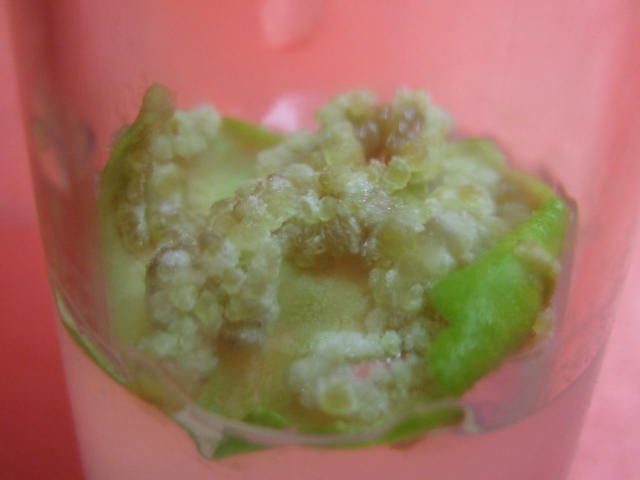
2828                                                                     AAMIR ALI ET AL.,

     Stevia can regenerate by crown division, trampled of stem into the ground and from
seeds. As the seeds of Stevia are very small in size and infertile, large scale mechanized
production of Stevia through seeds is not fruitful (Savita et al., 2004). Plant tissue culture
or micropropagation can be used for rapid propagation and conservation of such valuable
and endangered plant species (Nalawade et al., 2002), which are difficult to propagate by
conventional methods. This technique allows rapid multiplication, lack of seasonal
restriction, provides sufficient number of plants in very short span of time, self
incompatible inbred lines can be maintained. Micropropagation ensures the production of
disease free, high yielding and premium quality planting material for automation
(Chawla, 2000).
     When plants are grown In vitro they come under stress due to accumulation of
ammonia in culture vial, which sometimes leads towards somaclonal variation. To
enhance tolerance under stress conditions, the levels of low molecular weight antioxidant
and activity of antioxidant enzymes, such as guaiacol peroxidase, superoxide dismutase,
catalase, ascorbate peroxidase and glutathione reductase, is generally increased in plants
(Foyer et al., 1997). Therefore it is very important to note the level of these enzyme at
different level of In vitro propagation particularly during different stages of callogenesis
and somatic embryogenesis where culture are maintained for longer time under In vitro
conditions.
     The present investigation describes procedures for microprpagation and callogenesis
of Stevia rebaudiana. It also describes the changes in peroxidases contents during
different stags of callus growth and regeneration.

Materials and Methods

     In vitro grown Stevia rebaudiana plants were taken from the Seed Center, University
of the Punjab, Lahore, Pakistan. For sterilization, explant was first washed with running tap
water, and then treated with house hold detergent for five minutes. This was followed by
second washing with tap water to remove all the traces of detergent. The explant was then
treated with 10% Sodium hypochlorite solution for 15 minutes. After discarding Sodium
hypochlorite, the explants were washed three times with sterilized distilled water to remove
all the traces of Sodium hypochlorite. The sterilized explants were then inoculated by
proper dissecting and sizing of the about 4-5 mm were excised from stevia plants.
     Explants of shoot apical and nodal meristem were inoculated in liquid as well as
solid MS media (Murashige & Skoog, 1962) supplemented with different concentrations
of auxins and cytokinins either alone or in combination with each other. pH of the
medium was adjusted to 5.7 with 0.1 N solution of NaOH or HCl and 0.7% agar was used
for solidification of medium. The medium was autoclaved at 121ºC and 15 Ibs/Inch2
pressure for 15 minutes. Cultures were maintained under fluorescent light having 2500
lux light intensity. The incubation temperature was 26oC ± 1oC with 16 hour light and 8
hour dark period in every 24 hour cycle. The data was recorded for days for shoot
formation, number of shoot per culture vial, days for shoot multiplication, days for root
induction and for number of roots per plant.
     For hardening well developed In vitro plants were transferred into pots containing
different media (autoclaved sand, sand+soil and sand+soil+peat in the ratio of 1:1, 1:1:1
respectively). Potted plants were brought out from green house into open sun light after three
week of hardening and eventually these plants were shifted into the field for further growth.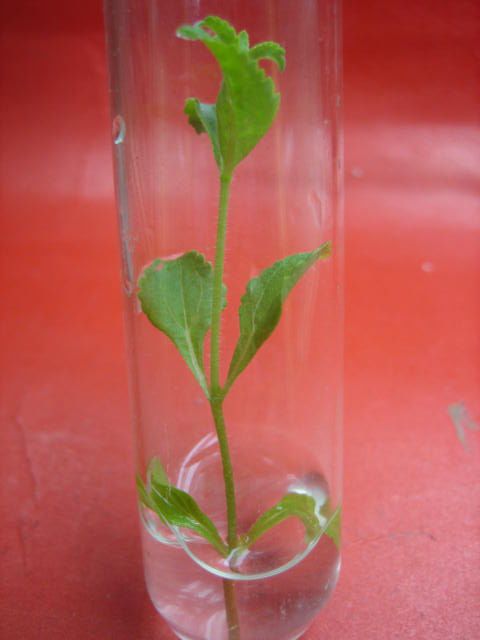
IN VITRO PROPAGATION OF STEVIA REBAUDIANA                                                       2829
     To determine the total soluble protein contents and peroxidase activity, 2.0 grams of
plant material was crushed in the ice chilled pestle and mortor containing 0.2 g of PVP
(polyvinylpyrrolidine) with 8 ml of 0.1M phosphate buffer at 4°C. For the extraction of
total soluble protein contents, slurry was centrifuged at 10,000 rpm for 10 mins at 4°C
while for the extraction of peroxidases, the slurry was centrifuged at 14,000 rpm for 10
min at 4°C. The supernatant was used for further analysis. Biuret method of Racusen &
Johnstone (1961) was adopted for the estimation of total soluble protein contents. For the
estimation of peroxidases, method proposed by Racusen & Foote (1965) was used.
     The experimental design was completely randomized with ten replicate cultures for
each hormonal treatment and each experiment was repeated thrice. Analysis of variance
(ANOVA) depicting significance among means was calculated by Duncan,s new multiple
range test (Steel & Torrie, 1980).

Results and Discussion
Micropropagation: For shoot formation in Stevia rebaudiana, MS medium containing
BAP (1.0 mg l-1) provided best result (90%) both from shoot apical meristem within 11.6
days of explant inoculation (Table 1). Slavova et al., (2003) also used only BAP for shoot
formation in Stevia rebaudiana. In case of kinetin mediated MS medium, 0.25 mg l-1 of
Kinetin provided good results from apical meristem. Tadhani et al., (2006) reported that
4.0 mg l-1 of kinetin showed maximum shoot formation response. Nuutila et al., (2002)
and Ali & Afghan (2003) reported that cultivation of same species may differ drastically
in their requirement for essential medium components. In the case of nodal meristem, rate
of shoot formation was high but it took more time than shoot apical meristem.
     When BAP was used in combination with kinetin, the maximum shoot formation
response was found to be 73% at 2.0 mg l-1 BAP with 0.25 mg l-1 Kinetin. Sivaram &
Mukundan, (2003) observed maximum shoot induction from shoot apex, nodal and leaf
explants on Murashige & Skoog (MS) medium supplemented with 6-benzyladenine (BA;
8.87 µM ) and indole-3-acetic acid (5.71µM ). Smitha et al., (2005) reported that when
0.05 mg l-1 kinetin was added in MS mediun which already had 1.0 mg l-1 of BAP, the
production of dark green healthy shoot enhanced.

  Table 1. Shoot induction from shoot apical and nodal meristem of Stevia rebaudiana cultured on MS
            medium supplemented with different growth hormones alone and in combination.
                         Composition      No. of shoot   Average shoot length        Rate of shoot
 Growth regulators
                            (mg l-1)      per culture      after 20 days (cm)     multiplication (%)
 MS medium                    -          1.6 ± 0.274d         2.0 ± 0.471d               50
                                                   bcd                     cd
                            0.50        3.6 ± 0.982          3.0 ± 0.471                 76
                                                       a                   a
                            1.00         8.6 ± 0.721          6.0 ± 0.471                90
 MS medium + BAP                                      bc                   bc
                            1.50         5.6 ± 0.721         4.0 ± 0.471                 86
                                                       cd               bcd
                            2.00         3.3 ± 0.720         3.3 ± 0.720                 80
                            0.50         3.0 ± 0.471d        2.3 ± 0.272cd               70
                            1.00        4.0 ± 0.471bcd       5.0 ± 0.471ab               73
 MS medium + Kinetin
                            1.50         6.0 ± 0.471b        6.0 ± 0.471a                76
                            2.00         2.6 ± 0.274d        4.0 ± 0.471bc               70
 Means followed by different letters in the same column differ significantly at p=0.05 according
 to Duncan’s new Multiple Range Test.
 The results were calculated from three replicated experiments for each treatment, each with 10
 explants per treatment.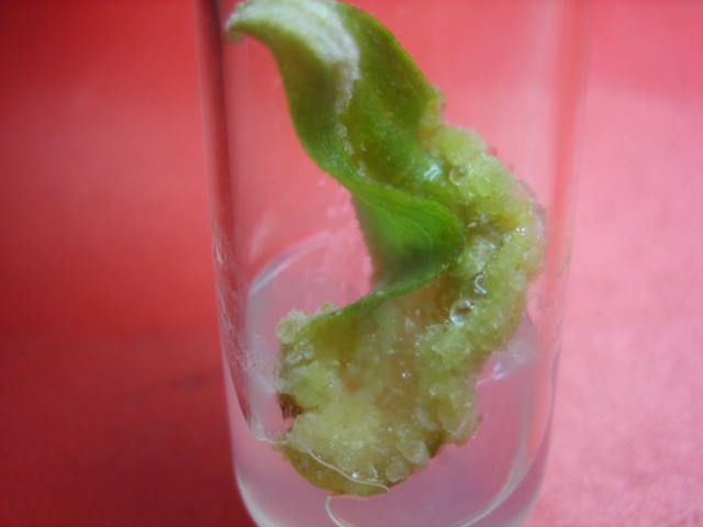
2830                                                                     AAMIR ALI ET AL.,

Shoot multiplication: The maximum number of shoots i.e., 8.6 shoots per culture vial
with average shoot length of 6.0 cm was obtained after 20 days of inoculation, on MS
basal medium having 1.0 mg l-1 of BAP ( Fig. 1d, Table 1). In case of Kinetin mediated
MS medium, 7.6 shoots per culture vials were obtained at 1.5 mg l-1 of kinetin. Tadhani
et al., (2006) reported maximum number of shoots on MS medium supplemented with
0.6 mg l-1 of BA. Smitha et al., (2005) induced multiple shoots from shoot buds of Stevia
rebaudiana with multiplication rate up to 1: 25 on a modified MS medium supplemented
with 1.0 mg l-1 of benzyladenin. In this study it was also found that explant showed better
shoot formation response in liquid medium as compared to the solid medium (Fig. 1a,1b;
data not given). In liquid medium, the close contact of the tissue with the medium may
facilitate the uptake of nutrients and phytohormones, leading to better shoot growth
(Sandal et al., 2001).

Rooting: Root induction was observed in hormone free MS basal medium also, but best
rooting response (96% with 7 roots per plant within 5 days of inoculation on rooting
medium) was obtained on full strength MS medium supplemented with 1.0 mg l-1 of
NAA (Fig. 1e; Table 3). Slavova et al., (2003) obtained 84% to 99% rooting on MS
medium supplemented with NAA. Tadhani et al., (2003) reported initiation of rooting
with in 6-7 days and obtained maximum number of roots on medium supplemented with
1.0 mg l-1 of IBA. Ferreira & Handro, (1988) reported that addition of auxin to the
rooting medium (especially 0.1 mg l-1 IBA) favored root formation in Stevia rebaudiana.
Smitha et al., (2005) recorded ≥ 90% rooting in modified MS medium supplemented with
1.5 mg l-1 indole-3-butyric acid.

Hardening and acclimatization: Well developed In vitro plants were shifted in different
media for hardening acclimatization of micropropagated plants of Stevia rebaudiana in
the glass house was achieved with 90 % survival rate in medium having autoclaved sand
+ soil + peat in 1:1:1 ratio ( Fig. 1f; Table 4).

Callogenesis: During the In vitro initiation of callus, the cell differentiation and
specialization that occurs in parent plant is reserved and cells of the explant become
dedifferentiated (Evan et al., 2003).
     In the present study best response for callogenesis was obtained from leaf followed
by nodal explants, while internodal explants showed poor response. Din et al., (2006)
found internodal segments as a best explant for callus induction, followed by leaf explant
and poorest response by nodal explant.
     Callus formation was observed, when auxin 2, 4-D or NAA were used alone or
supplemented with small amount of cytokinin (BAP). There was a wide range of variation
in days for callus initiation response and percentage of callus formation. The genes
affecting structure and type of plant development, also influences callus formation. This
depicts the involvement of inheritance in callus growth (Turhan, 2004). Among all
treatments, the highest rate of callus from leaf explants (96%) within 11 days of inoculation
was observed on MS basal medium containing 3.0 mg l-1 of 2,4-D (Table 5). When the 2,4-
D was used in combination with BAP, increase in rate of callus induction was noticed in
nodal and internodal explants as compared to 2,4-D alone (Table 6). The nodal and
internodal explants showed maximum callus induction response i.e., 90 % (within 9 days of
inoculation) and 73 % (within 7.3 days of inoculation) respectively on the MS medium
supplemented with 3.0 mg l-1 NAA + 1.0 mg l-1 BAP (Table 7). Din et al., (2006) observed
the highest amount of callus on MS medium containing 3.0 mg l-1 2,4-D (Fig. 2c).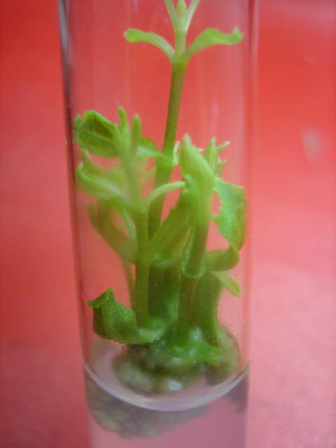
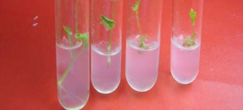
IN VITRO PROPAGATION OF STEVIA REBAUDIANA                                                   2831

 a                                                b

 c                                                d

 e                                                f
Fig. 1. Micropropagation of Stevia. (a),(b) Shoot formation from apical shoot meristemin solid and
liquid MS medium supplemented with 1.0 mg l-1 of BAP; (c) Different stages of shoot formation
from apical shoot meristem (after 7, 14, 20, and 25 days) inoculated in MS medium with 1.0 mg l-1
of BAP; (d) Shoot multiplication in liquid MS basal medium supplem- ented with 1.0 mg l-1 of
BAP; (e) In vitro rooting in MS basal medium supplemented with 1.5 mg l-1 IAA; (f) Hardening of
In vitro grown Stevia plant in sand.
2832 AAMIR ALI ET AL.,
a b
c
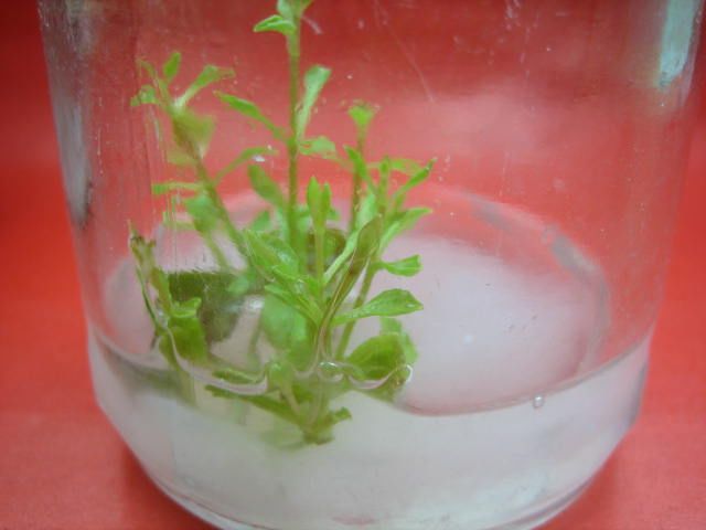
Fig. 2. Callogenesis in Stevia. (a), (b) Callus induction in leaf explant in light and dark respectively
inoculation in MS medium supplemented with 3.0 mg l-1 of 2, 4-D; (c ) Callus induction in nodal
explant inoculated in MS medium supplemented with 3.0 mg l-1 of NAA with 1.0 mg l-1 of BAP.
Table 2. In vitro shoot multiplication of Stevia rebaudiana cultured on MS medium supplemented
with different concentrations of BAP and Kinetin.
Shoot apical meristem Nodal meristem
Composition
Growth regulators Days for shoot Rate of shoot Days for shoot Rate of shoot
(mg l-1)
formation formation (%) formation formation (%)
MS medium - 21 ± 0.942a 50 22.2 ± 0.544a 53
bc b
0.25 15 ± 0.471 56 18.3 ± 0.981 60
0.50 14.3 ± 0.72bc 63 15.3 ± 0.720bcde 66
BAP
1.00 11.6 ± 0.721c 90 12.6 ± 0.982e 90
1.50 15.6 ± 0.721b 60 17.6 ± 0.721bc 63
0.25 13.3 ± 0.981bc 86 15.6 ± 0.721bcde 90
bc
0.50 15.0 ± 1.247 70 17.3 ± 0.720bcd 73
Kinetin
1.50 13.0 ± 0.942bc 66 14.3 ± 0.720de 66
bc
2.00 18.6 ± 0.721 50 15.0 ± 0.471cde 53
0.5+0.25 12.6 ± 0.721bc 46 13.6 ± 0.721e 56
bc
1.0+0.25 13.0 ± 0.942 50 14.0 ± 0.471e 60
BAP + Kinetin
1.5+0.25 13.6 ± 0.982bc 56 14.6 ± 1.186cde 66
2.0+0.25 12.3 ± 0.720bc 73 13.0 ± 0.471e 76
Means followed by different letters in the same column differ significantly at p=0.05 according to Duncan’s new
Multiple Range Test.
The results were calculated from three replicated experiments for each treatment, each with 10 explants per treatment.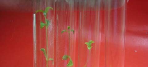
IN VITRO PROPAGATION OF STEVIA REBAUDIANA                                                                       2833
          Table 3. Effect of different concentration of NAA and IAA on rooting of
                       In vitro developed shoots of Stevia rebaudiana.
                        Composition          Days for         No. of roots      Rate of
 Growth regulators                -1
                           (mg l )        root induction        per plant     rooting (%)
                                                        bc                 d
 Basal MS medium               -           14.3 ± 0.981       2.0 ± 0.471          73
                                                        cd                 cd
                              0.5          11.3 ± 0.981       2.6 ± 0.274          80
                              1.0           7.3 ± 0.720e      5.0 ± 0.471ab        96
 MS medium + NAA                                       de                abcd
                              1.5          9.3 ± 0.720       4.0 ± 0.471           76
                                                        de                bcd
                              2.0          10.0 ± 0.471      3.0 ± 0.471           73
                              0.5          15.3 ± 0.720b     3.0 ± 0.471bcd        73
                              1.0          16.3 ± 1.186b     3.6 ± 0.982abcd       83
 MS medium + IAA                                        bc
                              1.5          13.3 ± 0.981       5.3 ± 0.720a         93
                                                         a                abc
                              2.0          19.6 ± 0.721      4.3 ± 0.272           86
 Means followed by different letters in the same column differ significantly at p=0.05 according to Duncan’s
 new Multiple Range Test.
 The results were calculated from three replicated experiments for each treatment, each with 10 explants per
 treatment.

                Table 4. Hardening of well developed in vitro plants.
   Medium composition          Days for hardening       Rate of plant survival (%)
                                              a
 Autoclaved sand                    40 ± 0.632                        70
                                              a
 Sand + Soil                        40 ± 0.456                        70
                                                b
 Sand + Soil + Peat               31.8 ± 0.334                        90
 Means followed by different letters in the same column differ significantly at p=0.05 according
 to Duncan’s new multiple range test.
 The results were calculated from three replicated experiments for each treatment, each with 10
 explants per treatment.

                   Table 5. Effect of different concentrations of 2, 4-D on callus induction
   MS medium +                        Days for callus induction                 Rate of callus induction (%)
    2,4-D (mg l-1)          Leaf               Node             Internode        Leaf     Node Internode
         0.0               0.0 ± 0c           0.0 ± 0c           0.0 ± 0c         0.0       0.0        0.0
         1.0           14.33 ± 0.274 a 15.66 ± 0.720 a       20.66 ± 1.274 a      73        46         30
         2.0           11.66 ± 0.982 b 13.33 ± 0.272 b       19.33 ± 0.272 ab     80        50         30
                                    b                    b                b
         3.0            11 ± 0.471        12.66 ± 0.274        17 ± 0.471         96        73         50
         4.0            12 ± 0.274 ab       13 ± 0.471 b     19.33 ± 0.272 ab     70        60         43
Means followed by different letters in the same column differ significantly at p=0.05 according to Duncan’s
new Multiple Range Test.
The results were calculated from three replicated experiments for each treatment, each with 10 explants per
treatment.

            Table 6. Effect of different concentrations of 2, 4-D with BAP on callus induction.
  MS medium +                          Days for callus induction                     Rate of callus induction (%)
   2,4-D (mg l-1)             Leaf               Node              Internode          Leaf      Node Internode
                                     a                    ab                 ab
      2.0 + 0.5           12 ± 0.471        15.66 ± 0.981         20 ± 0.471           90         63           43
      2.0 + 1.0         12.33 ± 0.720a        19 ± 0.942a       20.33 ± 0.720a         80         50           40
      3.0 + 0.5         11.33 ± 0.720a 15.66 ± 0.720ab           18b ± 0.471bc         83         53           43
                                       a                  b
      3.0 + 1.0         10.33 ± 0.272       14.33 ± 0.720       16.66 ± 0.272c         83         73           60
Means followed by different letters in the same column differ significantly at p=0.05 according to Duncan’s new
Multiple Range Test.
The results were calculated from three replicated experiments for each treatment, each with 10 explants per treatment.
2834 AAMIR ALI ET AL.,
Table 7. Effect of different concentrations of NAA with BAP on callus induction.
MS medium + Days for callus induction Rate of callus induction (%)
2,4-D (mg l-1) Leaf Node Internode Leaf Node Internode
1.0 + 0.5 12.6 ± 0.721ab 13 ± 0.942a 14 ± 0.471a 60 70 56
a a a
2.0 + 0.5 14 ± 0.471 13.6 ± 0.982 14.3 ± 0.981 53 60 50
bc ab a
3.0 + 0.5 10.6 ± 0.721 11 ± 0.471 13.6 ± 0.721 56 73 60
c b a
3.0 + 1.0 10 ± 0.471 9 ± 0.471 12 ± 0.471 70 90 73
Means followed by different letters in the same column differ significantly at p=0.05 according to Duncan’s
new Multiple Range Tests.
The results were calculated from three replicated experiments for each treatment, each with 10 explants per
treatment.
Estimation of total protein contents in calli of
different ages
Inte rno de
6
contents (mg/g of tissue)
No de
Total soluble protein
5 Le a f
4
3
2
1
0
0 4 6 8 10 12
Age of callus
Fig. 3. Estimation of total protein contents (mg g-1 of tissue) in callus cultures of different ages.
Specific activity of peroxidases at different stages of
callus growth
Leaf
0.02 Node
Specific activity of peroxidase
Internode
(unit/mg of protein)
0.015
0.01
0.005
0
0 4 6 8 10 12
Age of callus
Fig. 4. Specific activity of peroxidases at different stages of callus growth.IN VITRO PROPAGATION OF STEVIA REBAUDIANA 2835
Tadhani, et al., (2006) used leaves of Stevia for the production of callus on MS
medium supplemented with 2.0 mg l-1 NAA and 0.3 mg l-1 of BA (6-benzyladenine).
Sivaram & Mukundan, (2003) derived callus from on different combination of auxin and
cytokinins like BA with IAA, IBA or 2,4-D and kinetin with NAA, IAA, IBA or 2,4-D.
Fitho & Haltori, (1997) observed maximum callus formation response of Stevia on MS
basal medium supplemented with 0.05 M 2,4-D. Swanson et al., (1992) observed the
friable callus cultures from leaf explants of Stevia rebaudiana Bertoni cultured on MS
medium supplemented with naphthalene acetic acid (NAA, 0.5 mg l-1) and
benzylaminopurin (BAP, 0.5 mg l-1).
Many factors determine the ability of a specific tissue to form callus. Among these
chemical factors, include mineral nutrition and plant growth regulators, environmental
factors, such as light, temperature and humidity. At 3.0 mg l-1 of 2,4-D callus from
different explants i.e., leaf, node and internode initiated earlier in darkens than in light,
but the callus was compact and whitish brown in darkness while smooth and yellowish
green in light (Fig. 2a,2b; data not given).
The results of our investigation reflect that the explants obtained from different parts
of the same plant behave differently in the same culture media. The experimental result
regarding callus induction and growth had shown that callus generating capacity varies
with explants and media composition.
Biochemical investigations: Among different explants, the highest amount of total
soluble protein contents were found in leaf explant (1.35 mg g-1 of tissue) while node and
internode have 0.625 mg g-1 and 0.525 mg g-1 of tissue respectively. In comparison,
callus of any age has more total soluble protein contents than any part of plant. The
highest amount of total soluble protein contents (2.25mg g-1 of callus) was present in 6.0
week old callus cultures of leaf explant (Fig. 3).
In the present study, specific activity of peroxidases in different explants and in the
calli of different ages was also estimated. Among different explants, leaf (callus at 0
week) had the highest specific activity (0.011 units mg-1 of protein) of peroxdases, and in
callus cultures, the highest specific activity (0.0173 units mg-1 of protein) of peroxidases
was estimated in 6.0 week old callus derived from leaf, after that there was a gradual
decrease in the activity of peroxidases (Fig. 4). Kairong et al., (1999) reported that
peroxidase activity is high in callus and rapidly decreases in the early days of culture
differentiation. When plants are grown under In vitro conditions and exogenous growth
regulators (auxins and cytokinins) are also present in growth medium, calli exhibit high
ethylene production (Csiszar et al., 2003). In the result of ethylene production, defense
mechanisms at a transcriptional level and generation of active oxygen species including
H2O2 are activated, which result in increased peroxidase activities (Levins et al., 1995). In
the calli of tea plant cultured on MS medium containing 2, 4-D (10 mg l-1), 2.4 fold
increase in the activity of peroxidase was observed relative to the calli grown in the
absence of any phytohormone (Aoshima & Takemoto, 2006).
Peroxidases participates in lignin biosynthesis (Quiroga et al., 2000), in cross-
linking of cell wall components in plants (Hatfield et al., 1999) and restrict the expansion
of the cells (Andrews et al., 2002).
Conclusion
In present investigation protocols for In vitro propagation of Stevia rebaudiana have
successfully been standardized and changes in total soluble protein contents and
peroxidase activity during different stages of callus growth have been estimated. As this2836 AAMIR ALI ET AL.,
sweet herb, is an important medicinal plant, it is becoming an endangered species due to
its infertile and small sized seed. The methods of vegetative propagation are not efficient
to save this rare plant. Therefore protocols developed in present investigation are not only
useful for its mass scale propagation but also conservation of germplasm.
References
Aitchison, P.A., A.J. MacLeod and M.M. Yeoman. 1978. Growth patterns in tissue(callus) cultures.
In: Plant Tissue and Cell Culture, (Ed.): H.E. Street Blackwell Sci. Pub. Oxford 267-306.
Ali, K and S. Afghan. 2003. Effect of auxins and cytokinins for In vitro shoot and root development of
different sugarcane (Saccharum officinarum) clones. Pak. Sugar. J., 18: 33-36.
Bhosle, S. 2004. Commercial cultivation of Stevia rebaudiana. Agrobios Newsletter., 3: 43-45.
Biondis. 1986. Practical application of In vitro propagation: Present situation and future prospects.
Giorn. Bot. Ital., 120: 29-42.
Chawla, H.S. 2000. Introduction to Plant Biotechnology. Sci Pub. Inc. Enfield NH USA.
Csiszar, C., M. Szabo, L. Erdei, L. Marton, F. Horvath and I. Tari. 2003. Auxin autotrophic tobacco
callus tissue resists oxidative stress: the importance of glutathione S-transferase and
glutathione peroxidase activities in auxin heterotrophic and autotrophic calli. J. Plant.
Physiol., 161: 691-699.
Din, M.S.U., M.S. Chowdhury, M.M.H. Khan, M.B.U. Din, R. Ahmed and M.A. Baten. 2006. In
vitro propagation of Stevia rebaudiana Bert in Bangladesh. Afri J. of Biotech., 5: 1238-1240.
Evans, D.A., W.R. Sharp and C.E. Flick. 2003. Growth and behaviour of cell culture:
embryogenesis and organogenesis. In: Plant Tissue Culture: Methods and Applications in
Agriculture, 55-65.
Ferreira, C.M and W. Handro. 1988. Micropropagation of Stevia rebaudiana through leaf explants
from adult plants. Planta. Med., 54: 157-160.
Ferri, L.A., W. Alves-Do-Prado, S.S. Yamada, S. Gazola, M.R. Batista and R.B. Bazotte. 2006.
Investigation of the antihypertensive effect of oral crude stevioside in patients with mild
essential hypertension. Phytother. Res., 20: 732-736.
Fitho, J.C.B and K. Hattori. 1997. Embryogenic callus formation and histological studies from
Stevia rebaudiana (BERT.) Bertoni floret explants. R. Bras. Fisiol. Veg., 9: 185-188.
Foyer, C.H., H. Lopez-Delgado, J.F. Dat and I.M. Scott. 1997. Hydrogen peroxide and glutathione-
associated mechanisms of acclamatory stress tolerance and signaling. Physiol. Plant., 100:
241-254.
Frederico, A.P., P.M. Ruas, M.A. Marinmorlaes, C.F. Ruas and J.N. Nakajima. 1996. Chromosome
studies in some Stevia (Compositae) species from southern Brazil. Braz. J. Genet., 19: 605-609.
Gregersen, S., P.B. Jeppesen, J.J. Holst and K. Hermansen. 2004. Antihyperglycemi effects of
stevioside in type 2 diabetic subjects. Metabolism, 53: 73-106.
Hu, C.Y. and P.J. Wang. 1983. Meristem, shoot-tip and bud culture. Handbook of Plant Cell
Culture., Vol 1 MacMillan, New York, pp. 177-277.
Kairong, C., X. Gengsheng, L. Xinmin, X. Gengmei and W. Yafu. 1999. Effect of hydrogen
peroxide on somatic embryogenesis of Lycium barbarum L. J. of Plant Sci., 146: 9-16.
Levins, G., A. Valcina and D. Ozola. 1995. Induction of ascorbate peroxidase activity in stressed
pine (Pinus sylvestris L.) needles: a putative role for ethylene. Plant Sci., 112: 167-173.
Murashige, T and F. Skoog. 1962. A revised medium for rapid growth and bioassay with tobacco
tissue cultures. Physiol. Plant, 15: 473-487.
Nalawade, S.M., P.S. Abhay, L. Chen-Yue, K. Chao-Lin and T. Hsin-Sheng. 2002. Studies on
tissue culture of Chinese medicinal plant resources in Taiwan and their sustainable utilization.
Bot. Bull Acad. Sin., 44: 79-98.
Nuutila, A.M., C. Villiger and K.M. Oksman-Caldentey. 2002. Embryogenesis and regeneration of
green plantlets from oat (Avena sativa L.) leaf-base segments: influence of nitrogen balance,
sugar and auxin. Plant Cell Rep., 20: 1156-1161.IN VITRO PROPAGATION OF STEVIA REBAUDIANA 2837
Sandal, I., A. Bhattacharya and A.S. Ahuja. 2001. An efficient liquid culture system for tea shoot
proliferation. Plant Cell Tissue Organ Cult., 65: 75-80.
Savita, S.M., K. Sheela, A.G.S. Sharan Sunanda and P. Ramakrishna. 2004. Stevia rebaudiana – A
Functional Component for Food Industry. J. Hum. Ecol., 15: 261-264.
Sivaram, L and U. Mukhundan. 2003. In vitro culture studies on Stevia rebaudiana. In Vitro
cellular and developmental biology. Plant., 39: 520-523.
Slavova, Y., D. Nenkova and I. Ivanova. 2003. Study on the influence of the substance of benzymidazol
upon Stevia rebaudiana Bertoni, cultivated In vitro. Bulg. J. Agric. Sci., 9: 225-228.
Smitha, P.S., P.A. Nazeem, J. Thomas, R. Keshavachandran and D. Girija. 2005. Micropropagation
for mass multiplication of the important medicinal sweet herb-Stevia rebaudiana. J. of Medi.
and Aromt. Plant Sci., 27: 247-252.
Soejarto, D.D., A.D. Kinghorn and N.R. Fransworth. 1982. Potential sweetening agents of plant
origin. J. Nat. Prod., 45: 590-599.
Steel, R.G.D. and J.H. Torrie. 1980. Principles and procedures of statistics, 2nd edn. McGraw Hill
Book Co. Inc., New York. 232-249.
Swanson, S.M., G.B. Mahady and W.W.B. Christopher. 1992. Stevioside biosynthesis by callus,
root, shoot and rooted-shoot cultures In vitro. Plant Cell Tissue and Org. Cult., 28: 151-157.
Tadhani, M.B., V.H. Patel and R. Subhash. 2006. In vitro antioxidant activities of Stevia
rebaudiana leaves and callus. J. Food Compos. Anal., 20: 223-229.
Tanaka, O. 1997. Improvement of taste of natural sweeteners. Pure Appl. Chem., 69: 675-683.
Turhan, H. 2004. Callus induction and growth in transgenic potato genotypes. Afri. J. of Biotech.,
3: 375-378.
Yasukawa, K., S. Kitanaka and S. Seo. 2002. Inhibitory effect of stevioside on tumor promotion by
12-O-tetradecanoylphorbol-13-acetate in two-stage carcinogenesis in mouse skin. Biol.
Pharm. Bull., 25: 1488-1490.
Zaidan, L.B.P., S.M.C. Dietrich and G.M. Felippe. 1980. Effect of photoperiod on flowering and
stevioside content in plants of Stevia rebaudiana Bertoni. Jap. J. Crop Sci., 49: 569-574.
(Received for publication 10 December 2009)You can also read