The role of calcitonin gene-related peptide in migraine prevention by botulinum toxin type A
←
→
Page content transcription
If your browser does not render page correctly, please read the page content below
Neurology Asia 2018; 23(1) : 45 – 53

The role of calcitonin gene-related peptide in migraine
prevention by botulinum toxin type A
1
  Juntima Pleumsamran, 1,2Apisate Pleumsamran, 3Supang Maneesri-le Grand,
2,4
   Siwaporn Chankrachang, 5Fuminori Yamaguchi, 5Kazuyo Kamitori, 5Akram Hossain,
5
  Chisato Noguchi, 5Li Sui, 5Ayako Katagi, 5Youi Dong, 5Masaaki Tokuda
1
  Department of Physiology & 2The Northern Neuroscience Center, Faculty of Medicine, Chiang Mai
University, Chiang Mai; 3Department of Pathology, Faculty of Medicine, Chulalongkorn University,
Bangkok; 4Deparment of Internal Medicine, Faculty of Medicine, Chiang Mai University, Thailand;
5
  Department of Cell Physiology, Faculty of Medicine, Kagawa University, Kagawa, Japan

Abstract

Objectives: Calcitonin gene-related peptide (CGRP) is currently considered to be a major contributing
factor in migraine headache. Botulinum toxin type A (BTXA) was found to be effective in migraine
prevention. However, the mechanism of action in patients was unknown. Using injection as in
clinical setting, the study aimed to determine whether BTXA could decrease the sensitization of the
trigeminovascular nociceptive system through the reduction of CGRP action. Methods: Adult male
Wistar rats were pretreated with normal saline solution or BTXA before KCl application to induce
cortical spreading depression (CSD) or NaCl application as a control. Regional cerebral blood flow
at parietal cortex was measured for 90 min after KCl or NaCl application. Tissues from trigeminal
ganglion (TG) and trigeminal nucleus caudalis (TNC) were then collected for CGRP and c-Fos
measurement respectively. Results: BTXA pretreatment significantly decreased the cumulative blood
flow and number of hyperemic peaks induced by KCl. Numbers of CGRP positive cells at TG and
c-Fos positive cells at TNC were also reduced by BTXA.
Conclusion: BTXA pretreatment reduced CGRP production and release from the TG leading to lessen
CSD production and persistent activation of TNC which played a major role in migraine headache.

Keywords: Migraine, Calcitonin gene-related peptide (CGRP), botulinum toxin

INTRODUCTION                                                              headache severity increased and did not response
                                                                          to conventional treatment.5 Thus, development of
Migraine affects 17% of female and 8% of male
                                                                          preventive anti-migraine drugs that do not lead
population in Europe.1 It was estimated that in
                                                                          to medication overuse becomes important.
Europe 18.5 billion Euros per year was lost due
                                                                             Botulinum toxin is produced by Clostridium
to migraine.2 World Health Organization named
                                                                          botulinum and acts as a neurotoxin. Purified
migraine as 1 of 20 most disabling diseases.3
                                                                          botulinum toxin type A (BTXA) was the first
Therefore, migraine is a public health problem
                                                                          bacterial toxin to be approved by US Food and
with great impact on individual and society.
                                                                          Drug Administration (FDA) under the trade name
   There are 2 types of migraine treatment:
                                                                          Botox® for use in muscular disorders and cosmetic
abortive and preventive treatment. Abortive
                                                                          treatments.6 Unexpectedly, it was found that
treatment is used for alleviating acute headache
                                                                          migraine patients who received Botox® injection
attack; preventive treatment, for reducing
                                                                          for cosmetic reasons had reduced headache
headache frequency and severity. Preventive
                                                                          frequency and severity.7 In 2010, Botox® injection
treatment is often used in patients with attack
                                                                          was approved for headache prevention in patients
frequency more than 15 episodes per month or
                                                                          with chronic migraine.8 However, the sites and
patients with chronic migraine.4
                                                                          the mechanisms by which BTXA reduces and
   Preventive treatment has gained interests in
                                                                          prevents migraine headache in vivo are unknown.
recent years after it was found that frequent use
                                                                             Several lines of evidence supported the role of
of medications for abortive treatment could cause
                                                                          CGRP in migraine pathophysiology. Blood level of
a condition known as medication overuse when
Address correspondence to: Prof. Masaaki Tokuda, Department of Cell Physiology, Faculty of Medicine, Kagawa University, Kagawa, Japan. E-mail:
tokuda@med.kagawa-u.ac.jp

                                                                                                                                          45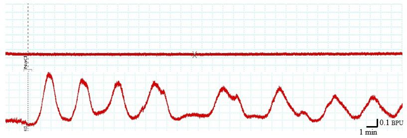
Neurology Asia                                                                                March 2018

CGRP was found to be increased during migraine         allowed free access to food pellets and tap water.
attack in patients.9 Intravenous injection of CGRP     Rats were allowed to acclimatize to housing
was found to often cause migraine-like headaches       conditions for 1 week before the experiment.
in patients with migraine. 10-12 Additionally,         All experiments were conducted in accordance
CGRP-induced migraine-like headaches could             with the approved standard guidelines for animal
be reduced by a triptan. 12 CGRP receptor              experimentation of the Kagawa University and
antagonists such as olcegepant and telcagepant         the Ethical Principles and Guidelines for the Use
were also found to reduce vasodilation, neurogenic     of Animals for Scientific Purposes by National
inflammation and migraine headache. Clinical           Research Council of Thailand 1999.
trials of CGRP blocking antibodies and CGRP               Animals were pretreated for 3 days with either
receptor blockers demonstrated promising results       NSS or BTXA at 3 different dosages before CSD
for effective migraine therapy.13 Additionally,        induction. During the blood flow measurement,
onabotulinumtoxinA treatment for 1 month in            NSS-pretreated rats received either NaCl (n = 8)
chronic migraine patients could reduce interictal      or KCl (n = 8). Similarly, BTXA-pretreated rats
plasma CGRP level in a responders group.14             received either NaCl (n = 24) as a control or
    In animal studies, cortical spreading depression   KCl (n = 24) crystal to induce cortical spreading
(CSD) generated a calcium-dependent release of         depression (CSD) which is considered to be the
endogenous CGRP from rat neocortical slices.15         basis for migraine generation.20
The CSD could be inhibited by CGRP receptor               In the BTXA-pretreated group, rats were
antagonists suggesting the role of CGRP in             injected with 40 µl of BTXA (Botox®, Allergan
initiating and maintaining CSD. Drugs with             Inc.) using microliter syringe (Hamilton Company,
preventive anti-migraine activities were shown         USA) at 3, 10 and 30 units/kg body weight at
to reduce CSD while antiepileptic drugs without        eyebrows of the rats on both sides (20 µl each
the efficacy on migraine such as oxcarbazepine         side) 3 days prior to NaCl or KCl application. As
had no effect on CSD.15,16                             a negative control, normal saline solution (NSS)
    In this study CSD was induced in rats by           was administered instead of BTXA.
direct application of KCl crystal on the surface
of cerebral cortex which produced typical              Induction of migraine-like phenomena
changes in cerebral blood flow.17 Thus, changes
                                                       Migraine headache is associated with a generation
in cerebral blood flow could be used to signify
                                                       of CSD.21 Several methods including application of
corresponding changes in the CSD. Additionally,
                                                       KCl can be used to induce CSD in animal model.16
KCl application could also cause a release of
                                                       Rats were anesthetized with intraperitoneal
CGRP from trigeminal ganglia in primary cell
                                                       injection of pentobarbital sodium at a dose of 50
culture.18
                                                       mg/kg body weight and ventilated with a positive
    Activation of the cranial pain pathway by CSD
                                                       pressure ventilator (Rodent ventilator model
in migraine headache produces an increase in
                                                       683, Harvard Apparatus, South Natick, USA)
neuronal activity of the pain pathway including
                                                       through tracheotomy openings. Blood pressure
TNC which receives input from the trigeminal
                                                       at femoral artery was monitored continuously
ganglion. To determine the level of TNC activation
                                                       with an intra-arterial pressure transducer (Gould
by CSD, the expression of c-Fos was measured.19
                                                       P23 Statham, USA). Blood pressure data was
    The objective of this study was to determine
                                                       digitized and recorded using a data acquisition
how BTXA affect the trigeminovascular
                                                       system for further off-line analysis (PowerLab,
nociceptive system (TVNS) using CSD model
                                                       ADInstruments, CO, USA). After tracheotomy
in rats. As an effective migraine preventive
                                                       and cannulation, rats were placed on a surgical
medication, BTXA should be able to reduce
                                                       frame and their heads fixed to the head holder. A
CSD production and CGRP synthesis by the
                                                       craniotomy of 2 mm in diameter was performed
trigeminovascular nociceptive system.
                                                       on the parietal bone at 7 mm posterior and 1 mm
                                                       lateral to the bregma. Dura mater was cut to expose
METHODS
                                                       the cerebral cortical surface. To prevent drying and
Male Wistar rats between the ages of 8-10 weeks        hypothermia, the cortical surface was superfused
and the body weights of 300-350 g were purchased       with warm artificial cerebrospinal fluid (CSF).17
from National Laboratory Animal Center, Mahidol        To induce CSD, the artificial CSF perfusion was
University, Nakhon Pathom, Thailand. Animals           halted then a 3-mg solid KCl was placed directly
were housed under standard conditions and              on the surface of the parietal cortex. The crystal

46
was allowed to dissolve completely on the cortical paraformaldehyde in 0.1 M PBS pH 7.4 and
surface without reapplication of the artificial CSF. then paraffin processed and transverse 3 µm
NaCl crystal was used instead of KCl crystal for thick sections were cut and deparaffinized
rats in control group.22 before immunostaining. TG paraffin sections
were stained for CGRP by immunoperoxidase
Measurement of focal cerebral hyperemia staining as described by Chatchaisak D.23 The
immunostaining TG sections were viewed under a
To measure the cerebral blood flow, an anterior
confocal microscope and CGRP-immunopositive
craniotomy of 2 mm in diameter was performed
neurons were manually counted. Every fifth section
on the parietal bone at 1 mm anterior and 1 mm
of TG from each rat was chosen for counting of
lateral to the bregma. To prevent drying and
both positive and negative immunoreactive (IR)
hypothermia, the cortical surface was superfused
TG neurons.
with artificial CSF.17 A fiber optic pencil probe of
the Blood Flow Meter (Powerlab, ADInstruments,
Statistical analysis
CO, USA) was placed perpendicularly with a
distance of 2 mm above the cortical surface. The results are expressed as mean+S.E.M. Data
The 780 nm wavelength laser beam was used to was evaluated using Student’s t-test or one-way
measure changes in blood flow under the probe. ANOVA followed by the Student-Newman-Keuls
Data was recorded for 90 min and the maximum test. P-values less than 0.05 were considered
amplitude of each hyperemic peak was measured statistically significant.
for all peaks observed during a 60-min period after
KCl application. Amplitudes for every hyperemic RESULTS
peak during the entire 60 min period were summed
to produce cumulative blood flow in 1 hour. After Changes in Cerebral Blood Flow by KCl
cerebral blood flow measurements, rats were
KCl application to the cerebral cortex produced
transcardially perfused with 250 ml of ice-cold
repeated cycles of cerebral hyperemia while
phosphate buffered saline (PBS) with a pH of 7.4.
NaCl application did not produce any hyperemic
The cervical part of spinal cord and trigeminal
peaks (Figure 1). For NSS-pretreated group, KCl
ganglions (TG) were removed and immersed in
induced an increased in cerebral blood flow with
4% paraformaldehyde in 0.1 M PBS, pH 7.4 for
the average cumulative blood flow amplitude of
subsequent measurement of c-Fos and CGRP.
16.72±2.77x10³ Blood Perfusion Unit (BPU)
(Figure 2). The average number of peaks in 1
Measurement of c-Fos expression at trigeminal
hour of the NSS-pretreated group was 11.80±1.11
nucleus caudalis
peaks/h (ranging from 10 to 14 peaks) (Figure 3).
The caudal medulla (3 mm caudal to the obex) The results confirmed that only KCl but not NaCl
to the first cervical cord were fixed overnight in could induce CSD resulting in episodic increases
4% paraformaldehyde in 0.1 M PBS pH 7.4, then of cerebral blood flow (Figure 1).
placed in a cryoprotective solution consisting of
30% sucrose in 0.1 M PBS, pH 7.4. The tissue Effect of BTXA on Cerebral Blood Flow
was cut into transverse serial sections at 30 µm
After BTXA injection, no apparent facial muscle
thickness using a cryostat at -20°C (Leica CM
weakness or ptosis were observed. Drooling and
1580, Germany). Serial sections were collected
dyspnea were also absent even after the highest
1 in every 5 sections and then rinsed in 0.1
dose of BTXA. The averaged cumulative blood
M PBS. All sections were prepared for c-Fos
flow amplitude of the BTXA-pretreated groups at
immunochemistry staining as described by
the dosage of 3, 10 and 30 units/kg body weight
Supornsilchai W.22 The number of c-Fos positive
were 6.28±1.70, 4.68±0.87 and 1.16±0.31x10³
cells in lamina I and II of trigeminal nucleus
BPU, respectively (Figure 2). These blood flow
caudalis was determined using image analysis
amplitudes of BTXA pretreated groups were
software (ImagePro® Plus; Media Cybernetics
significantly less than that of the NSS-pretreated
Inc., Bethesda, Maryland, USA).
group. Compared to NSS-pretreated group, BTXA
at the dosage of 3, 10 and 30 units/kg body weight
Measurement of CGRP expression at trigeminal
significantly reduced the averaged hyperemic
ganglion
peak numbers per hour to 9.20±0.20, 6.40±0.51
TG tissue were fixed overnight in 4% and 3.00±0.32 peaks/h, respectively (Figure 3).
47Neurology Asia March 2018
(A)
(B)
Figure 1. Changes in cerebral blood flow. Application of 3 mg KCl (B) but not 3 mg NaCl (A) produced cyclical
increase in local cerebral blood flow. BPU, Blood Perfusion Unit.
Figure 2. Total (cumulative) cerebral blood flow during 60 min of recording. BTXA reduced the total increase in
blood flow induced by KCl. Data are expressed as mean+S.E.M; n = 8 in each groups. All differences
are significant at P < 0.05. Asterisk (*) indicates significant difference between control (NSS) group and
other groups.
Figure 3. The average number of hyperemic peaks. BTXA reduced the number of hyperemic peaks during the
recording period. Data are expressed as mean+S.E.M; n = 8 in each groups. All differences are significant
at P < 0.05. Asterisk (*) indicates significant difference between control group and other groups.
48Figure 4. Expression of c-Fos at the TNC. (A) Increased c-Fos expression induced by KCl. (B) Reduction of c-Fos
expression by BTXA pre-treatment. Marker scale bar indicates a length of 100 µm.
Therefore, BTXA decreased cerebral blood as indicated by an increased number of c-Fos
flow induced by CSD through a reduction in positive cells at TNC.
cumulative flow amplitude and number of flow
peaks. Effect of BTXA on the Number of c-Fos Positive
Cells Induced by KCl at Trigeminal Nucleus
Effect of KCl on the Number of c-Fos Positive Caudalis
Cells at Trigeminal Nucleus Caudalis
BTXA pretreatment significantly reduce the
KCl increased the number of c-Fos positive cell numbers of c-Fos positive cell at TNC to
at lamina I and II of TNC (Figure 4 and 5). The 3.04+0.59 cells when compared to the number
average number of c-Fos positive cells in KCl of cells in NSS-pretreated group (Figure 5). The
group (16.21+2.23 cells) was significantly higher result indicated that BTXA could reduce the
than that of the NaCl group (1.50+0.76 cells). activity of TNC neurons leading to a reduction in
Therefore, KCl application on the cortical surface pain transmission through the trigeminovascular
increased activity of the cranial pain pathway nociceptive pathway.
Figure 5. Number of c-Fos positive cells in lamina I and II of TNC. BTXA reduced the number of c-Fos positive
cells activated by KCl. Data are expressed as mean+S.E.M; n = 8 in each group. All differences are
significant at P< 0.05. Hash mark (#) indicates significant difference between NaCl group and KCl group;
Asterisk (*), significant difference between KCl group and BTXA-pretreated groups.
49Neurology Asia March 2018
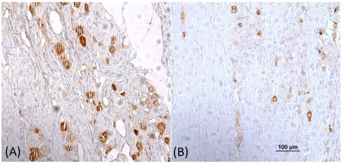
Figure 6. CGRP expression in TG. (A) Increased CGRP expression induced by KCl; (B) Reduction of CGRP
expression by BTXA pretreatment. Marker scale bar indicates a length of 100 µm.
Effect of KCl on the Number of Cells Expressing number of total cells at the trigeminal ganglion
CGRP mRNA at Trigeminal Ganglion to 35.71+3.82 % when compared with that of
NSS-pretreated group (Figure 7).
When compared with the NaCl group, KCl
produced a significant increase in number of
DISCUSSION
CGRP-positive cells per number of total cells ratio
at the trigeminal ganglion (Figure 6). The averaged This is the first study to investigate the effects
CGRP-positive cells per total cells of KCl control of BTXA, administered on the facial area
group and of NaCl group were 57.81+2.22% and in a similar manner as in standard clinical
39.11+0.49% respectively (Figure 7). setting, on parameters related to the migraine
pathophysiology using rat CSD model. For
Effect of BTXA on the CGRP-positive Cells treatment of chronic migraine in human, the
Induced by KCl at Trigeminal Ganglion recommended total dosage by FDA and Allergan
is 155 units given intramuscularly24 which may
BTXA at 30 units/kg dosage significantly reduced
be comparable to the dosage of 3 units/kg in
the percentage of CGRP-positive cells per
Figure 7. Percent of CGRP-positive cells at TG. BTXA decrease the percent of CGRP-positive cells induced
by KCl. Data are expressed as mean+S.E.M; n = 8 in each group. All differences are significant at
P < 0.05. Hash mark (#) indicated significant difference between NaCl group and KCl group; Asterisk
(*), significant difference between KCl group and BTXA-pretreated groups.
50this study. Additionally, our preliminary study such as acetylcholine at the neuromuscular
demonstrated that the maximum effect of BTXA is junction resulting in muscle relaxation.27
reached 3 days following the initial administration Moreover, results from animal and in vitro
which is in agreement with a previous study by experiments showed that BTXA reduced pain
Antonucci et al.25 In summary, results from this by decreasing the release of neuropeptide
study confirmed that BTXA at dosages comparable related to pain modulation including CGRP,
to the clinically recommended dosage could glutamate, substance P and bradykinin.28,29 In
reduce changes associated with CSD production a rat model, subcutaneous BTXA injection
and migraine headache. suppressed nitroglycerin-induced CGRP and SP-
In rat, application of KCl induced CSD that like immunoreactivity in the jugular plasma and
stimulated the trigeminovascular nociceptive in the medulla oblongata.30 BTXA could also be
system (TVNS) as indicated by increased c-Fos retrogradely transported along the axon to affect
positive cells at the trigeminal nucleus caudalis SNAP-25 protein at distant sites in the central
(TNC).17 It was proposed that CSD caused a nervous system.25
release of many neuropeptides such as substance Current study showed that BTXA reduced
P, nitric oxide (NO) and calcitonin gene-related both the number and the amplitude of hyperemic
peptide (CGRP) from nerve terminals to adjacent peaks indicating a parallel reduction in CSD and
meninges and nerves resulting in vasodilation, hyperemia, respectively. It is most likely that
neurogenic inflammation and activation of the injected BTXA is retrogradely transported along
TVNS.26 ophthalmic division (VI) of the trigeminal nerve
In this study changes in cerebral blood flow to affect SNAP-25 protein at both the peripheral
signify corresponding changes in CSD. CSD, and central branches of the axon.25 The reduction
recorded as direct current (DC) shift, produced of CGRP release at the peripheral branches of
corresponding changes in regional cerebral TG neurons surrounding cerebral blood vessels
blood flow (rCBF).22 Hyperemic peak frequency by BTXA reduced vasodilatation and cerebral
(number of peaks/h) was found to be directly hyperemia as indicated by decreased number of
correlated with the CSD frequency. However, CGRP-positive cells at the trigeminal ganglia.
the amplitude of increased blood flow was not Centrally, BTXA most likely reduced
directly correlated with the amplitude of DC shift. neurotransmitter glutamate, NO and/or CGRP
Additionally, paracetamol was shown to reduce release from the presynaptic TG nerve terminal
the hyperemic amplitude but not the DC-shift or causing a decreased activation of the TNC as
CSD amplitude.22 Therefore, individual hyperemic supported by the reduction of c-Fos expression and
peak amplitude was not determined by CSD but the number of cells positive for c-Fos at lamina I
by vasodilators such as CGRP released from and II of TNC. As a result, transmission of pain
perivascular nerves in response to CSD. signal along the TVNS was reduced leading to
In the current study, BTXA reduced both decreased central sensitization.31
the number of hyperemic peaks per hour and Because the number of hyperemic peaks was
the cumulative amplitude of hyperemic peaks. reduced by BTXA, it was also possible that
Therefore, it can be concluded that BTXA could BTXA could directly inhibit the production of
both suppress CSD production as determined by CSD thereby decreasing the stimulation of the
reduced number of hyperemic peaks per hour and TVNS and subsequent production of c-Fos,
decreased cerebral vasodilatation as determined nNOS and CGRP induced by CSD. However, no
by reduced cumulative blood flow. Because of direct evidence from this study or from previous
the direct relationship between hyperemic peak studies that demonstrated direct inhibition of CSD
frequency and CSD wave frequency, it can be production by BTXA. Future experiments are
concluded that BTXA suppresses CSD production required to determine the direct effect of BTXA
and eventually migraine headache. on CSD generation. Although the mechanism
BTXA mainly acts as a proteolytic enzyme to of CSD generation in migraine patients is not
degrade the SNAP-25 protein, a type of SNARE known, it has been shown that CSD could
protein. The SNAP-25 protein is required for activate meningeal nociceptors.32,33 that BTXA
vesicle fusion that releases neurotransmitters from reduces the stimulatory effects of CSD on the
the nerve endings. BTXA specifically cleaves trigeminovascular nociceptive pathway. However,
these SNAREs, so prevents secretory vesicles the possibility of BTXA inhibition of upstream
from docking/fusing with the nerve synapse events such as the accumulation of glutamate
plasma membrane and releasing neurotransmitters that could trigger the generation of CSD and the
51Neurology Asia March 2018
release of CGRP and other peptides could not be 11. Hansen JM, Hauge AW, Olesen J and Ashina M.
ruled out. Calcitonin gene-related peptide triggers migraine-
In conclusion, BTXA pretreatment decreased like attacks in patients with migraine with aura.
Cephalalgia 2010; 30: 1179-86.
CGRP production and release by TG neurons 12. Asghar MS, Hansen AE, Amin FM, et al. Evidence
at the perivascular nerves leading a reduction for a vascular factor in migraine. Ann Neurol 2011;
in CSD production as indicated by a decrease 69: 635-45.
in the number of cerebral hyperemic peaks per 13. Edvinsson L. CGRP receptor antagonists and
hour. Centrally, BTXA also reduced activation of antibodies against CGRP and its receptor in migraine
the TNC as indicated by decreased the number treatment. Br J Clin Pharmacol 2015; 80: 193-9.
14. Cernuda-Morollon E, Ramon C, Martinez-Camblor
of neurons expressing c-FOS. By acting both
P, Serrano-Pertierra E, Larrosa D, Pascual J.
at the peripheral and central locations, BTXA OnabotulinumtoxinA decreases interictal CGRP
prevent the sensitization of the trigeminovascular plasma levels in patients with chronic migraine. Pain
nociceptive pathway which is an underlying 2015; 156: 820-4.
basis of migraine headache. Results from the 15. Tozzi A, de Iure A, Di Filippo M, et al. Critical role
current study demonstrated the important role of of calcitonin gene-related peptide receptors in cortical
CGRP in migraine pathogenesis and in the future spreading depression. Proceedings of the National
Academy of Sciences of the United States of America
development of preventive migraine therapy. 2012; 109: 18985-90.
16. Costa C TA, Rainero I, Cupini LM, Calabresi P,
ACKNOWLEDGEMENT Ayata C, Sarchielli P. Cortical spreading depression
as a target for anti-migraine agents. The journal of
This work is supported by the Japan Society for headache and pain 2013; 14: 62.
the Promotion of Science [grant number R10926]. 17. Maneesri S, Patamanont J, Patumraj S and
Srikiatkhachorn A. Cortical spreading depression,
DISCLOSURE meningeal inflammation and trigeminal nociception.
Neuroreport 2004; 15: 1623-7.
Conflict of interest: None. 18. Durham PL, Niemann C and Cady R. Repression of
stimulated calcitonin gene-related peptide secretion
REFERENCES by topiramate. Headache 2006; 46: 1291-5.
19. Ingvardsen BK, Laursen H, Olsen UB and Hansen
1. Stovner LJ and Hagen K. Prevalence, burden, and AJ. Possible mechanism of c-fos expression in
cost of headache disorders. Curr Opin Neurol 2006; trigeminal nucleus caudalis following cortical
19: 281-5. spreading depression. Pain 1997; 72: 407-15.
2. Olesen J, Gustavsson A, Svensson M, Wittchen HU 20. Supronsinchai W, Hoffmann J, Akermann S and
and Jonsson B. The economic cost of brain disorders Goadsby PJ. KCl-induced repetitive cortical
in Europe. Eur J Neurol 2012; 19: 155-62. spreading depression inhibits trigeminal neuronal
3. Matak I, Riederer P and Lackovic Z. Botulinum firing mediated by 5-HT1B/1D and opioid receptor.
toxin’s axonal transport from periphery to the spinal The journal of headache and pain 2013; 14: P69-P.
cord. Neurochem Int 2012; 61: 236-9. 21. Ayata C. Cortical spreading depression triggers
4. Pringsheim T, Davenport WJ and Becker WJ. migraine attack: pro. Headache 2010; 50: 725-30.
Prophylaxis of migraine headache. CMAJ 2010; 182: 16. 22. Supornsilpchai W, le Grand SM and Srikiatkhachorn
5. Eross EJ. Patient page. Chronic migraine and A. Cortical hyperexcitability and mechanism of
medication-overuse headache. Neurology 2006; 66: medication-overuse headache. Cephalalgia 2010;
E43-4. 30: 1101-9.
6. Whitcup SM, Turkel CC, DeGryse RE and Brin MF. 23. Chatchaisak D, Srikiatkhachorn A, Maneesri-le
Development of onabotulinumtoxinA for chronic Grand S, Govitrapong P and Chetsawang B. The role
migraine. Ann NY Acad Sci 2014; 1329: 67-80. of calcitonin gene-related peptide on the increase
7. Silberstein S, Mathew N, Saper J and Jenkins S. in transient receptor potential vanilloid-1 levels in
Botulinum toxin type A as a migraine preventive trigeminal ganglion and trigeminal nucleus caudalis
treatment. For the BOTOX Migraine Clinical activation of rat. J Chem Neuroanat 2013; 47: 50-6.
Research Group. Headache 2000; 40: 445-50. 24. Ashkenazi A and Blumenfeld A. OnabotulinumtoxinA
8. Jackson JL, Kuriyama A and Hayashino Y. Botulinum for the treatment of headache. Headache 2013; 2:
toxin A for prophylactic treatment of migraine and 54-61.
tension headaches in adults: a meta-analysis. JAMA 25. Antonucci F, Rossi C, Gianfranceschi L, Rossetto
2012; 307: 1736-45. O and Caleo M. Long-distance retrograde effects
9. Goadsby PJ, Edvinsson L and Ekman R. Vasoactive of botulinum neurotoxin A. J Neurosci 2008; 28:
peptide release in the extracerebral circulation of 3689-96.
humans during migraine headache. Ann Neurol 1990; 26. Moskowitz MA. Genes, proteases, cortical spreading
28: 183-7. depression and migraine: impact on pathophysiology
10. Lassen LH, Haderslev PA, Jacobsen VB, Iversen HK, and treatment. Funct Neurol 2007; 22: 133-6.
Sperling B and Olesen J. CGRP may play a causative 27. Arezzo JC. Possible mechanisms for the effects
role in migraine. Cephalalgia 2002; 22: 54-61.
52of botulinum toxin on pain. Clin J Pain 2002; 18:
S125-32.
28. Peng Chen Z, Morris JG, Jr., Rodriguez RL,
Shukla AW, Tapia-Nunez J, Okun MS. Emerging
opportunities for serotypes of botulinum neurotoxins.
Toxins 2012; 4: 1196-222.
29. Caputi CA. Effectiveness of BoNT-A in the treatment
of migraine and its ability to repress CGRP release.
Headache 2004; 44: 837-8.
30. Shao Y-F, Zhang Y, Zhao P, et al. Botulinum Toxin
Type A Therapy in Migraine: Preclinical and Clinical
Trials. Iran Red Crescent Med J 2013; 15: e7704.
31. Pradhan AA, Smith ML, McGuire B, Tarash I, Evans
CJ, Charles A. Characterization of a novel model of
chronic migraine. Pain 2014; 155: 269-74.
32. Zhang X LD, Noseda R, Kainz V, Jakubowski M,
Burstein R. Activation of meningeal nociceptors
by cortical spreading depression: implications for
migraine with aura. J Neurosci 2010; 30: 8807-14.
33. Zhao J, Levy D. Modulation of intracranial meningeal
nociceptor activity by cortical spreading depression: a
reassessment. J Neurophysiol 2015; 113(17):2778-85.
53You can also read