Clear Cell Carcinoma of the Abdominal Wall as a Rare Complication of General Obstetric and Gynecologic Surgeries: 15 Years of Experience at a ...
←
→
Page content transcription
If your browser does not render page correctly, please read the page content below
International Journal of
           Environmental Research
           and Public Health

Article
Clear Cell Carcinoma of the Abdominal Wall as
a Rare Complication of General Obstetric and
Gynecologic Surgeries: 15 Years of Experience
at a Large Academic Institution
Yen-Ling Lai 1 , Heng-Cheng Hsu 2,3 , Kuan-Ting Kuo 4 , Yu-Li Chen 1, *, Chi-An Chen 1 and
Wen-Fang Cheng 1,2,5
 1    Department of Obstetrics and Gynecology; National Taiwan University Hospital, Taipei 100, Taiwan;
      adina@kimo.com (Y.-L.L.); chianchen@ntu.edu.tw (C.-A.C.); wenfangcheng@yahoo.com (W.-F.C.)
 2    Graduate Institute of Clinical Medicine, College of Medicine, National Taiwan University, Taipei 100,
      Taiwan; b101092037@gmail.com
 3    Department of Obstetrics and Gynecology, National Taiwan University Hospital Hsin-Chu Branch,
      Hsin-Chu 300, Taiwan
 4    Department of Pathology and Graduate Institute of Pathology, College of Medicine, National Taiwan
      University, Taipei 100, Taiwan; kuokt@ntu.edu.tw
 5    Department of Graduate Institute of Oncology, College of Medicine, National Taiwan University,
      Taipei 100, Taiwan
 *    Correspondence: uly1007@yahoo.com.tw; Tel.: +886-2-2312-3456 (ext. 71964); Fax: +886-2-2311-4965

 Received: 13 January 2019; Accepted: 12 February 2019; Published: 14 February 2019

 Abstract: The objective of this article was to report the clinicopathological characteristics, treatment
 modalities, and outcomes of patients with clear cell carcinoma (CCC) of the abdominal wall. Medical
 records of six patients diagnosed with CCC of the abdominal wall between May 2003 and May
 2018 at the National Taiwan University Hospital were reviewed. All patients had prior obstetric
 or gynecologic surgeries. The primary clinical presentation was enlarging abdominal masses at
 previous surgical scars. Four patients underwent initial/primary surgeries with/without adjuvant
 chemotherapy. One patient received neoadjuvant chemotherapy followed by surgical intervention
 and adjuvant chemotherapy, the other received chemotherapy and sequential radiotherapy without
 any surgical intervention. Two of four patients undergoing initial/primary surgeries had disease
 recurrence and the remaining two cases without initial surgery experienced disease progression
 during primary treatment. Inguinal lymph nodes were the most frequent sites of recurrence. In
 conclusion, previous obstetric or gynecologic surgery can be a risk factor for CCC of the abdominal
 wall. Complete resection of abdominal wall tumor and suspected intra-abdominal lesions with
 hysterectomy and bilateral inguinal lymph nodes dissection may be the primary treatment. Adjuvant
 chemotherapy would be considered for potential benefits. For patients without bilateral inguinal
 lymph nodes dissection, careful inguinal lymph node palpation during postoperative surveillance is
 necessary. More cases are still needed to elucidate the clinical management of this disease.

 Keywords: endometriosis; malignant transformation; clear cell carcinoma; abdominal wall; inguinal
 lymph node; cesarean section

1. Introduction
    It is estimated that cancers arise in approximately 1% of woman with ovarian endometriosis [1].
Clear cell and endometrioid carcinomas are the malignancies most commonly seen in ovaries
containing endometriosis [2]. Malignant transformation of extra-ovarian endometriosis is rare and

Int. J. Environ. Res. Public Health 2019, 16, 552; doi:10.3390/ijerph16040552       www.mdpi.com/journal/ijerph
Int. J. Environ. Res. Public Health 2019, 16, 552                                                   2 of 12

the most frequently reported sites are the rectovaginal septum, colon, and vagina [1]. Implantations
of ectopic endometrium in the scar tissue of the anterior abdominal wall after operations, especially
cesarean sections, laparoscopic surgeries, and hernia repairs, is referred to as abdominal wall
endometriosis [3]. It is plausible that abdominal wall endometriosis could undergo malignant changes
resulting in clear cell carcinoma (CCC) of the abdominal wall [4]. CCC originating in the abdominal
wall is an extremely rare form of cancer. After the first case documented in 1986 [5], only few cases
have been reported to date.
     In 1925, Sampson described the first case of malignant transformation of ovarian endometriosis,
and defined three criteria for the diagnosis of malignant transformation of endometriosis:
(1) Demonstration of both neoplastic tissue and endometriosis within the tumor, (2) histological
appearance resembling endometrial stroma surrounding characteristic glands, and (3) no other primary
tumor site [6]. In addition, Scott added the fourth criterion of metaplasia between benign endometriosis
and carcinoma in 1953 [7]. However, about 44% of reported cases did not correlate well with all these
four histological criteria [8–15].
     The management of CCC of the abdominal wall is not well established because of its extremely
low incidence rate. Therefore, we comprehensively reviewed the characteristics of six cases with this
disease in our institution over a 15-year time period. To our knowledge, the current case series is the
largest one to date. It is noteworthy that this is the first study reporting the recurrence pattern of CCC
of the abdominal wall, which may have potential to modulate the primary treatment strategy for the
improvement of clinical outcomes for these patients.

2. Materials and Methods
      Following approval by the National Taiwan University Hospital Institutional Review Board,
a retrospective review was conducted (201805083RIND). All medical records of 6 patients treated for
CCC of the abdominal wall in the National Taiwan University Hospital between May 2003 and May
2018 were reviewed. These cases were confirmed via pathological diagnosis. The medical parameters,
including basic characteristics, primary treatment modalities, pathologic characteristics, and clinical
outcomes, were comprehensively reviewed.
      The basic characteristics included age at diagnosis, body mass index (BMI), parity, previous
obstetric and gynecologic surgery histories, the interval between the most recent obstetric or
gynecologic surgeries and disease diagnosis, tumor size, presenting symptoms, pretreatment serum
cancer antigen 125 (CA-125) level, and findings of pretreatment computed tomography (CT). The
treatment strategies included the methods of surgery, timing of administration of chemotherapy,
and use of radiotherapy. The pathologic characteristics included histology, status of surgical margin,
co-existing endometriosis, and existence of granuloma caused by suture materials of the abdominal
wall tumor. When the removal of gynecologic organs was performed, pathologic examination for CCC
and endometriosis in these specimens were recorded.
      The clinical outcomes included recurrent sites, types of salvage treatment, disease-free survival
(DFS), overall survival (OS), and current status of these patients. Periodic examinations during
follow-up included history-taking, pelvic and rectal examinations, and regional lymph node palpation
every 3 months for 3 years, and every 6 months thereafter after completion of the primary treatment.
Levels of serum CA-125 was determined on each visit. Smears of the vaginal cuff were done annually.
CT or magnetic resonance imaging (MRI) was done for suspected recurrence. CA-125 levels ≥2-fold
the upper limit of normal in two consecutive tests with 2-week intervals, abnormal results of imaging
studies or tissue proven from a biopsy was considered as recurrence. DFS was defined as the length
of time after completion of the primary treatment until the date of disease relapse or last follow-up.
OS was calculated as the time from initial diagnosis of CCC of the abdominal wall until the date of
death or last follow-up.
Int. J. Environ. Res. Public
Int. J. Environ. Health
Res. Public 2019,
Health 16,16,
2019, 552x 3 of 123 of 12
3. Results
3. Results
3.1. Prior Obstetric or Gynecologic Surgery with the Abdominal Wall Contacted with the Endometrium or
3.1. Prior ObstetricTissue
Endometriotic or Gynecologic Surgery
may be a Risk Factor with
for thethe AbdominalofWall
Development CCCContacted with the
of the Abdominal Endometrium or
Wall
Endometriotic Tissue may be a Risk Factor for the Development of CCC of the Abdominal Wall
Table 1 shows the clinicopathologic characteristics of the six patients. The mean patient age at
the time
Table of diagnosis
1 shows was 52.7 years. characteristics
the clinicopathologic The mean BMI of wasthe22.6
sixkg/m 2 (range, 17.0–28.8 kg/m2). All
patients. The mean patient age at the
patients received obstetric or gynecological surgeries. Five 2
patients
time of diagnosis was 52.7 years. The mean BMI was 22.6 kg/m (range, 17.0–28.8 had cesarean section
kg/m 2 ). Alland
histories patients
one had laparoscopic surgery for ovarian endometriosis. The mean time
received obstetric or gynecological surgeries. Five patients had cesarean section historiesfrom the most recent
and one
obstetric or gynecologic surgery to diagnosis of CCC of the abdominal wall was 20.2 years (range,
had laparoscopic surgery for ovarian endometriosis. The mean time from the most recent obstetric or
4.0–33.0 years). The shortest interval (4.0 years) was noted in the patient with laparoscopic surgery
gynecologic surgery to diagnosis of CCC of the abdominal wall was 20.2 years (range, 4.0–33.0 years).
(case 3). The mean tumor size was 10.1 cm (range, 4.8–17.5 cm). Five patients with cesarean section
The shortest interval (4.0 years) was noted in the patient with laparoscopic surgery (case 3). The mean
histories presented with an enlarging lower abdominal wall mass containing scars of a cesarean
tumorsection
size was 10.11A),
(Figure cmand
(range, 4.8–17.5
the other cm).
patient Five patients
complained of an with cesarean
enlarging section
left lower histories
quadrant masspresented
near
with an enlarging lower abdominal wall mass containing scars of a cesarean section
a laparoscopic trocar wound. The mean pretreatment serum carcinoma antigen 125 (CA-125) level (Figure 1A), and
the other patient complained of an enlarging left lower
was 28.3 U/mL (range, 20.1–38.9 U/mL; normal value < 35.0 U/mL).quadrant mass near a laparoscopic trocar
wound. The mean pretreatment serum carcinoma antigen 125 (CA-125) level was 28.3 U/mL (range,
20.1–38.9 U/mL; normal value < 35.0 U/mL).
Figure 1. Representative
Figure 1. Representative gross, histology,
gross, histology,and
andpreoperative computed
preoperative computed tomography
tomography (CT)(CT)
scanscan of clear
of clear
cell carcinoma
cell carcinoma of theofabdominal
the abdominal
wall. wall.
(A) A(A) Acm
17.5 17.5 cm lesion
mass mass lesion
in thein the lower
lower abdomenabdomen with
with an an
ulcerative
ulcerative
surface (case 1).surface (case 1).examination
(B) Pathologic (B) Pathologic examination
of the abdominal of wall
the tumor
abdominal wall tubulocystic
showed tumor showed growth
tubulocystic
patterns growth patterns
lined by cuboidal, hobnaillined
cells,by
andcuboidal, hobnail
clear cells. Focalcells, and clear
papillary, cells. Focal and
micropapillary, papillary,
cribriform
micropapillary, and cribriform patterns were also present (case 4, H&E stain, 200X). (C) Granuloma
patterns were also present (case 4, H&E stain, 200X). (C) Granuloma caused by suture material was
caused by suture material was noted in the tumor sample (case 4, H&E stain, 40X). (D) A 13.0 cm
noted in the tumor sample (case 4, H&E stain, 40X). (D) A 13.0 cm lobulated heterogeneous tumor (star)
lobulated heterogeneous tumor (star) was located at the anterior lower abdominal wall with
was located at the anterior lower abdominal wall with peritoneal involvement (arrows) (case 4). (E) No
peritoneal involvement (arrows) (case 4). (E) No definite lesions in the uterus, ovaries, and fallopian
definite lesions
tubes were in the uterus,
noted (case 4).ovaries, and fallopian tubes
(F) Lymphadenopathy alongwere noted
the right (case 4).
inferior (F) Lymphadenopathy
epigastric vessels and
alongexternal
the right inferior
iliac vesselsepigastric
was noted vessels
(arrow) and
(caseexternal
2). iliac vessels was noted (arrow) (case 2).Int. J. Environ. Res. Public Health 2019, 16, 552 4 of 12
Table 1. Clinicopathologic characteristics of the six patients with clear cell carcinoma of the abdominal wall.
Case 1 Case 2 Case 3 Case 4 Case 5 Case 6
Basic Characteristics
Age (year) 52 56 52 56 55 45
BMI (kg/m2 ) 28.8 26.0 17.0 20.8 22.6 20.6
Parity 2 2 0 1 3 3
Previous Obs/Gyn surgeries Cesarean section Cesarean section LSC left oophorectomy Cesarean section Cesarean section Cesarean section
Time to onset (year) a 19.0 33.0 4.0 21.0 24.0 20.0
Tumor size (cm) 17.5 6.5 7.0 12.0 12.5 4.8
Lower abd. wall mass Painful lower abd. wall
Presenting symptoms Lower abd. wall mass LLQ mass Lower abd. wall mass Lower abd. wall mass
with ulceration ulceration
Pretreatment serum CA-125 level
20.1 22.3 38.5 23.0 26.7 38.9
(U/mL)
1. A lower abd. wall tumor
1. A lower abd. wall 1. A lower abd. wall 1. A lower abd. wall tumor 1. Multiple lower abd. 1. A lower abd. wall
2. Small LN along right
tumor tumor with peritoneal involvement wall tumors tumor
Preoperative CT scanning findings external iliac vessels
2. A 6.5 cm left adnexal 2. No definite focal lesion 2. No definite focal lesion in 2. No definite focal lesion 2. No definite focal lesion
3. No definite focal lesion in
tumor in the Gyn organs the Gyn organs in the Gyn organs in the Gyn organs
the Gyn organs
Treatment Strategies
Tumor excision + + + + + -
TAH, BSO, omentectomy, TAH, BSO, omentectomy
Gyn surgeries TAH, BSO TAH, BSO TAH, BSO, BPLND -
BPLND (after NACT)
Neoadjuvant (preoperative) C/T - - - - +b -
Primary C/T - - - - - +c
Adjuvant (postoperative) C/T - +d +e +f +g -
Radiotherapy - - - - - +hInt. J. Environ. Res. Public Health 2019, 16, 552 5 of 12
Table 1. Cont.
Case 1 Case 2 Case 3 Case 4 Case 5 Case 6
Pathology
Abd. wall tumor CCC CCC CCC CCC CCC CCC i
Surgical margin Free Free Free Free Involved -i
Co-existing endometriosis in abd.
- - + - + -
wall tumorj
Histological appearance showing
endometrial stroma and glands in - - - - - -
abd. wall tumor
Other primary tumor sites - - - - - -
CCC of gyn organs Negative Negative k Negative Negative Negative -
Presence of suture granuloma Yes No No Yes No No
Adenomyosis
Intraabbdominal endometriosis No Adenomyosis No Adenomyosis No
Endometrioma
Note. BMI, body mass index; Obs/Gyn, obstetric and gynecologic; C-section, Cesarean section; LSC, laparoscopic; LLQ, left lower quadrant; TAH, total abdominal hysterectomy; BSO,
bilateral salpingo-oophorectomy; BPLND, bilateral pelvic lymph node dissection; RSO, right salpingo-oophorectomy; NACT, neoadjuvant chemotherapy; CCC, clear cell carcinoma; C/T,
chemotherapy; abd., abdominal; CA-125, cancer antigen 125; CT, computed tomography; LN, lymph node; + means “with” and – means “without”. a Time to onset was defined as the
interval between the most recent Obs/Gyn surgeries and diagnosis of CCC of abdominal wall. b Five cycles of triweekly paclitaxel (175 mg/m2) and carboplatin (AUC = 5) combined
with bevacizumab (7.5 mg/kg) plus one cycle of gemcitabine (800 mg/m2 ) and carboplatin (AUC = 5). c Seven cycles of paclitaxel (175 mg/m2 ) and carboplatin (AUC = 5) plus one
cycle of liposomal doxorubicin (40mg/m2 ) and carboplatin (AUC = 5). d Eight cycles of triweekly paclitaxel (175 mg/m2 ) and carboplatin (AUC = 5). e Six cycles of triweekly paclitaxel
(175 mg/m2 ) and carboplatin (AUC=5). f The patient is under adjuvant chemotherapy now with triweekly paclitaxel (175 mg/m2) and carboplatin (AUC = 5). g Three cycles of gemcitabine
(800 mg/m) and carboplatin (AUC = 5) combined with bevacizumab (7.5 mg/kg). h Three-dimensional conformal radiation therapy (3DRT) with 4400 cGy delivered in 22 fractions. i CCC
was diagnosed via tumor biopsy without tumor resection, thus the involvement of surgical margin could not be assessed in this case. j Co-existing endometriosis refers to the endometriosis
existing associated with CCC of the abdominal wall. k Metastasis to the right pelvic lymph node was noted, although there was no evidence of gynecologic organs involvement.Int. J. Environ. Res. Public Health 2019, 16, 552 6 of 12
3.2. Modalities of Primary Treatment
The primary treatment strategies of six patients for CCC of the abdominal wall are demonstrated
in Table 1. One patient received tumor excision plus total abdominal hysterectomy (TAH) and bilateral
salpingo-oophorectomy (BSO) without adjuvant treatment (case 1). Three patients received tumor
excision plus intra-abdominal surgeries (case 2, TAH, BSO, bilateral pelvic lymph node dissection
(BPLND), and infracolic omentectomy; case 3, TAH and BSO; case 4, TAH, BSO, and BPLND) followed
by adjuvant chemotherapy. The regimen of adjuvant chemotherapy was paclitaxel (175 mg/m2 ) and
carboplatin (AUC = 5 or 6).
Case 5 initially underwent two regimens of neoadjuvant chemotherapy: (1) Paclitaxel (175 mg/m2 )
and carboplatin (AUC = 5) combined with bevacizumab (7.5 mg/kg), and (2) gemcitabine (800 mg/m2 )
and carboplatin (AUC = 5). Surgical intervention with tumor excision, TAH, BSO, and infracolic
omentectomy was performed for tumor progression. Postoperative adjuvant chemotherapy was
prescribed using gemcitabine (800 mg/m2 ) and carboplatin (AUC = 5) combined with bevacizumab
(7.5 mg/kg). However, the disease progression was still noted. Case 6 received tumor biopsy other
than resection for disease diagnosis. This patient received two regimens of chemotherapy: (1) paclitaxel
(175 mg/m2 ) and carboplatin (AUC = 5), and (2) liposomal doxorubicin (40 mg/m2 ) and carboplatin
(AUC = 5). In addition, sequential radiotherapy with a total dose of 4400 cGy was administered after
completing primary chemotherapy. However, the tumor progression still could not be controlled.
3.3. Malignancies without Clinical Manifestations of Image Studies are not Pathologically Confirmed in
Pelvic Organs
The pathologic characteristics of surgical specimens are shown in Table 1. The histology of
the abdominal wall tumors was confirmed as CCC (Figure 1B). Co-existing endometriosis in these
abdominal wall tumors was identified in two cases (case 3 and case 5). In addition, granulomas
caused by suture materials could be found in two tumor tissues (case 1 and case 4) in the histological
examination (Figure 1C). Free margins with a distance more than 2 cm from the abdominal wall tumors
were noted in four cases (case 1–4), but the surgical margin was involved with tumor cells in case 5.
In addition to the resection of the abdominal wall tumors, the removal of gynecologic organs,
including uterus, bilateral ovaries, and fallopian tubes, was performed in five patients (case 1–5,
Table 1). Intra-abdominal endometriosis by pathologic examination was found in three cases (case 1, 3,
and 5), but results for malignancy were all negative in the gynecologic organs. The pathologic findings
were compatible with those of preoperative CT scans, which showed abdominal wall tumors without
the involvement of gynecologic organs (Table 1, Figure 1D,E). However, in case 2, the preoperative CT
scans demonstrated right pelvic lymphadenopathy (Figure 1F), which was pathologically confirmed
as metastasis of the right pelvic lymph nodes.
3.4. Inguinal Lymph Nodes are the Most Common Site of Disease Relapse
Clinical outcomes of the six patients are shown in Table 2. After completing primary treatment,
three patients had a period of DFS (case 1 = 10.0 months; case 2 = 3.0 months; case 3 = 93.0
months). No evidence of disease relapse was detected in case 3. One patient (case 4) was under
the treatment of postoperative adjuvant chemotherapy. The remaining two patients (case 5 and 6)
experienced progressive enlargement of abdominal wall tumors even under the different primary
treatment modalities.
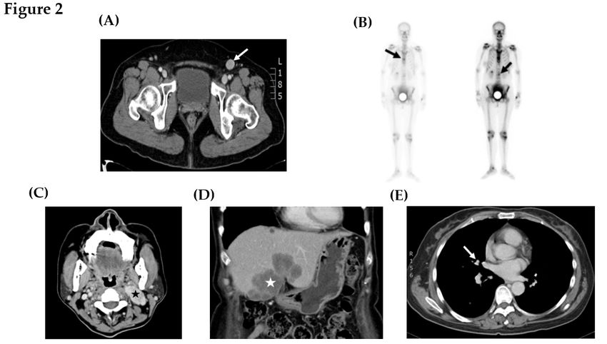
Sites of tumor recurrence and metastasis are shown in Table 2 and Figure 2. The inguinal lymph
node (Figure 2A) was the most common site of disease recurrence and progression (case 1, 2, 5, and
6). Other metastases, including bone (Figure 2B, case 1), neck lymph node (Figure 2C, case 5), liver
(Figure 2D, case 5), and lung (Figure 2E, case 5), were also observed. In addition, the management of
disease relapse is recorded in Table 2. In case 1, disease recurrence was detected in the inguinal lymph
node and bone. She did not receive any salvage treatment because the disease progressed rapidly and
her hepatic function was poor and caused by severe fatty liver disease. In case 2, tumor relapse wasInt. J. Environ. Res. Public Health 2019, 16, 552 7 of 12
Int. J. Environ. Res. Public Health 2019, 16, x 7 of 12
only located in the inguinal lymph node. Resection of this lesion with a free margin was performed.
node and bone. She did not receive any salvage treatment because the disease progressed rapidly
No evidenceand ofher
disease was noted during follow-up.
hepatic function was poor and caused by severe fatty liver disease. In case 2, tumor relapse
In casewas
5, the
onlyprogression of the abdominal
located in the inguinal wall tumor
lymph node. Resection of thiswith
lesionmetastasis to thewas
with a free margin inguinal lymph
performed.
nodes was noted No evidence
during of disease waschemotherapy.
the neoadjuvant noted during follow-up.
Then, resection of this progressive lesion with
In case 5, the progression of the abdominal wall tumor with metastasis to the inguinal lymph
the involvednodes
margin was performed. Multiple distant metastases, including neck lymphadenopathy,
was noted during the neoadjuvant chemotherapy. Then, resection of this progressive lesion
liver, and lung,
with were detected
the involved whilewas
margin receiving
performed.adjuvant
Multiplechemotherapy.
distant metastases, This patientneck
including received salvage
chemotherapy lymphadenopathy, liver, and lung,
and radiotherapy, but were
dieddetected while23
with OS receiving
months. adjuvant
In chemotherapy.
case 6, the Thisabdominal wall
patient received salvage chemotherapy and radiotherapy, but died with OS 23 months. In case 6, the
tumor progression with inguinal lymphadenopathy was noted under chemotherapy and sequential
abdominal wall tumor progression with inguinal lymphadenopathy was noted under chemotherapy
radiotherapy.and However, no salvage
sequential radiotherapy. treatment
However, wastreatment
no salvage arranged was for progressive
arranged disease
for progressive diseasebecause of the
because
poor condition of of thepatient.
this poor condition
Theofmean
this patient.
OS of The mean six
these OS ofpatients
these six patients
was 26.1was 26.1 months,with
months, with a range of 5.0
a range of 5.0 to 97.0 months.
to 97.0 months.
Figure 2. Representative
Figure 2. Representative image image studiesofofrecurrent
studies recurrent tumors.
tumors.(A) CT scanCT
(A) showed
scana showed
2.0 cm enlarged
a 2.0 cm enlarged
Commented [SP11]: Please define
lymphadenopathy (arrow) in the left inguinal area (case 2). (B) Whole body bone scan demonstrated
lymphadenopathy (arrow) in the left inguinal area (case 2). (B) Whole body bone scan demonstrated appropriate.
hot areas at the thoracic and lumbar spine (arrows) (case 1). (C) CT scan showed a 4.0 cm necrotic
hot areas atmass
thesuggesting
thoracicmetastases
and lumbar spine
at the left neck (arrows) (case
area (star) (case 1).CT(C)
5). (D) CT
scan scana showed
showed 6.0 cm tumora at
4.0 cm necrotic
Commented [賴彥伶12R11]: We h
mass suggesting metastases
the lateral and medial at the left
segments neck
of the liverarea
(star)(star) (case
(case 5). (E) CT5). (D)
scan CT scan
exhibited a 0.7showed a 6.0 cm tumor at
cm pulmonary
nodule at the superior segment of the right lower lobe (arrow) (case 5). “CT” in Materials and Methods pr
the lateral and medial segments of the liver (star) (case 5). (E) CT scan exhibited a 0.7 cm pulmonary
nodule at the superior segment
Table 2. Clinical of the
outcomes of theright lowerwith
six patients lobe (arrow)
clear (caseof5).
cell carcinoma the abdominal wall.
Case 1 Case 2 Case 3 Case 4 Case 5 Case 6
2. Clinical outcomes
TableRecurrent/met Inguinal LN,
of Inguinal
the sixLNpatients with clearNA
No recurrence
cell
a
carcinoma of the abdominal
Abd. wall, Abd. wall,
wall.
astatic site bone inguinal LN, inguinal LN
Case 1 Case 2 Case 3 Case neck
4 LN, liver,Case 5 Case 6
lung
Recurrent/metastatic site Inguinal LN, Inguinal LN No NA a Abd. wall, Abd. wall,
Salvage - Tumor - - Chemotherapy b -
bone recurrence inguinal LN, inguinal LN
treatment excision +
neck LN,
Radiotherapy c liver, lung
Salvage treatment b
DFS (m) 10 - 3 Tumor 93 - NAa - Progression
Chemotherapy
Progression -
OS (m) 14 11 excision 97 5 23 + 7
c
Current status Recurrence NED NED NED DODRadiotherapy
Progression
DFS (m) 10 lymph node; NED,3 no evidence of93 a
Note. Abd., abdominal; LN, disease; DOD, NA Progression
died of disease; NA, not available; Progression
OS (m) 14 OS, overall survival.
DFS, disease-free survival; 11 a The woman 97
is under adjuvant5 chemotherapy. After
23removing 7
Current status Recurrence NED NED NED DOD Progression
the tumor with free margins, no evidence of disease is noted. Cisplatin (35 mg/m ) on D1, D8, 5-FU (2600 mg/m )
b 2 2
Note. Abd., abdominal; LN, lymph node; NED, no evidence of disease; DOD, died of disease; NA, not available;
DFS, disease-free survival; OS, overall survival. a The woman is under adjuvant chemotherapy. After removing the
tumor with free margins, no evidence of disease is noted. b Cisplatin (35 mg/m2 ) on D1, D8, 5-FU (2600 mg/m2 ) on
D1, D8, D15 and leucovorin (300 mg/m2 ) on D1, D8, D15 combined with bevacizumab (7.5 mg/kg) on D1 every
three weeks for six cycles, and two cycles of doxorubicin liposomal (40 mg/m2 ) combined with bevacizumab (7.0
mg/kg). c Intensity-modulated radiotherapy (IMRT) with 5500 cGy was delivered in 25 fractions for metastatic
lesion at the abdominal wall and inguinal.
4. Discussion
CCC of the abdominal wall is extremely rare. Taburiaux et al. reviewed existing reports in the
English language literature on cancer arising from abdominal wall endometriosis from September
1986 to August 2014 [4]. A total of 27 cases were identified, and 18 patients were reported to haveInt. J. Environ. Res. Public Health 2019, 16, 552 8 of 12
clear cell carcinomas. Our retrospective review described six cases of pathologically confirmed
clear cell carcinoma of the abdominal wall in detail over 15 years at our institution. In this study,
previous obstetric or gynecologic surgery having abdominal wall contact with the endometrium or
endometriotic tissue may be a risk factor for the development of CCC of the abdominal wall. Tumor
resection with a free margin may be the essential treatment modality for this disease. The inguinal
lymph nodes were the most common location of disease recurrence.
In our case series, all patients had a history of cesarean section or laparoscopic surgery for
endometriosis, consistent with previous reports [5,8–23]. A previous surgical procedure with
endometrial cavity opening or endometriosis may be a risk factor for developing CCC of the abdominal
wall, which could be supported by the granulomas caused by suture materials found in tumor samples
(Figure 1C). Dissemination of endometrial tissue or endometriosis at the time of surgery is biologically
plausible because there is an opportunity for the inoculation of endometrial cells from hysterotomy or
endometrioma to the peritoneum or abdominal wall [3,4].
Several studies have demonstrated that endometriosis can be the precursor and have the
potential for malignant transformation to CCC of the ovaries [2,24–26]. In the tumors coexisting
with endometriosis, a benign endometriotic gland can be observed to merge with atypical and overtly
malignant glands [6,24]. Taburiaux et al. reported that 56% of patients with CCC of the abdominal wall
had co-existing endometriosis. In our series, only two patients had co-existing endometriosis found
in the abdominal wall tumors (case 3 and case 5). Underreporting of the association of malignancy
with endometriosis could be attributed to several factors [1]. First, the sampling technique may
not be adequate to find a small focus of endometriosis adjacent to a malignant tumor. Second, a
cancer may destroy the endometriotic tissue from which it arose. Third, the endometriosis may be
considered to be a minor component. Not all CCC of the abdominal wall were endometriosis-associated
malignancies. Therefore, the elucidation of the pathogenesis is essential for the prevention and
treatment of this disease.
The mean interval between the last surgery and the diagnosis of CCC of the abdominal wall
was long (20.2 years), which suggested a slow evolution of the tumorigenesis (Table 1). The typical
manifestation was an abdominal wall mass adjacent to previous surgical scars. All the serum CA-125
levels were not greater than 40 U/mL in our case series, suggesting that CA-125 may not be an
appropriate tumor marker for CCC of the abdominal wall.
Primary wide tumor resection combined with total abdominal hysterectomy and bilateral
salpingo-oophorectomy or cancer staging surgery was the preferred modality of primary treatment
at our institution. For complete removal of all visible tumor tissue, a synthetic mesh or flap may
be required for abdominal wall reconstruction. All the specimens without malignant suspicion on
preoperative CT scanning histologically showed no evidence of clear cell carcinoma. Only right
pelvic lymphadenopathy on preoperative images was finally confirmed as metastasis by pathologic
examination (Figure 1F). Thus, when considering surgical management, grossly total tumor resection
may be appropriate for diagnostic and therapeutic purposes. As for gynecologic organs, given the
fact that endometrium contact may be the risk factor for CCC of the abdominal wall, it still can be
reasonable to perform hysterectomy routinely even though no evidence of malignancy in gynecologic
organs was detected in our cohort.
Disease control of primary surgery with or without adjuvant chemotherapy was good. In our
study, 50% of patients (3/6, case 1–3) had disease remission after primary operation. One patient (1/6,
case 4) without evidence of disease was under postoperative chemotherapy. Two patients (2/6, case 5
and 6) who did not receive initial tumor resection with a free margin experienced uncontrolled disease
progression. Therefore, initial complete tumor resection with/without adjuvant chemotherapy (case
1–4) seemed to have better disease control in comparison with other primary treatment modalities,
such as neoadjuvant chemotherapy followed by operation and adjuvant chemotherapy (case 5), or
chemotherapy and sequential radiotherapy (case 6). The role of adjuvant chemotherapy was not
established. In our cohort, one patient not receiving adjuvant treatment eventually encounteredInt. J. Environ. Res. Public Health 2019, 16, 552 9 of 12
disease recurrence (case 1). Four patients received adjuvant chemotherapy (case 2–5), two of them
remained without evidence of disease (case 3 and 4), and the other two experienced disease recurrence
(case 2 and 5). Overall, it would be difficult to draw a conclusion based on such limited cases.
However, the results seemed to support the potential benefits of adjuvant chemotherapy. Therefore,
administration of adjuvant chemotherapy after primary operation could be considered. At our
institution, platinum-based chemotherapy was the most used.
The survival intervals and prognostic factors are difficult to investigate due to the limited number
of cases with standard management to treat CCC of the abdominal wall [27]. Taburiaux et al. reported
a median survival time of 30.0 months [4]. In our study, the mean OS of the studied population was
26.1 months. In addition, no associated risk factor for poor prognosis can be identified because of the
disease rarity.
Patterns of disease recurrence/metastasis have not been reported in the literature before. The most
common site of recurrence and metastasis was the inguinal lymph node. Inguinal lymphadenopathy
can be detected in all cases with disease recurrence or progression (Table 2). The reason may be that
superficial lymphatics of the abdominal wall located below the umbilicus run in an inferior direction
towards the superficial inguinal lymph nodes [28]. Other recurrent and metastatic locations, including
bone, neck lymph node, liver, and lung, were also noted in certain cases. Considering the effective
treatment modality and common recurrent/metastatic site of CCC of the abdominal wall, complete
resection of the abdominal wall tumor and intra-abdominal suspected lesions on preoperative image
studies with hysterectomy and bilateral inguinal lymph node dissection may be suggested as the first
step to treat these patients (Figure 3).Int. J. Environ. Res. Public Health 2019, 16, 552 10 of 12
Figure
Figure 3. Flowchart
3. Flowchart of management
of management for patients
for patients withcell
with clear clear cell carcinoma
carcinoma (CCC) of(CCC) of abdominal
abdominal wall.
wall. Note: Note:
CCC, CCC,
clear clear cell carcinoma;
cell carcinoma; NACT: neoadjuvant
NACT: neoadjuvant chemotherapy;
C/T:chemotherapy;
chemotherapy; C/T: chemotherapy;
R/T: R/T: radiotherapy.
radiotherapy.Int. J. Environ. Res. Public Health 2019, 16, 552 11 of 12
5. Conclusions
CCC of the abdominal wall is a rare and distinct entity. Our study addressed comprehensive
clinical courses and outcomes of these cases in a single institution. Complete resection of the abdominal
wall tumor and intra-abdominal suspected lesions on preoperative image studies with hysterectomy
and bilateral inguinal lymph node dissection may be suggested as the first step to treat CCC of the
abdominal wall. Adjuvant chemotherapy would be recommended for potential benefits. For those
patients without bilateral inguinal lymph node dissection, careful inguinal lymph node palpation
during postoperative surveillance is needed. More such cases are still needed to elucidate the survival
benefits of this surgical procedure, the role of adjuvant chemotherapy or radiotherapy, and the protocol
of disease follow-up.
Author Contributions: Study conception and design: Y.-L.L., and Y.-L.C.; Acquisition of data: Y.-L.L., and H.-C.H.;
Pathology review: K.-T.K.; Analysis and interpretation of data: Y.-L.C., Y.-L.L., W.-F.C., and C.-A.C.; Drafting of
manuscript: Y.-L.L. and Y.-L.C.; Critical revision: Y.-L.L., Y.-L.C., and W.-F.C.
Conflicts of Interest: All authors declare no conflict of interest.
References
1. Stern, R.C.; Dash, R.; Bentley, R.C.; Snyder, M.J.; Haney, A.F.; Robboy, S.J. Malignancy in endometriosis:
Frequency and comparison of ovarian and extraovarian types. Int. J. Gynecol. Pathol. 2001, 20, 133–139.
[CrossRef] [PubMed]
2. Saavalainen, L.; Lassus, H.; But, A.; Tiitinen, A.; Härkki, P.; Gissler, M.; Pukkala, E.; Heikinheimo, O. Risk
of Gynecologic Cancer According to the Type of Endometriosis. Obstet. Gynecol. 2018, 131, 1095–1102.
[CrossRef] [PubMed]
3. Ecker, A.M.; Donnellan, N.M.; Shepherd, J.P.; Lee, T.T. Abdominal wall endometriosis: 12 years of experience
at a large academic institution. Am. J. Obs. Gynecol. 2014, 211, 363.e1–363.e5. [CrossRef] [PubMed]
4. Taburiaux, L.; Pluchino, N.; Petignat, P.; Wenger, J.M. Endometriosis-Associated Abdominal Wall Cancer: A
Poor Prognosis? Int. J. Gynecol. Cancer 2015, 25, 1633–1638. [CrossRef] [PubMed]
5. Schnieber, D.; Agner-Kolb, D. Malignant transformation of extragenital endometriosis. Geburtshilfe Frauenheilkd
1986, 46, 658–659. [CrossRef] [PubMed]
6. Sampson, J.A. Endometrial carcinoma of the ovary arising in endometrial tissue in that organ. Arch. Surg.
1925, 10, 1–72. [CrossRef]
7. Scott, R.B. Malignant changes in endometriosis. Obs. Gynecol. 1953, 2, 283–289.
8. Ishida, G.M.; Motoyama, T.; Watanabe, T.; Emura, I. Clear cell carcinoma arising in a cesarean section scar.
Report of a case with fine needle aspiration cytology. Acta Cytol. 2003, 47, 1095–1098. [CrossRef]
9. Sergent, F.; Baron, M.; Le Cornec, J.B.; Scotté, M.; Mace, P.; Marpeau, L. Malignant transformation of
abdominal wall endometriosis: A new case report. J. Gynecol. Obs. Biol. Reprod. 2006, 35, 186–190. [CrossRef]
10. Alberto, V.O.; Lynch, M.; Labbei, F.N.; Jeffers, M. Primary abdominalwall clear cell carcinoma arising in a
caesarean section scar endometriosis. Ir. J. Med. Sci. 2006, 175, 69–71. [CrossRef]
11. Harry, V.N.; Shanbhag, S.; Lyall, M.; Narayansingh, G.V.; Parkin, D.E. Isolated clear cell adenocarcinoma in
scar endometriosis mimicking an incisional hernia. Obs. Gynecol. 2007, 110, 469–471. [CrossRef] [PubMed]
12. Achach, T.; Rammeh, S.; Trabelsi, A.; Ltaief, R.; Ben Abdelkrim, S.; Mokni, M.; Korbi, S. Clear cell
adenocarcinoma arising from abdominal wall endometriosis. J. Oncol. 2008, 2008, 478325. [CrossRef]
13. Williams, C.; Petignat, P.; Belisle, A.; Drouin, P. Primary abdominal wall clear cell carcinoma: Case report
and review of literature. Anticancer Res. 2009, 29, 1591–1594. [PubMed]
14. Mert, I.; Semaan, A.; Kim, S.; Ali-Fehmi, R.; Morris, R.T. Clear cell carcinoma arising in the abdominal wall:
Two case reports and literature review. Am. J. Obstet. Gynecol. 2012, 207, e7–e9. [CrossRef] [PubMed]
15. Li, X.; Yang, J.; Cao, D.; Lang, J.; Chen, J.; Shen, K. Clear-cell carcinoma of the abdominal wall after cesarean
delivery. Obs. Gynecol. 2012, 120, 445–448. [CrossRef] [PubMed]
16. Hitti, I.F.; Glasberg, S.S.; Lubicz, S. Clear cell carcinoma arising in extraovarian endometriosis: Report of
three cases and review of the literature. Gynecol Oncol. 1990, 39, 314–320. [CrossRef]
17. Miller, D.M.; Schouls, J.J.; Ehlen, T.G. Clear cell carcinoma arising in extragonadal endometriosis in a
caesarean section scar during pregnancy. Gynecol Oncol. 1998, 70, 127–130. [CrossRef] [PubMed]Int. J. Environ. Res. Public Health 2019, 16, 552 12 of 12
18. Park, S.W.; Hong, S.M.; Wu, H.G.; Ha, S.W. Clear cell carcinoma arising in a cesarean section scar
endometriosis: A case report. J. Korean Med. Sci. 1999, 14, 217–219. [CrossRef]
19. Bats, A.S.; Zafrani, Y.; Pautier, P.; Duvillard, P.; Morice, P. Malignant transformation of abdominal wall
endometriosis to clear cell carcinoma: Case report and review of the literature. Fertil Steril. 2008, 90,
1197.e13–1197.e16. [CrossRef]
20. Bourdel, N.; Durand, M.; Gimbergues, P.; Dauplat, J.; Canis, M. Exclusive nodal recurrence after treatment of
degenerated parietal endometriosis. Fertil. Steril. 2010, 93, 2074.e1–2074.e6. [CrossRef]
21. Yan, Y.; Li, L.; Guo, J.; Dauplat, J.; Canis, M. Malignant transformation of an endometriotic lesion derived
from an abdominal wall scar. Brief. Commun. Int. J. Gynaecol. Obs. 2011, 115, 202–203. [CrossRef] [PubMed]
22. Shalin, S.C.; Haws, A.L.; Carter, D.G.; Zarrin-Khameh, N. Clear cell adenocarcinoma arising from
endometriosis in abdominal wall cesarean section scar: A case report and review of the literature.
J. Cutan. Pathol. 2012, 39, 1035–1041. [CrossRef]
23. Razzouk, K.; Roman, H.; Chanavaz-Lacheray, I.; Scotté, M.; Verspyck, E.; Marpeau, L. Mixed clear cell
and endometrioid carcinoma arising in parietal endometriosis. Gynecol. Obs. Investig. 2007, 63, 140–142.
[CrossRef]
24. Modesitt, S.C.; Tortolero-Luna, G.; Robinson, J.B.; Gershenson, D.M.; Wolf, J.K. Ovarian and extraovarian
endometriosis-associated cancer. Obs. Gynecol. 2002, 100, 788–795.
25. Jimbo, H.; Yoshikawa, H.; Onda, T.; Yasugi, T.; Sakamoto, A.; Taketani, Y. Prevalence of ovarian endometriosis
in epithelial ovarian cancer. Int. J. Gynaecol. Obs. 1997, 59, 245–250. [CrossRef]
26. Kobayashi, H. Ovarian cancer in endometriosis: Epidemiology, natural history, and clinical diagnosis. Int. J.
Clin. Oncol. 2009, 14, 378–382. [CrossRef] [PubMed]
27. Mihailovici, A.; Rottenstreich, M.; Kovel, S.; Wassermann, I.; Smorgick, N.; Vaknin, Z. Endometriosis-associated
malignant transformation in abdominal surgical scar: A PRISMA-compliant systematic review.
Medicine (Baltimore) 2017, 96, e9136. [CrossRef] [PubMed]
28. Lengelé, B.; Nyssen-Behets, C.; Scalliet, P. Anatomical bases for the radiological delineation of lymph node
areas. Upper limbs, chest and abdomen. Radiother. Oncol. 2007, 84, 335–347.
© 2019 by the authors. Licensee MDPI, Basel, Switzerland. This article is an open access
article distributed under the terms and conditions of the Creative Commons Attribution
(CC BY) license (http://creativecommons.org/licenses/by/4.0/).You can also read