The Role of Four-Dimensional Automatic Right Ventricular Quantification Technology to Determine RV Function and Hemodynamics in Patients With ...
←
→
Page content transcription
If your browser does not render page correctly, please read the page content below
ORIGINAL RESEARCH
                                                                                                                                                published: 14 July 2021
                                                                                                                                       doi: 10.3389/fcvm.2021.628610

                                            The Role of Four-Dimensional
                                            Automatic Right Ventricular
                                            Quantification Technology to
                                            Determine RV Function and
                                            Hemodynamics in Patients With
                             Edited by:
                                            Pulmonary Hypertension Compared
                     Patrick W. Serruys,
               Imperial College London,
                                            With Right Heart Catheterization
                        United Kingdom
                       Reviewed by:
                                            Weichun Wu 1† , Bingyang Liu 2† , Min Huang 3† , David H. Hsi 4 , LiLi Niu 1 , Yue Tian 1 ,
                      Aphrodite Tzifa,      Jingru Lin 1 , Jiangtao Wang 5 , Shuai Yang 1,6 , Hongquan Lu 1 , Changming Xiong 2*,
              Mitera Hospital, Greece       Zhenhui Zhu 1* and Hao Wang 1*
                      Runqing Huang,        1
                                             State Key Laboratory of Cardiovascular Disease, Department of Echocardiography, National Center for Cardiovascular
            Mayo Clinic, United States
                                            Diseases, Fuwai Hospital, Chinese Academy of Medical Sciences and Peking Union Medical College, Beijing, China, 2 State
                   *Correspondence:         Key Laboratory of Cardiovascular Disease, Department of Cardiology, Pulmonary Vascular Disease Center, National Center
                            Hao Wang        for Cardiovascular Diseases, Fuwai Hospital, Chinese Academy of Medical Sciences and Peking Union Medical College,
               amystamford@163.com          Beijing, China, 3 Department of Ultrasound, Meishan People’s Hospital, Meishan, China, 4 Heart and Vascular Institute,
                           Zhenhui Zhu      Stamford Hospital, Stamford, CT, United States, 5 General Electric Healthcare, Beijing, China, 6 Department of Cardiology,
                      dr.zhu@139.com        Capital Institute of Pediatrics, Beijing, China
                     Changming Xiong
                 xiongcmfw@163.com
                                            Background: Four-dimensional automatic right ventricular quantification technology
     † These  authors have contributed
    equally to this work and share first
                                            (4D auto-RVQ) is a new method that can simultaneously measure right ventricular
                            authorship      (RV) structure and strain. The role of 4D auto-RVQ in determining RV function and
                                            hemodynamics is not clear. The role of 4D auto-RVQ in determining RV function and
                    Specialty section:
          This article was submitted to
                                            hemodynamics is not clear. We assessed the 4D auto-RVQ to measure right heart
               Cardiovascular Imaging,      structure, function, and hemodynamics in patients with pulmonary hypertension (PHTN)
                a section of the journal
                                            correlated with right heart catheterization (RHC).
  Frontiers in Cardiovascular Medicine

       Received: 12 November 2020           Methods: We enrolled a prospective cohort of 103 patients with PHTN and 25
           Accepted: 10 June 2021           healthy controls between September 2017 and December 2018. All patients with
           Published: 14 July 2021
                                            PHTN underwent echocardiography and RHC. Patients were included if they underwent
                             Citation:
                                            two-dimensional (2D) and 4D auto-RVQ echocardiographic sequences on the same
Wu W, Liu B, Huang M, Hsi DH, Niu L,
 Tian Y, Lin J, Wang J, Yang S, Lu H,       day as RHC. We analyzed RV functional indices using 2D and 4D auto-RVQ analyses.
  Xiong C, Zhu Z and Wang H (2021)          We divided patients with PHTN into three groups according to echocardiographic
        The Role of Four-Dimensional
           Automatic Right Ventricular      image quality as follows: high (n = 24), average (n = 48), and poor (n = 4).
          Quantification Technology to      Hemodynamic parameters were measured using RHC, including mean right atrial
           Determine RV Function and
                                            pressure, mean pulmonary arterial pressure, RV cardiac index (RV-CI), and pulmonary
      Hemodynamics in Patients With
 Pulmonary Hypertension Compared            vascular resistance.
     With Right Heart Catheterization.
  Front. Cardiovasc. Med. 8:628610.
                                            Results: There were significant differences in most 2D and 4D auto-RVQ parameters
     doi: 10.3389/fcvm.2021.628610          between patients with PHTN and healthy controls. Interobserver variability showed

Frontiers in Cardiovascular Medicine | www.frontiersin.org                         1                                              July 2021 | Volume 8 | Article 628610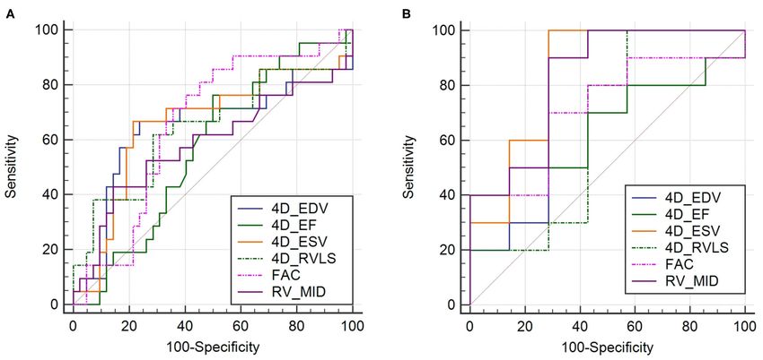
Wu et al. 4D-Echocardiographic in Pulmonary Hypertension
significant agreement with 4D auto-RVQ for most measurements except for 4D
end-diastolic volume. Indices measured by auto 4D-RVQ in the high-quality image group
had a good correlation with RHC but not in the average- and poor-quality image group.
Mid-RV diameter showed the best predictive power for the right RV-CI [area under the
curve (AUC) 0.935; 95% confidence interval (CI), 0.714–0.997; p < 0.001]. RV end-
systolic volume >121.50 mL had a 71.43% sensitivity and a 100% specificity to predict
right RV-CI (AUC, 0.890; 95% CI, 0.654–0.986; p < 0.001).
Conclusions: 4D auto-RVQ may be used to estimate RV function and some
hemodynamic changes compared with RHC in PHTN patients with high image quality.
Furthermore, a large sample of the study is needed to evaluate RV function by 4D
auto-RVQ in PHTN patients with average image quality.
Keywords: pulmonary hypertension, 4D-echocardiography, strain, right heart catheterization, right heart function
INTRODUCTION to use this technology to estimate right heart function and
important hemodynamic indices, as well as to obtain other
Echocardiography is the most commonly used imaging technique indirect information on right heart structure and function in
for the study of right ventricular (RV) morphology, volume, patients with PHTN.
function, and tissue characterization (1, 2). The accuracy of
2DE is inferior to cardiac magnetic resonance imaging (3).
Because of the complex RV geometry, 2DE cannot capture RV
MATERIALS AND METHODS
inflow and outflow in the same image acquisition. Real-time Study Population
three-dimensional echocardiography (3DE), also named four- Our study was a prospective cross-sectional project by design.
dimensional echocardiography (4DE), is a more accurate and Adult inpatients with PHTN and healthy volunteers were
quicker method to assess RV volume and function than 2DE, enrolled from September 2017 to December 2018. All patients
but in some cases, this approach still poses some technical with PHTN underwent echocardiography and RHC. Patients
difficulties (4). with PHTN were included if they had a diagnosis of idiopathic
The right ventricle has a unique crescent shape and complex PAH, chronic thromboembolic PAH, connective tissue disease
muscle moment, which influences the accurate evaluation of RV PAH, or residual PAH after surgery for congenital heart disease
function. In patients with pulmonary hypertension (PHTN), the by RHC with a mean PAP of >25 mm Hg (7). Patients
right heart chambers are significantly enlarged, and the scanning were included if they underwent 2D and 4D auto-RVQ on
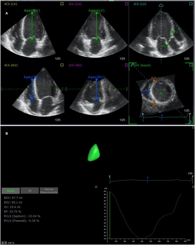
width of the echocardiography is insufficient to cover the entire the same day. Patients were excluded if they wereWu et al. 4D-Echocardiographic in Pulmonary Hypertension FIGURE 1 | End-systolic verification and editing of endocardial RV borders using TomTec 4D RV Function 2.0 (A) and 4D auto-RVQ measurement (B). RV, right ventricular; EDV, end-diastolic volume; ESV, end-systolic volume; EF, ejection fraction; RVLS (septum), right ventricular longitudinal strain of the septum; RVLS (free wall), right ventricular longitudinal strain of the free wall; TAPSE, tricuspid annular plane systolic excursion. Frontiers in Cardiovascular Medicine | www.frontiersin.org 3 July 2021 | Volume 8 | Article 628610
Wu et al.                                                                                                      4D-Echocardiographic in Pulmonary Hypertension

cardiac cycles were recorded during breath-holding with stable            TABLE 1 | Clinical, 2DE, and 4D auto-RVQ characteristics of 76 patients with
electrocardiography tracing. All patients underwent standard              pulmonary arterial hypertension.

2DE and Doppler echocardiography examinations with detailed               Variables                         PHTN (n = 76)        Healthy control           p-value
evaluation of right heart function. A comprehensive evaluation                                                                      (n = 25)
of the right ventricle by 2DE obtained six standardized views,
including the parasternal long-axis, RV inflow, parasternal short-        Age (years)                       37.13 ± 13.46         36.28 ± 12.56             >0.05
axis, apical four-chamber, and subcostal views.                           Gender (male)                       24 (31.6%)             9 (21.4%)              >0.05
   Offline analysis was then performed on digitally stored images.        BSA (m2 )                           1.57 ± 0.26
Indices of 2DE included TAPSE and RV fractional area change.              NT-proBNP (pg/mL)                 932.85 (322.2,
Right heart size was quantified as RV end-diastolic area at the                                               2,155.5)
end of the electrocardiogram (ECG) T-wave. RV chamber size                Hemodynamics (n = 76)
was assessed at the apex of the ECG R-wave in the apical four-            RAP (mm Hg)                         4.56 ± 4.01
chamber view with a focus on the right ventricle. We measured             mPAP (mm Hg)                      53.22 ± 14.58
pulmonary systolic pressure by Doppler tricuspid regurgitation            RV-CI (L/min/m2 )                2.95 (2.38, 3.50)
peak velocity plus estimated right atrial pressure.                       PVR (dyn·s·cm−5 )                940.29 (771.29,
                                                                                                             1,236.50)
4D Auto-RVQ Acquisition and Analysis                                      SvO2 (%)                           69.39 ± 5.75
Digital data were analyzed offline (EchoPAC 7 Workstation                 6 MWD (m)                        404.82 ± 98.74
version 201, GE Healthcare; TomTec 4D RV Function 2.0).                   Clinical classification
Measurements using 4D auto-RVQ were acquired using the 4-                 IPAH                                40 (52.6%)
V matrix-array transducer on the cardiac ultrasound GE system.            CTEPHTN                             15 (19.7%)
Images were acquired using single beats with frame rates of               CTD-PAH                            10 (13.2.0%)
≥12 FPS. Apical RV-focused four-chamber views were acquired               PAH after operation of CHD            5 (6.6%)
with patients in the lateral decubitus position. The transducer           2DE RV characteristics
position was modified for optimal simultaneous visualization              RVD (mm)                         30.0 (25.0, 36.0) 22.0 (19.3, 24.0)
Wu et al. 4D-Echocardiographic in Pulmonary Hypertension
between 4D auto-RVQ and strain, as well as those parameters 27 patients on whom RHC was not performed on the same day
measured using the reference method. Interobserver agreement as echocardiography. Finally, 76 patients diagnosed with PHTN
for qualitative analysis score using 4D auto-RVQ manual by RHC were recruited. We divided patients with PHTN (n = 76)
and software assessments was calculated using the intraclass into three groups according to echocardiographic image quality
correlation coefficient. Receiver operating characteristic (ROC) as follows: high image quality (n = 24), average image quality (n
curves were used to investigate and compare the predictive ability = 48), and poor image quality (n = 4). Clinical and conventional
of 4D auto-RVQ parameters to evaluate right heart function. A echocardiographic and 4D auto-RVQ characteristics of adult
p-value of 50.8 mm
RV, right ventricular; EDV, end-diastolic volume; ESV, end-systolic volume; EF, ejection had an 85.7% sensitivity and a 90.9% specificity. A RV-ESV of
fraction; RVLS (s, %), right ventricular septum longitudinal strain; RVLS (fw, %), right >121.5 mL had a 71.4% sensitivity and a 100% specificity, as well
ventricular free wall longitudinal strain; TAPSE, tricuspid annular plane systolic excursion;
FAC, fractional area change; RVD (basal/mid/long), right ventricular diameter (basal,
as a Youden index of 0.714 to predict RV-CI (AUC, 0.890; 95%
middle, longitudinal). CI, 0.654–0.986; p < 0.001).
TABLE 3 | Correlation between RHC and 4D auto-RVQ indices in all PHTN cases (n = 76).
RV-CI (L/min/m2 ) mRAP (mm Hg) mPAP (mm Hg) PVR (dyn·s·cm–5 )
4D indices R P R P R P R P
RV-EDV (mL) −0.131 0.268 0.221 0.061 0.223 0.056 0.080 0.496
RV-ESV (mL) −0.153 0.196 0.225 0.055 0.269 0.020 0.124 0.294
RV-SV (mL) −0.030 0.801 0.162 0.170 0.051 0.664 −0.045 0.700
RV-EF (%) 0.162 0.171 −0.135 0.257 −0.24 0.035 −0.195 0.096
RVLS (s, %) 0.037 0.758 −0.250 0.033 0.143 0.223 0.101 0.392
RVLS (fw, %) −0.095 0.425 −0.126 0.289 0.140 0.235 0.168 0.152
RVD (basal, mm) −0.266 0.031 0.275 0.025 0.081 0.513 0.063 0.612
RVD (mid, mm) −0.073 0.541 0.103 0.388 −0.01 -0.078 0.030 0.802
RVD (long, mm) 0.029 0.806 0.182 0.126 0.008 0.948 −0.164 0.166
TAPSE (mm) 0.226 0.056 0.072 0.546 −0.078 0.511 −0.268 0.022
RV-FAC (%) 0.152 0.203 −0.099 0.406 −0.165 0.162 −0.203 0.085
RV, right ventricular; EDV, end-diastolic volume; ESV, end-systolic volume; EF, ejection fraction; RVLS (s, %), right ventricular septum longitudinal strain; RVLS (fw, %), right ventricular
free wall longitudinal strain; TAPSE, tricuspid annular plane systolic excursion; FAC, fractional area change; RVD (basal/mid/long), right ventricular diameter (basal, middle, longitudinal);
RV-CI, right ventricular cardiac index; mRAP, mean right atrial pressure; mPAP, mean pulmonary arterial pressure; PVR, pulmonary vascular resistance.
Frontiers in Cardiovascular Medicine | www.frontiersin.org 5 July 2021 | Volume 8 | Article 628610Wu et al. 4D-Echocardiographic in Pulmonary Hypertension
TABLE 4 | Correlation between RHC and 4D auto-RVQ indices in the high-quality image PHTN group (n = 24).
RV-CI (L/min/m2 ) mRAP (mm Hg) mPAP (mm Hg) PVR (dyn·s·cm–5 )
4D indices R P R P R P R P
RV-EDV (mL) −0.579 0.007 0.548 0.012 0.503 0.024 0.444 0.050
RV-ESV (mL) −0.642 0.002 0.529 0.016 0.501 0.024 0.476 0.034
RV-SV (mL) −0.126 0.596 0.358 0.122 0.281 0.230 0.145 0.541
RV-EF (%) 0.529 0.016 −0.211 0.371 −0.169 0.477 −0.293 0.211
RVLS (s, %) −0.172 0.469 −0.225 0.340 0.351 0.129 0.565 0.009
RVLS (fw, %) −0.519 0.019 −0.032 0.893 0.120 0.615 0.506 0.023
RVD (basal, mm) −0.691 0.001 0.435 0.063 0.621 0.005 0.606 0.006
RVD (mid, mm) −0.501 0.025 0.312 0.180 0.290 0.229 0.641 0.002
RVD (long, mm) −0.204 0.402 0.678 0.001 0.720 0.0001 −0.056 0.819
TAPSE (mm) 0.070 0.775 0.357 0.133 −0.038 0.879 −0.324 0.176
RV-FAC (%) 0.510 0.026 −0.109 0.657 −0.163 0.504 −0.372 0.117
RV, right ventricular; EDV, end-diastolic volume; ESV, end-systolic volume; EF, ejection fraction; RVLS (s, %), right ventricular septum longitudinal strain; RVLS (fw, %), right ventricular
free wall longitudinal strain; TAPSE, tricuspid annular plane systolic excursion; FAC, fractional area change; RVD (basal/mid/long), right ventricular diameter (basal, middle, longitudinal);
RV-CI, right ventricular cardiac index; mRAP, mean right atrial pressure; mPAP, mean pulmonary arterial pressure; PVR, pulmonary vascular resistance.
TABLE 5 | All PHTN cases (n = 76) of 4D auto-RVQ areas of receiver operating characteristic (ROC) curves of various right ventricular function parameters.
ROC curve area 95% CI p-value Cutoff Sensitivity (%) Specificity (%) Youden index
RV-EDV (mL) 0.651 0.522–0.766 0.070 > 153.30 66.67 76.74 0.434
RV-ESV (mL) 0.664 0.535–0.777 0.040 > 117.50 66.67 79.07 0.457
RV-SV (mL) 0.561 0.431–0.685 0.485 > 40.20 42.86 81.40 0.242
RV-EF (%) 0.580 0.450–0.703 0.270 ≤25.63 76.19 51.16 0.273
RVLS (s, %) 0.525 0.396–0.651 0.770 > −9.39 71.43 11.63 0.169
RVLS (fw, %) 0.646 0.516–0.761 0.070 > −9.78 61.90 72.09 0.340
RVD (basal, mm) 0.643 0.505–0.766 0.080 > 39.4 70.59 57.50 0.281
RVD (mid, mm) 0.585 0.455–0.707 0.320 > 55.20 42.86 86.05 0.289
RVD (long, mm) 0.566 0.435–0.690 0.440 > 76.4 42.86 85.71 0.286
TAPSE (mm) 0.618 0.487–0.738 0.140 ≤6.5 33.33 90.48 0.238
RV-FAC (%) 0.658 0.527–0.773 0.029 ≤19.17 71.43 64.29 0.357
RV, right ventricular; EDV, end-diastolic volume; ESV, end-systolic volume; EF, ejection fraction; RVLS (s, %), right ventricular septum longitudinal strain; RVLS (fw, %), right ventricular
free wall longitudinal strain; TAPSE, tricuspid annular plane systolic excursion; FAC, fractional area change; RVD (basal/mid/long), right ventricular diameter (basal, middle, longitudinal).
DISCUSSION This may be due to the large RV volume in patients with PHTN
so the right ventricle could not be completely included in some
Despite its clinical importance, the study of RV function is cases. Furthermore, 4D auto-RVQ can possibly detect differences
technically challenging, in patients with PHTN. It is usually very in RV diameter, volume, and function. RV volume and basal
difficult to evaluate RV morphology and function using 2DE. and middle diameters were significantly enlarged in PHTN,
Recently, 3DE and 4DE have been introduced and are reportedly but the longitudinal diameter was not significantly changed.
feasible and clinically applicable for RV volumetric quantification This may be due to lateral expansion of the RV in patients
in patients with acquired RV pressure or volume overload (11). with PHTN. In terms of the strain indices measured by 4D
At present, the advantages of 4DE could better reflect RV size auto-RVQ, the longitudinal strains of the septum and free wall
and function. As a new technology, 4D auto-RVQ can combine were significantly lower in patients with PHTN compared with
the 3D echocardiographic morphological, functional, and strain the control group (15, 16). Similar to our findings, some reports
characteristics for comprehensive RV evaluation. It has the found that the longitudinal strain of the septum and free wall
potential to reflect changes in RV function and pulmonary artery had the potential to independently predict intermediate- to
hemodynamics (9, 12). high-risk features in PAH patients (17). Smith also showed that
Our results showed that 4D auto-RVQ, like 3D RV longitudinal strain of the free wall (hazard ratio, 7.63; 95% CI,
echocardiography, can accurately detect RV volume 1.76–10.27; p < 0.001) was a significant determinant of all-cause
measurements in both PHTN patients and healthy control mortality (18).
with good interobserver reproducibility (13, 14). However, the When analyzing the data from RHC and 4D auto-
intraclass correlation coefficient of 4D-EDV was suboptimal. RVQ indices, we observed a good correlation between 4D
Frontiers in Cardiovascular Medicine | www.frontiersin.org 6 July 2021 | Volume 8 | Article 628610Wu et al. 4D-Echocardiographic in Pulmonary Hypertension
FIGURE 2 | Receiver operating characteristic curves in all patient groups of PHTN (n = 76) of 4D auto-RVQ areas of (A) and in the high-quality image group of PHTN
(n = 24) (B). 4D_EDV, four-dimensional end-diastolic volume; 4D_ESV, four-dimensional end-systolic volume; 4D_EF, four-dimensional ejection fraction; 4D-RVLS,
right ventricular longitudinal strain of the free wall; FAC, fractional area change; RV_MID, mid–right ventricular diameter.
TABLE 6 | High-quality image group of PHTNs (n = 24) of 4D auto-RVQ areas of receiver operating characteristic (ROC) curves of various right ventricular function
parameters.
ROC curve area 95% CI p-value Cutoff Sensitivity (%) Specificity (%) Youden index
RV-EDV (mL) 0.831 0.583–0.963 0.007 > 153.94 71.43 100 0.714
RV-ESV (mL) 0.890 0.654–0.986 0.001 > 121.50 71.43 100 0.714
RV-SV (mL) 0.636 0.381–0.845 0.421 > 41.45 57.14 90.91 0.481
RV-EF (%) 0.779 0.525–0.937 0.002 ≤26.21 100 54.55 0.545
RVLS (s, %) 0.506 0.266–0.745 0.960 > −5.73 42.86 81.82 0.245
RVLS (fw, %) 0.773 0.518–0.993 0.019 > −9.95 71.43 81.82 0.532
RVD (basal, mm) 0.831 0.583–0.963 0.001 > 34.15 85.71 81.82 0.675
RVD (mid, mm) 0.935 0.714–0.997 0.000 > 50.8 85.71 90.91 0.766
RVD (long, mm) 0.600 0.340–0.824 0.540 > 78.6 42.86 100 0.428
TAPSE (mm) 0.557 0.302–0.792 0.714 ≤11.3 71.43 60 0.314
RV-FAC (%) 0.800 0.539–0.951 0.011 ≤14.66 71.43 90 0.614
RV, right ventricular; EDV, end-diastolic volume; ESV, end-systolic volume; EF, ejection fraction; RVLS (s, %), right ventricular septum longitudinal strain; RVLS (fw, %), right ventricular
free wall longitudinal strain; TAPSE, tricuspid annular plane systolic excursion; FAC, fractional area change; RVD (basal/mid/long), right ventricular diameter (basal, middle, longitudinal).
quantification and RHC in patients with high-quality images, but We did notice that some authors previously used RV-EF as
not in the other two groups with suboptimal imaging quality. an index compared with cardiac magnetic resonance imaging
RV-ESV by 4D auto-RVQ tracked well with RV-CI, mean right (19, 20).
atrial pressure, mean PAP, and PVR measurements with RHC. In summary, 4D auto-RVQ is a possibly useful quantitative
We propose that RV-ESV may be an effective indicator of tool to measure RV function with validation by RHC and
right heart function and hemodynamic changes in patients with provide meaningful data reflecting RV function. It is
PHTN. RV function analysis needs to take into consideration important to obtain high-quality RV images and to include
of its afterload including PVR. Measurement of 4D-ESV, 4D- the entire RV within the scanning sector. Previous studies
RV strain, and RV basal and middle diameters had a good have shown that in patients with chronic PHTN, 3D and
correlation with PVR. In the high-quality image group, all six 3D speckle-tracking echocardiography parameters for global
4DE parameters predicted right heart function. The indices with and regional RV dysfunction were better than conventional
the best predictive power were RV middle diameter and 4D-ESV. echocardiographic indices (21, 22). The software for 4D
Frontiers in Cardiovascular Medicine | www.frontiersin.org 7 July 2021 | Volume 8 | Article 628610Wu et al. 4D-Echocardiographic in Pulmonary Hypertension
auto-RVQ generates functional parameters from 4D datasets, DATA AVAILABILITY STATEMENT
making it an easy tool to implement in clinical practice
and reducing the amount of time spent using multiple The raw data supporting the conclusions of this article will be
software parameters to simultaneously analyze 4D datasets made available by the authors, without undue reservation.
and speckle-tracking echocardiography.
RHC remains the gold standard for RV hemodynamic ETHICS STATEMENT
evaluation, but 4D auto-RVQ has the potential to be
included in routine clinical applications because it provides The studies involving human participants were reviewed and
a relatively quantitative evaluation method for RV function approved by the Ethics Committee of Fuwai Hospital (No. 2018-
in PHTN patients if high-quality echocardiographic images 1063). Written informed consent to participate in this study was
were acquired. provided by the participants’ legal guardian/next of kin.
LIMITATIONS AUTHOR CONTRIBUTIONS
There are several limitations to our data and results. As a WW, BL, and MH drafted the manuscript. LN, YT, JL, JW, SY,
single-center study in a cardiac referral center, we might have and HL contributed to data collection and statistical analysis.
had selection bias. Among 103 patients, 27 patients did not HW, DH, CX, and ZZ provided supervision and revised the
simultaneously undergo ultrasonography and RHC and were manuscript. CX contributed to the conception, design, and
excluded from the final analysis. World Health Organization supervision of the study. All authors contributed to the article
functional class IV patients were not enrolled in the study. and approved the submitted version.
Our data are mainly applicable to patients with high-quality
echocardiographic images. Our sample size was relatively small FUNDING
for an ROC analysis. Finally, we collected only the RHC data
of PHTN without the results of cardiac magnetic resonance The present study was supported by a grant from Capital
imaging. Thus, it is difficult to evaluate changes in cardiac volume Health Development and Scientific Research Projects
in real time. (Grant No. 2016-2-4036). Fund from Construction of Key
Laboratory (Cultivation) of Chinese Academy of Medical
Sciences (2019PT310025).
CONCLUSION
In conclusion, 4D auto-RVQ is a new method to ACKNOWLEDGMENTS
estimate RV function and hemodynamic changes
compared with gold-standard RHC in patients with We thank Emily Woodhouse, Ph.D., from Liwen Bianji, Edanz
PHTN only if high-quality echocardiographic images Editing China (www.liwenbianji.cn/ac), for editing the English
were acquired. text of a draft of this manuscript.
REFERENCES guideline from the British Society of Echocardiography. Echo Res Pract. (2020)
7:G19–41. doi: 10.1530/ERP-19-0051
1. Dandel M, Hetzer R. Evaluation of the right ventricle by echocardiography: 7. Galiè N, Humbert M, Vachiery J-L, Gibbs S, Lang I, Torbicki A, et al.
particularities and major challenges. Expert Rev Cardiovasc Ther. (2018) 2015 ESC/ERS Guidelines for the diagnosis and treatment of pulmonary
16:259–75. doi: 10.1080/14779072.2018.1449646 hypertension: The Joint Task Force for the Diagnosis and Treatment of
2. Venkatachalam S, Wu G, Ahmad M. Echocardiographic assessment of Pulmonary Hypertension of the European Society of Cardiology (ESC)
the right ventricle in the current era: application in clinical practice. and the European Respiratory Society (ERS): Endorsed by: Association
Echocardiography. (2017) 34:1930–47. doi: 10.1111/echo.13651 for European Paediatric and Congenital Cardiology (AEPC), International
3. Gürdogan M, Ustabaşioglu FE, Kula O, Korkmaz S. Cardiac magnetic Society for Heart and Lung Transplantation (ISHLT). Eur Respir J. (2015)
resonance imaging and transthoracic echocardiography: investigation of 46:903–75. doi: 10.1183/13993003.01032-2015
concordance between the two methods for measurement of the cardiac 8. Miller WL, Grill DE, Borlaug BA. Clinical features, hemodynamics, and
chamber. Medicina. (2019) 55:260. doi: 10.3390/medicina55060260 outcomes of pulmonary hypertension due to chronic heart failure with
4. Kjaergaard J, Petersen CL, Kjaer A, Schaadt BK, Oh JK, Hassager C. reduced ejection fraction: pulmonary hypertension and heart failure. JACC
Evaluation of right ventricular volume and function by 2D and 3D Heart Fail. (2013) 1:290–9. doi: 10.1016/j.jchf.2013.05.001
echocardiography compared to MRI. Eur J Echocardiogr. (2006) 7:430–8. 9. Aubert R, Venner C, Huttin O, Haine D, Filippetti L, Guillaumot A,
doi: 10.1016/j.euje.2005.10.009 et al. Three-dimensional echocardiography for the assessment of right
5. Lang RM, Badano LP, Mor-Avi V, Afilalo J, Armstrong A, Ernande L, et al. ventriculo-arterial coupling. J Am Soc Echocardiogr. (2018) 31:905–15.
Recommendations for cardiac chamber quantification by echocardiography doi: 10.1016/j.echo.2018.04.013
in adults: an update from the American Society of Echocardiography and 10. Rudski LG, Lai WW, Afilalo J, Hua L, Handschumacher MD, Chandrasekaran
the European Association of Cardiovascular Imaging. Eur Heart J Cardiovasc K, et al. Guidelines for the echocardiographic assessment of the right
Imaging. (2015) 16:233–70. doi: 10.1093/ehjci/jev014 heart in adults: a report from the American Society of Echocardiography
6. Zaidi A, Knight DS, Augustine DX, Harkness A, Oxborough D, Pearce K, endorsed by the European Association of Echocardiography, a registered
et al. Echocardiographic assessment of the right heart in adults: a practical branch of the European Society of Cardiology, and the Canadian
Frontiers in Cardiovascular Medicine | www.frontiersin.org 8 July 2021 | Volume 8 | Article 628610Wu et al. 4D-Echocardiographic in Pulmonary Hypertension
Society of Echocardiography. J Am Soc Echocardiogr. (2010) 23:685–713. in pulmonary hypertension. J Am Coll Cardiol. (2014) 64:41–51.
doi: 10.1016/j.echo.2010.05.010 doi: 10.1016/j.jacc.2014.01.084
11. Knight DS, Grasso AE, Quail MA, Muthurangu V, Taylor AM, Toumpanakis 19. Kidawa M, Chizyński K, Zielińska M, Kasprzak JD, Krzeminska-Pakula M.
C, et al. Accuracy and reproducibility of right ventricular quantification Real-time 3D echocardiography and tissue Doppler echocardiography in the
in patients with pressure and volume overload using single-beat three- assessment of right ventricle systolic function in patients with right ventricular
dimensional echocardiography. J Am Soc Echocardiogr. (2015) 28:363–74. myocardial infarction. Eur Heart J Cardiovasc Imaging. (2013) 14:1002–9.
doi: 10.1016/j.echo.2014.10.012 doi: 10.1093/ehjci/jes321
12. Zhang XM, Zhuang Q, Yang MH, Wang W, Zheng Y, Qiao ZQ, et al. 20. Gopal AS, Chukwu EO, Iwuchukwu CJ, Katz AS, Toole RS, Schapiro W,
[Value of four-dimensional echocardiography combined with speckle tracking et al. Normal values of right ventricular size and function by real-time 3-
technique on the assessment of right heart function and prognosis in patients dimensional echocardiography: comparison with cardiac magnetic resonance
with pulmonary arterial hypertension]. Zhonghua Xin Xue Guan Bing Za Zhi. imaging. J Am Soc Echocardiogr. (2007) 20:445–55. doi: 10.1016/j.echo.2006.
(2018) 46:965–71. doi: 10.3760/cma.j.issn.0253-3758.2018.12.007 10.027
13. Nagata Y, Wu VC-C, Kado Y, Otani K, Lin F-C, Otsuji Y, et al. Prognostic 21. Vitarelli A, Mangieri E, Terzano C, Gaudio C, Salsano F, Rosato
value of right ventricular ejection fraction assessed by transthoracic E, et al. Three-dimensional echocardiography and 2D-3D speckle-
3D echocardiography. Circ Cardiovasc Imaging. (2017) 10:e005384. tracking imaging in chronic pulmonary hypertension: diagnostic
doi: 10.1161/CIRCIMAGING.116.005384 accuracy in detecting hemodynamic signs of right ventricular (RV)
14. Moceri P, Duchateau N, Baudouy D, Schouver E-D, Leroy S, Squara F, et al. failure. J Am Heart Assoc. (2015) 4:e001584. doi: 10.1161/JAHA.114.0
Three-dimensional right-ventricular regional deformation and survival in 01584
pulmonary hypertension. Eur Heart J Cardiovasc Imaging. (2018) 19:450–8. 22. Lu KJ, Chen JXC, Profitis K, Kearney LG, DeSilva D, Smith G, et al.
doi: 10.1093/ehjci/jex163 Right ventricular global longitudinal strain is an independent predictor
15. Jone P-N, Schäfer M, Pan Z, Bremen C, Ivy DD. 3D echocardiographic of right ventricular function: a multimodality study of cardiac magnetic
evaluation of right ventricular function and strain: a prognostic study in resonance imaging, real time three-dimensional echocardiography and
paediatric pulmonary hypertension. Eur Heart J Cardiovasc Imaging. (2018) speckle tracking echocardiography. Echocardiography. (2015) 32:966–74.
19:1026–33. doi: 10.1093/ehjci/jex205 doi: 10.1111/echo.12783
16. Leong DP, Grover S, Molaee P, Chakrabarty A, Shirazi M, Cheng
YH, et al. Nonvolumetric echocardiographic indices of right ventricular Conflict of Interest: The authors declare that the research was conducted in the
systolic function: validation with cardiovascular magnetic resonance and absence of any commercial or financial relationships that could be construed as a
relationship with functional capacity. Echocardiography. (2012) 29:455–63. potential conflict of interest.
doi: 10.1111/j.1540-8175.2011.01594.x
17. Liu B-Y, Wu W-C, Zeng Q-X, Liu Z-H, Niu L-L, Tian Y, et al. The value Copyright © 2021 Wu, Liu, Huang, Hsi, Niu, Tian, Lin, Wang, Yang, Lu, Xiong, Zhu
of three-dimensional echocardiography in risk stratification in pulmonary and Wang. This is an open-access article distributed under the terms of the Creative
arterial hypertension: a cross-sectional study. Int J Cardiovasc Imaging. (2020) Commons Attribution License (CC BY). The use, distribution or reproduction in
36:577–84. doi: 10.1007/s10554-019-01743-1 other forums is permitted, provided the original author(s) and the copyright owner(s)
18. Smith BCF, Dobson G, Dawson D, Charalampopoulos A, Grapsa J, are credited and that the original publication in this journal is cited, in accordance
Nihoyannopoulos P. Three-dimensional speckle tracking of the right with accepted academic practice. No use, distribution or reproduction is permitted
ventricle: toward optimal quantification of right ventricular dysfunction which does not comply with these terms.
Frontiers in Cardiovascular Medicine | www.frontiersin.org 9 July 2021 | Volume 8 | Article 628610You can also read