A Pulmonary Artery-Vein Separation Algorithm Based on the Relationship between Subtrees Information
←
→
Page content transcription
If your browser does not render page correctly, please read the page content below
Hindawi
Journal of Healthcare Engineering
Volume 2021, Article ID 5550379, 8 pages
https://doi.org/10.1155/2021/5550379

Research Article
A Pulmonary Artery-Vein Separation Algorithm Based on the
Relationship between Subtrees Information

 Kun Yu,1,2 Ziming Zhang,1,3 Xiaoshuo Li,3 Pan Liu,3 Qinghua Zhou,3 and Wenjun Tan 1,3

 1
 Key Laboratory of Intelligent Computing in Medical Image, Ministry of Education, Shenyang 110189, China
 2
 College of Medicine and Biological Information Engineering, Northeastern University, Shenyang 110189, China
 3
 College of Computer Science and Engineering, Northeastern University, Shenyang 110189, China

 Correspondence should be addressed to Wenjun Tan; tanwenjun@cse.neu.edu.cn

 Received 19 January 2021; Revised 20 May 2021; Accepted 29 May 2021; Published 9 June 2021

 Academic Editor: Ayush Dogra

 Copyright © 2021 Kun Yu et al. This is an open access article distributed under the Creative Commons Attribution License, which
 permits unrestricted use, distribution, and reproduction in any medium, provided the original work is properly cited.
 Physicians need to distinguish between pulmonary arteries and veins when diagnosing diseases such as chronic obstructive
 pulmonary disease (COPD) and lung tumors. However, manual differentiation is difficult due to various factors such as
 equipment and body structure. Unlike previous geometric methods of manually selecting the points of seeds and using neural
 networks for separation, this paper proposes a combined algorithm for pulmonary artery-vein separation based on subtree
 relationship by implementing a completely new idea and combining global and local information, anatomical knowledge, and
 two-dimensional region growing method. The algorithm completes the reconstruction of the whole vascular structure and the
 separation of adhesion points from the tree-like structure characteristics of blood vessels, after which the automatic classification
 of arteries and veins is achieved by using anatomical knowledge, and the whole process is free from human intervention. After
 comparing all the experimental results with the gold standard, we obtained an average separation accuracy of 85%, which achieved
 effective separation. Meanwhile, the time range could be controlled between 40 s and 50 s, indicating that the algorithm has
 good stability.

1. Introduction of information for the development of vascular surgery
 strategies.
Pulmonary artery-vein (A/V) separation of CT images is At the beginning, some geometric and artificial methods
important to fit downstream medical tasks quantitatively in were proposed, but they have great limitations. Nakamura
clinic. In CT images, pulmonary arteries and veins overlap in et al. [3] have proposed a pulmonary vascular classification
many places. The discrimination of arterial from venous method based on the spatial arrangement of blood vessels
irrigation is challenge because of (1) the limited scanning using the distance of the bronchus from the blood vessels
resolution of CT image, (2) the relatively high spatial density and the distance of interlobar fissures from the closest
of pulmonary blood vessels, and (3) the part of the volume pulmonary blood vessels. However, there are only three
and size effect. These factors also lead to complexity in cases included in the study, which limited its generality. A
separating arteries and veins in pulmonary CT images [1]. tracking-based automated method for low-dose CT scans to
Meanwhile, the classification of arteries and veins is also of separate arteries from other surrounding structures has been
great practical significance for diagnosis of chronic ob- introduced by Wala et al. [4]. The algorithm starts from
structive pulmonary diseases (COPDs) and the detection of automatically detected seed points located in the basal
lung tumors, which attracts many efforts to the development pulmonary areas and continues by tracking the vessel and
of this area [2]. The separated arteries and veins can be more detecting bifurcations. A quantitative evaluation of results
visually presented to the physician, allowing for more ac- reported 64% sensitivity and 90% specificity values. Using
curate and effective patient care, as well as providing a wealth multiscale opening operators, Saha et al. [5] have also
2 Journal of Healthcare Engineering

separated the arteries and veins of different sizes and lo- 2. Materials and Methods
cations based on two sets of manually selected seeding
points. The disadvantage of the above two methods lies in the 2.1. Geometric Graph Representation and Separation. The
manual collection of seed points, which requires a high pulmonary vasculature can be abstracted as a feature tree
degree of professionalism and increases the workload of with a certain logical structure maintaining the original
doctors. Gao et al. [6] then have improved Saha’s semiau- vascular topology. Such trees can be bifurcated structures,
tomatic method by embedding a 2D-3D interconnected trinomial structures, and so on. The classification of vessels
graphical user interface to achieve 91%–95% accuracy, but essentially separates the adhesion points on the CT map so
they still have the effort consuming problem caused by user that the arteries and veins form two separate trees.
intervention. The method used by Park et al. [2] is to Due to the morphological complexity, pulmonary blood
construct minimum spanning trees and cut edges to separate vessels appear to be bifurcations, trifurcations, artery-vein
arteries and veins. Although it can effectively achieve the attachments, and false positives in CT images, making it
separation, manual optimization is still needed. difficult for a centerline algorithm to deal with them. On the
 Later automatic separation methods were proposed, and other hand, the centerline algorithm shows good structural
neural networks were used for A/V separation. Kitamura characteristics when the vessels are locally disturbed by noise
et al. [7] proposed an automatic A/V separation method. It or false positives [11]. Therefore, this paper starts with
uses energy minimization of high-order potentials and se- centerline algorithm, employing a geometric graph repre-
lects higher-order clusters based on prior knowledge of data sentation to roughly separate vascular branch structures and
and the desired shape. This method has been evaluated in 10 nonvascular structures.
sets of CT data. However, only the pulmonary vessels with Ideally, a normal bifurcated vessel will be a pixel con-
CT values greater than 200 HU have been considered, which nected with three adjacent pixels (Figure 1(a)). The relevant
to certain extent impairs its universality. A classification information about a center point (a node) and its 26
method based on a deep learning novel and powerful neighbors can be stored in a data structure as follows: idx
method for dealing with non-Euclidean data is presented, represents the location information of the pixels based on
while Convolutional Neural Network (CNN) framework has which adhesion point is calculated (some are more than
been proposed recently [8]. Using a 3D CNN method, small one); links represent the index matrix of a link to which the
blood vessels are extracted directly from noncontrast CT adhesion point is connected; conn represents the index
images to learn specific A/V characteristics without further matrix of a node adjacent to the adhesion point;
operation. However, the model is only evaluated for its com x, com y, com z represent the x, y, and z coordinates
ability to diagnose COPD in the article, which lacks a val- of the adhesion point, respectively; and ep is equal to either 0
idated evaluation of the accuracy for A/V separation. More or 1, where 0 represents the nonleaf nodes and 1 represents
recently, a new framework to approach the separation of the leaf nodes. The connectivity is defined within the 26
tree-like structures using local information along with a neighbors, and edges represent a voxel collection of two
specifically designed graph-cut methodology has been adjacent points, shown as a simple tubular structure.
proposed [9]. It employs a random forest (RF) preclassifier A link structure includes 3 parts as follows: n start
to exploit the local anatomical differences of arteries and represents the index value of the starting pixel; n end
veins. The experiments reveal a relevant improvement in the represents the index value of the end pixel;points represent
accuracy of the vessel classification with the proposed the index value of all pixels contained in the link. Each node
framework compared to using only local information. Graph and link can represent any morphological structure men-
Convolutional Networks (GCNs) are a powerful method for tioned above, i.e., bifurcations, trifurcations, attachments,
dealing with non-Euclidean data, while CNNs can learn end points, or false positive structures. Once a data structure
features from Euclidean data such as images [10]. In Zhai of a node is known, we can obtain its neighborhood topology
et al.’s work, they propose a novel method to combine CNNs through the link property information. As shown in
with GCNs (CNN-GCN), which can consider both Eu- Figure 1(b), we can easily identify tri- and multifurcation
clidean and non-Euclidean features, to separate the pul- through this transformation. The corresponding nodes are
monary vascular trees into arteries and veins (A/V) [10]. It shown as yellow circles. The rest of the circles are leaf nodes
shows a better performance compared with CNN method colored blue. Branches are divided into red and black colors,
but slightly worse results compared to observers, which where black represents branches connected to the leaf nodes,
needs a further improvement. and the rest are colored red.
 Here, we propose a novel algorithm for pulmonary When a length of a link (the value of the property
artery-vein (A/V) separation. In this method, local and “links”) is greater than 3, there will be four or more blood
global information, anatomic knowledge, and 2D area vessels connected around the certain node. This node is then
growth method are involved, aiming to solve the above- regarded as an adhesion point of arteries and veins. After
mentioned problems in pulmonary arteries and veins sep- detecting the adhesion points, an originally continuous link
aration. An average accuracy of 85% has been achieved in the will be split into two links. Take quadfurcations as an ex-
overall process. The processing time is ranging between 40 s ample, the coordinates of the central adhesion point are set
and 50 s, indicating good performance and stability of this to (x, y, z), and the coordinates of the two leaf nodes with an
algorithm. Our new method may provide a new idea for the unknown angle are set to A (x1, y1, z1) and B (x2, y2, z2),
A/V classification of CT images. respectively. The rearrangement of four links will be done by
Journal of Healthcare Engineering 3
(a) (b)
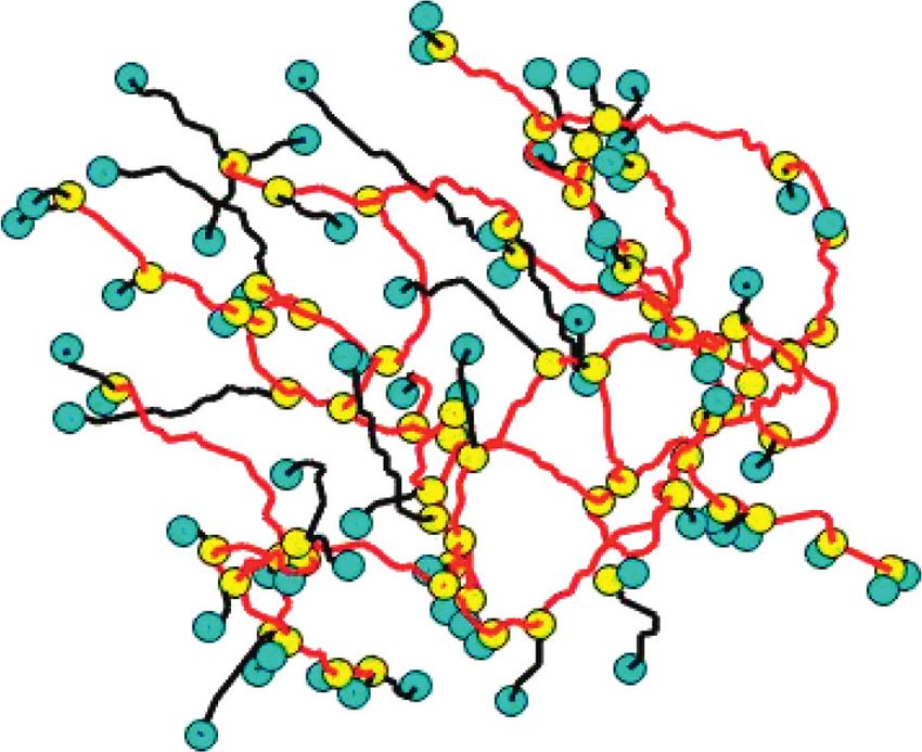
Figure 1: Schematic diagram of centerline to geometry: (a) centerlines of blood vessels; (b) geometric graph representation.
calculating the angles (θ) between branches with a single leaf relationship between subtrees. This method is based on
node and their adhesion point, according to the following anatomic knowledge that (1) arteries and veins meet at al-
equations: veolar sacs and (2) arteries and veins are approaching each
2 2 2 other rather than connected to each other [11, 12]. Thus, a
a1 � x1 − x + y1 − y + z1 − z , (1) one-to-one relationship can then be built based on the
spatial location of the leaf nodes from two subtrees without
2 2 2
b1 � x2 − x + y2 − y + z2 − z , (2) knowing the classes. By analyzing each leaf node of a subtree
and finding the closest leaf node from another subtree within
2 2 2 a Euclidean distance, peripheral matching defines the
c1 � x1 − x2 + y1 − y2 + z1 − z2 , (3)
matching strength between two subtrees (IMS) increasing by
a1 + b1 − c1 1. A higher IMS indicates two subtrees likely belonging to
pos � , (4) different classes of blood vessels. To reduce the coincidence
2 ∗ sqrt a1 ∗ sqrt b1
of establishing this relationship, a minimum value of IMS
180 between the two vascular subtrees is required (Figure 3).
θ � arccos(pos) ∗ . (5) Peripheral matching can describe the relationship be-
π tween two subtrees with the value of IMS. However, in some
According to the above quadfurcation, there will be three cases, it is still unknown whether there is a relationship
different combinations. The combination with the maxi- between two subtrees if they do not have an IMS value. For
mum value of θ will be adopted. Meanwhile, a new pair of this situation, we can establish indirect class relationships
links will be regenerated to represent the original four links. through the known relationships. For example, for three
Both the intrinsic property of the original adhesion node and subtrees S � {ψ A1 , ψ B2 , ψ C3 }, the exact A/V classes information
the connected links are then modified. For different links of A, B, and C is unknown. However, both subtrees of class A
passing through adhesion points, a separation is made to and C, and class B and C have a high IMS. It is easy to infer
ensure they belong to a different class. that A and B belong to a different class compared to C. Since
there are only two classes, subtree A and subtree B then
belong to the same class of subtrees. After the division of
2.2. Combination of Subtrees. Starting with the leaf nodes, relationships between subtree classes, the number of dif-
connected links are classified into many different classes. In ferent subtree types in the geometric graph can be further
practice, the sub_trees can only belong to either an arterial reduced.
class or a venous class. The reduction of links classification is
achieved by a flowchart shown in Figure 2. Starting with the
leaf nodes, connected links can be classified into the same 2.4. Restoration of Vascular Tree. Before distinguishing the
class. After this operation, the adhesion points in a subtree specific class of blood vessels, a restoration of vascular tree
can be removed, leaving only zero and trifurcations. Pul- based on geometric graph is conducted. Each pixel point is
monary blood vessels have been divided into a set of subtrees used as a seed point for a two-dimensional (2D) region
based on local information. growing. A new dataset is constructed and the corre-
sponding vascular subtree is exported until all data slides are
2.3. Relationships between Subtrees. In the next steps, we will processed. Through this restoration procedure, an intact
extract the relationship between the subtrees using global tubular structure of pulmonary blood vessels can be built
information. Peripheral matching is applied to establish the (Figure 4), which will facilitate the further classification step.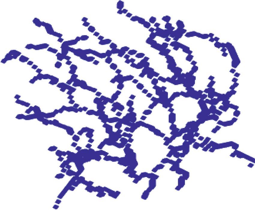
4 Journal of Healthcare Engineering

 Start

 Have the leaf nodes Yes
 End
 been traversed?
 Yes
 No

 Add 1 to the COUNT Has this node been
 property traversed?

 No
 Push this node into stack

 Yes Is the
 stack empty?

 No

 Is the CONN Change the COVER property
 Yes
 property of the of the element on the top of
 stack empty? the stack as ACCESSED

 No

 Push the node from the CONN
 matrix into the stack

 Figure 2: Flowchart of reducing subtree classification.

2.5. Classification of Blood Vessels. The final task is to 3. Results and Discussion
classify these vascular trees by the volume differences
between arteries and veins. Based on anatomical To evaluate the performance of our method, 10 sets of CT
knowledge, the overall volume of venous trees is higher image data from a top first-class hospital are used. The data
than arterial trees. Thus, the total volume of subtrees resolution is 512 × 512, and the number of data layers is
belonging to the same class is extracted from the vascular between 368 and 436. In order to verify the accuracy of
segmentation. A volume of each class per unit length is pulmonary A/V separation, the data of the extracted to-
used, where the class with the highest volume is classified pology of pulmonary blood vessels are used for experiments.
as veins and the other class as arteries. At this stage, the All experiments are conducted using MATLAB 2015b and
majority of subtrees have been classified. However, for
subtrees with only a few leaf nodes, there is not enough
 ® ™
 Visual Studio 2010 on a PC with Intel Core i5-6700 CPU
 @ 3.40 GHz processor, 8.00 GB memory, and 1 TB Western
peripheral information to establish a matching rela- Data hard drive.
tionship. This kind of subtrees cannot connect to any As shown in Figure 5, each set of data is converted into
others. To ensure the continuity of vascular labeling, each geometric graph representations (Figures 5(e)–5(h)) from
leaf node needs to connect to the root node through just centerlines (Figures 5(a)–5(d)). Examples of details from
one path. At each node in this path, the next direction is corresponding geometric graphs are shown in Figures 5(i)–
given by maximizing the branch angle and minimizing 5(l), where both nodes and branches are divided into two
the difference in the diameter of the blood vessels be- categories. Leaf nodes are represented in blue color, adhe-
tween the front and back edges. For some unlabeled sion nodes are colored yellow, branches connected to leaf
subtrees, there may be more than one path to the root nodes are colored black, and the rest of the branches are
node. They are classified by a majority vote of the already colored red. This representation will facilitate the acquisition
classified edges. If there are more subtrees in these paths of topological relationships between nodes and branches.
that belong to the veins, then the unlabeled subtree For the nodes at the adhesion points with more than 3
belongs to the venous class; otherwise, it belongs to the branches, a geometric graph separation process is carried
arterial class. out. These adhesion points are taken as central points, and

Journal of Healthcare Engineering 5

 Start

 No
 i
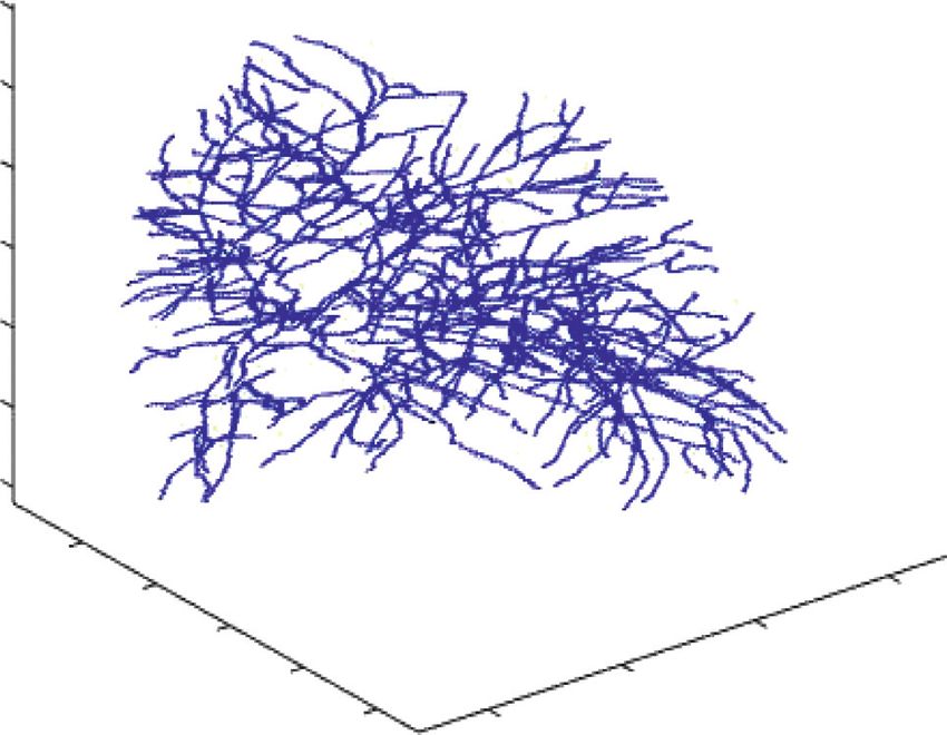
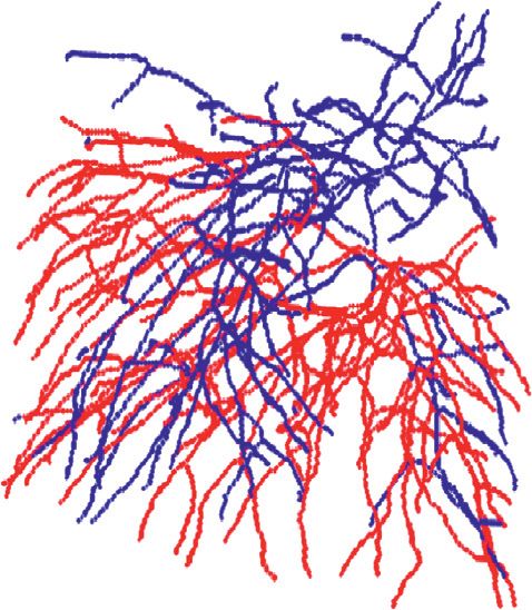
6 Journal of Healthcare Engineering

 (a) (b) (c) (d)

 (e) (f ) (g) (h)

 (i) (j) (k) (l)

 (m) (n) (o) (p)

Figure 5: Schematic diagram of the pulmonary vascular geometric graph representation: (a–d) centerlines reconstructed based on Dataset 1
(a, b) and Dataset 2 (c, d), among which a and c are right lungs, and b and d are left lungs; (e–h) horizontally corresponding geometric graph
representation; (i–l) graphic details of corresponding representations; (m–p) subtree separation results.

region growing method. As shown in Figure 6, pulmonary the branches. The accuracy of the separation of the right and
arteries are colored red, and pulmonary veins are colored left lungs was similar at about 85% at different levels.
blue. However, as the number of layers increased, the accuracy of
 We next evaluate the experimental performance. During both left and right lung separation decreased slightly and the
the whole process, some adhesion points with five or more running time increased slightly.
branches are not fully separated due to their structural Moreover, the final separation accuracy is obtained by
complexity, which results in a misjudgment of the A/V comparing the experimental results with the gold standard
separation. Here, a preliminary accuracy rate is obtained with an average accuracy of 85% (Table 1). The processing
from the ratio of misjudged branches to the total number of time of the whole process/algorithm is then recorded. The
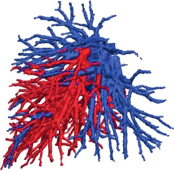
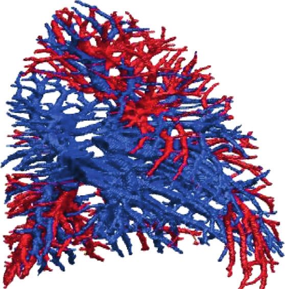
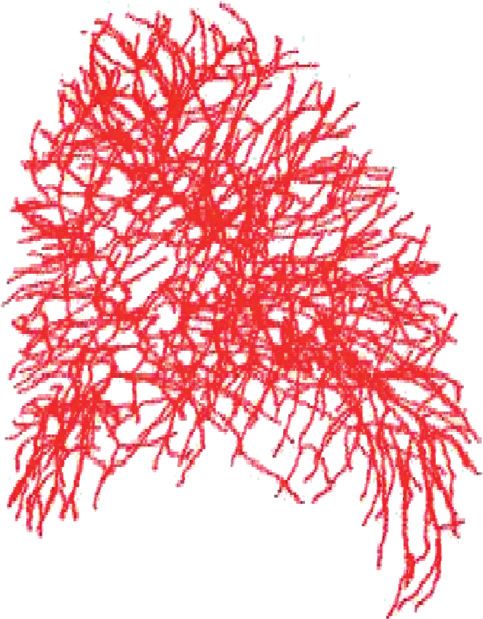
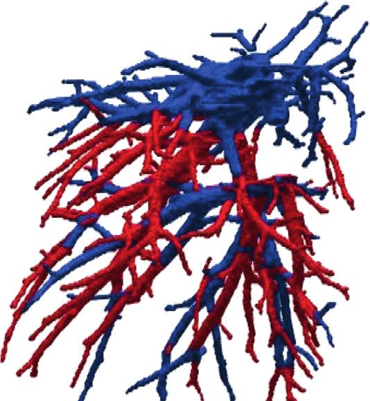
Journal of Healthcare Engineering 7

 (a) (b) (c) (d)

Figure 6: Schematic diagram of pulmonary arteriovenous vascular tree: (a) Dataset 1, right lung; (b) Dataset 1, left lung; (c) Dataset 2, right
lung; (d) Dataset 2, left lung.

 Table 1: Accuracy of pulmonary arteriovenous separation.
 Separation accuracy
 Statistic of branches
 (%)
 No. of Running
Dataset Total number of The number of Total number of The number of Acc of Acc of
 layers time (s)
 branches in the misjudged branches branches in the misjudged branches left lung right
 left lung in the left lung right lung in the right lung (%) lung (%)
1 368 943 123 532 59 86.76 85.74 46
2 386 498 68 506 70 86.34 86.03 49
3 378 624 84 587 85 85.74 85.12 52
4 403 486 79 619 95 83.79 84.64 48
5 436 536 89 472 74 83.40 84.32 52
 Average 85.20 85.17 49.4

running time of all five experiments described above exhibits Conflicts of Interest
a smooth line within the time range between 40 s and 50 s
(Table 1), indicating a good stability of this algorithm. The authors declare that there are no conflicts of interest
 regarding the publication of this paper.
4. Conclusions
This work achieves an automatic A/V separation by a
 Acknowledgments
method combining both global and local information. This work was supported by the Fundamental Research
Firstly, a geometric graph is built based on the centerlines; Funds for the Central Universities (N182410001 and
next, a separation of the adhesion points is performed by N2104008) and National Natural Science Foundation of
applying the information of the geometric graphs; subtrees China (61971118).
are then combined according to the local and global in-
formation; the combined subtrees are growing into vascular
trees by using 2D area growth method; and finally the ar- References
terial and venous vascular trees are classified based on the [1] A. Fabijańska, “Segmentation of pulmonary vascular tree from
knowledge of anatomy. The average accuracy of the overall 3D CT thorax scans,” Biocybernetics & Biomedical Engi-
process can reach 85% which is promising for a clinical neering, vol. 35, no. 2, pp. 106–119, 2015.
application. [2] S. Park, S. M. Lee, N. Kim, J. B Seo, and H. Shin, “Automatic
 Currently, there are relatively few studies that perform reconstruction of the arterial and venous trees on volumetric
automatic arteriovenous separation with both global and chest CT,” Medical Physics, vol. 40, no. 7, Article ID 71906,
local information. This study can serve as a guide in the 2013.
treatment of lung diseases such as pulmonary nodules and [3] S. Nakamura, Y. Mekada, I. Ide et al., “Pulmonary artery and
lung tumors. In future studies, we will investigate how we vein classification method using spatial arrangement features
can differentiate arterioles more quickly and accurately. from X-ray CT image,” International Congress Series,
 vol. 1281, p. 1403, 2005.
Data Availability [4] J. Wala, S. Fotin, and J. Lee, Automated Seg-Mentation of the
 Pulmonary Arteries in Low-Dose CT by Vessel Tracking,
The data used to support the findings of this study are Cornell University, Ithaca: Department of Electrical and
available from the corresponding author upon request. Computer Engineering, Ithaca, NY, USA, 2011.
8 Journal of Healthcare Engineering

 [5] P. K. Saha, Z. Zhiyun Gao, S. K. Alford, M. Sonka, and
 E. A. Hoffman, “Topomorphologic separation of fused iso-
 intensity objects via multiscale opening: separating arteries
 and veins in 3-D pulmonary CT,” IEEE Transactions on
 Medical Imaging, vol. 29, no. 3, pp. 840–851, 2010.
 [6] Z. Zhiyun Gao, R. W. Grout, C. Holtze, E. A. Hoffman, and
 P. K. Saha, “A new paradigm of interactive artery/vein sep-
 aration in noncontrast pulmonary CT imaging using multi-
 scale topomorphologic opening,” IEEE Transactions on
 Biomedical Engineering, vol. 59, no. 11, pp. 3016–3027, 2012.
 [7] Y. Kitamura, Y. Li, W. Ito, and H. Ishikawa, “Data-dependent
 higher-order clique selection for artery-vein segmentation by
 energy minimization,” International Journal of Computer
 Vision, vol. 117, no. 2, pp. 142–158, 2016.
 [8] P. Nardelli, D. Jimenez-Carretero, D. Bermejo-Pelaez et al.,
 “Pulmonary artery-vein classification in CT images using deep
 learning,” IEEE Transactions on Medical Imaging, vol. 37,
 no. 11, pp. 2428–2440, 2018.
 [9] D. Jimenez-Carretero, D. Bermejo-Peláez, P. Nardelli et al., “A
 graph-cut approach for pulmonary artery-vein segmentation
 in noncontrast CT images,” Medical Image Analysis, vol. 52,
 pp. 144–159, 2019.
[10] Z. Zhai, M. Staring, and X. Zhou, “Linking convolutional
 neural networks with graph convolutional networks: appli-
 cation in pulmonary artery-vein separation,” Graph Learning
 in Medical Imaging, vol. 3, 2019.
[11] J.-P. Charbonnier, M. Brink, F. Ciompi, E. T. Scholten,
 C. M. Schaefer-Prokop, and E. M. van Rikxoort, “Automatic
 pulmonary artery-vein separation and classification in com-
 puted tomography using tree partitioning and peripheral
 vessel matching,” IEEE Transactions on Medical Imaging,
 vol. 35, no. 3, pp. 882–892, 2016.
[12] C. Payer, M. Pienn, Z. Bálint et al., “Automated integer
 programming based separation of arteries and veins from
 thoracic CT images,” Medical Image Analysis, vol. 34,
 pp. 109–122, 2016.
You can also read