Sectoral changes of the peripapillary choroidal thickness in patients with unilateral branch retinal vein occlusion
←
→
Page content transcription
If your browser does not render page correctly, please read the page content below
Peripapillary choroidal thickness in BRVO

                                                                                                 ·Clinical Research·

Sectoral changes of the peripapillary choroidal thickness
in patients with unilateral branch retinal vein occlusion
Na Eun Lee1, Hae Min Kang1, Jeong Hoon Choi2, Hyoung Jun Koh3, Sung Chul Lee3
1
  Department of Ophthalmology, Catholic Kwandong               ● CONCLUSION: Sectoral analysis shows that the mean
University College of Medicine, International St. Mary’s       PCTs in all sectors are reduced significantly over 6mo
Hospital, Incheon 22711, Republic of Korea                     in the BRVO-affected eyes, but not in the non-affected
2
 Choikang Seoul Eye Clinic, Seoul 01110, Republic of Korea     contralateral eyes.
3
 Institute of Vision Research, Department of Ophthalmology,    ● KEYWORDS: branch retinal vein occlusion; choroid;
Yonsei University College of Medicine, Seoul 03722, Republic   choroidal thickness; peripapillary choroidal thickness
of Korea                                                       DOI:10.18240/ijo.2019.03.19
Correspondence to: Hae Min Kang. Department of
Ophthalmology, Catholic Kwandong University College of         Citation: Lee NE, Kang HM, Choi JH, Koh HJ, Lee SC. Sectoral changes of
Medicine, International St. Mary’s Hospital, 100 Simgok-ro,    the peripapillary choroidal thickness in patients with unilateral branch
Seo-gu, Incheon 22711, Republic of Korea. liebe05@naver.com    retinal vein occlusion. Int J Ophthalmol 2019;12(3):472-479
Received: 2018-06-20       Accepted: 2018-08-06
                                                               INTRODUCTION
Abstract
● AIM: To investigate sectoral changes in the mean
peripapillary choroidal thickness (PCT) in patients with
                                                               R      etinal vein occlusion (RVO) is a common retinal
                                                                      vascular disorder, and its estimated 15-year cumulative
                                                               incidence is 2.3% in the population, with a majority of cases
unilateral branch retinal vein occlusion (BRVO).               being branch retinal vein occlusion (BRVO) [1-2]. BRVO
● METHODS: This retrospective, interventional study            mostly occurs at arteriovenous crossings where a retinal
included 41 patients with acute, unilateral BRVO without       artery crosses over a retinal vein within a common adventitial
macular edema. All patients completed at least a 6-month       sheath[3]. Systemic vascular diseases such as hypertension and
follow-up period. The PCT was measured at eight locations      arteriosclerosis, which are associated with thickening of the
(temporal, superotemporal, superior, superonasal,              retinal artery, are risk factors for BRVO[3-12]. Other risk factors
nasal, inferonasal, inferior, and inferotemporal). In          include diabetes mellitus, smoking, hyperlipidemia, glaucoma,
addition to calculating the average of all locations, the      and ocular inflammatory diseases[3-12].
peripapillary choroidal area was divided into four sectors:    Several investigations support the pathophysiological
superior (average of superotemporal PCT, superior PCT,         correlation between BRVO and glaucoma. One study showed
and superonasal PCT), temporal, inferior (average of           that ocular blood flow was significantly lower in both BRVO-
inferotemporal PCT, inferior PCT, and inferonasal PCT),        affected eyes and non-affected contralateral eyes, suggesting
and nasal.                                                     that significant vascular pathogenesis exists in patients with
● RESULTS: In the BRVO-affected eyes, the mean PCT             BRVO, like central retinal vein occlusion (CRVO) and hemi-
was 177.7±69.8 μm (range, 70.1-396.0 μm) at baseline and       CRVO[13]. It has been reported that the laminar and prelaminar
127.8±54.8 μm (range, 56.4-312.1 μm) at 6mo (P
Int J Ophthalmol, Vol. 12, No. 3, Mar.18, 2019 www.ijo.cn
Tel: 8629-82245172 8629-82210956 Email: ijopress@163.com
They showed that BRVO occurred in eyes with more advanced a slit lamp examination, measurement of intraocular pressure
stages of glaucoma, and the patients who developed BRVO using a Goldmann tonometer, and a fundus examination after
showed more rapid glaucoma progression in the contralateral dilation. We used an autorefractor to measure refractive error
eyes without BRVO compared with glaucoma patients who did and converted the results to spherical equivalents [diopters
not develop BRVO. These studies suggest that there may be a (D)]. We used partial coherence interferometry (IOL Master;
pathophysiological correlation of BRVO and glaucoma. Carl Zeiss, Dublin, CA, USA) to measure the axial length of
One study evaluated regional changes of choroidal thickness each eye. We used a confocal scanning laser ophthalmoscope
in BRVO with macular edema[17]. They showed that the mean and the Heidelberg Retina Angiograph system (HRA-2;
choroidal thickness of the occlusive area was significantly Heidelberg Engineering, Dossenheim, Germany) to perform
thicker than that of the non-occlusive, subfoveal, and FA. In our clinical setting, we closely monitor patients with
corresponding areas in the contralateral eyes and normal control BRVO in the first 6mo after the diagnosis. We ask patients
eyes[17]. Because BRVO is a sectoral disease, there can also be with newly developed BRVO to visit the ophthalmology clinic
regional differences in the mean PCTs. However, our previous monthly during the first 6mo, and then adjusted the subsequent
study lacked sectoral analyses of PCTs in BRVO patients. follow-up intervals according to each patient’s status. Thus, we
Thus, in this study, we investigated sectoral changes in mean could investigate the patients’ data at baseline and at 1-, 3-, and
PCT values in patients with unilateral BRVO. In addition, we 6-month follow-up visits in this study.
investigated the possible effect of the occluded location on the Measurement of Peripapillary Choroidal Thickness We
changes of sectoral PCT changes. measured the PCT using spectral domain optical coherence
SUBJECTS AND METHODS tomography (OCT) with the enhanced depth imaging (EDI)
Ethical Approval The Institutional Review Board of modality (Spectralis; Heidelberg Engineering). A retinal
International St. Mary’s Hospital, Catholic Kwandong nerve fiber layer (RNFL) circle scan centered on the optic
University College of Medicine approved the study. The study nerve head (3.40 mm diameter) was used to measure the PCT.
design and protocols adhered to the tenets of the Declaration The scans consisted of 1024 A-scans with high definition
of Helsinki. We obtained written informed consent from each frame enhancement software. The instrument’s light source
patient at the time of fluorescein angiography (FA). emitted light at an 810-nm wavelength. To improve choroidal
Enrollment of Study Subjects For this retrospective, visualization, each image was created from 100 averaged
interventional study, we reviewed the medical records of B-scans in a single raster line scan. We defined the PCT
treatment-naïve patients who were diagnosed with acute as the perpendicular distance from the outer border of the
unilateral BRVO from January 2015 to September 2016 in the hyperreflective line representing the retinal pigment epithelium
Vitreoretinal Clinic at Catholic Kwandong University College to the chorioscleral interface. Two independent observers
of Medicine, Incheon, Republic of Korea. (Kang HM and Lee NE) who were blinded to the clinical
Inclusion criteria for this current study were as follows: 1) data of each patient measured the PCT using digital calipers
treatment-naïve acute, unilateral BRVO diagnosed using provided by the Heidelberg Spectralis OCT software, and the
FA, and 2) follow-up for at least 6mo after being diagnosed values were averaged. We used the mean PCT values measured
with BRVO. We excluded patients with one or more of the by Kang HM (senior retinal specialist) if the difference
following: 1) long-standing BRVO for greater than 3mo; between the measured values were less than 10 μm. If the
2) high intraocular pressure (≥22 mm Hg) as measured difference of the measured PCT values were different more
using a Goldmann contact tonometer; 3) increased cup- than 10 μm, the senior investigators (Koh HJ and Lee SC)
disc ratio (≥0.5); 4) any history of glaucoma before being made the final judgement.
diagnosed with BRVO; 5) axial length ≥26.5 mm and any We measured PCT at eight locations (i.e. superior, superonasal,
signs of pathological myopia (e.g. a lacquer crack or posterior nasal, inferonasal, inferior, inferotemporal, temporal, and
staphyloma); 6) concomitant ocular disease (e.g. central superotemporal) and then averaged the measurements to
serous chorioretinopathy, diabetic retinopathy, or epiretinal obtain the mean PCT value (Figure 1). We also calculated the
membrane); or 7) treatment for macular edema combined difference in PCT between baseline and 6mo (delta PCT; ΔPCT)
with BRVO because intravitreal agents could affect choroidal and the ratio of the change in PCT (percent PCT; %PCT) as
thickness[18-20]. follows: absolute value of ΔPCT/baseline PCT×100%.
The primary outcome measurement was the sectoral change in For sectoral analysis of PCT, we divided the peripapillary
mean PCT over 6mo in patients with unilateral BRVO. area into four sectors [i.e. superior (SPCT), temporal (TPCT),
Ocular Examination During the initial visit, we performed a inferior (IPCT), and nasal (NPCT)]. We calculated the mean
thorough baseline examination for each patient that included SPCT by averaging the PCT measurements obtained at the
473Peripapillary choroidal thickness in BRVO
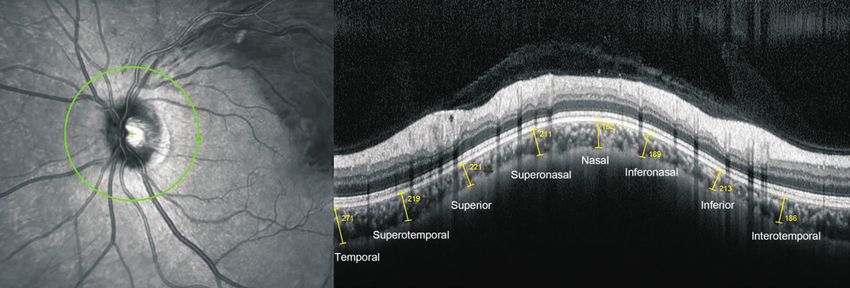
Figure 1 Representative image of PCT measurement in a 53-year-old male patient with unilateral BRVO in the left eye PCT was
measured using spectral domain OCT with EDI. A circular scan centered on the optic nerve head (3.40-mm diameter, “RNFL circle scan”) in the
peripapillary area. Choroidal thickness was defined as the perpendicular distance from the outer border of the hyperreflective line representing
the retinal pigment epithelium to the chorioscleral interface. PCT was measured at eight locations (yellow lines from left to right: temporal,
superotemporal, superior, superonasal, nasal, inferonasal, inferior, and inferotemporal), and then averaged for each eye. BRVO: Branch retinal
vein occlusion; PCT: Peripapillary choroidal thickness; OCT: Optical coherence tomography; EDI: Enhanced depth imaging.
superonasal, superior, and superotemporal locations. We Table 1 Comparison of baseline characteristics between the
averaged the PCT measurements obtained at the inferonasal, BRVO-affected eyes and non-affected contralateral eyes
inferior, and inferotemporal locations to calculate the mean BRVO- Non-affected
Parameters affected eyes contralateral Pb
IPCT. We also calculated the difference between baseline and (n=41) eyes (n=41)
6mo (ΔSPCT, ΔTPCT, ΔIPCT, and ΔNPCT) and the ratio Axial length (mm) 22.9.8±1.1 22.8±0.9 0.911
of the change (%SPCT, %TPCT, %IPCT, and %NPCT) for Refractive error (D) a
-0.2±1.1 -0.4±1.3 0.870
each sector. After the sectoral analysis was completed, we Cup-disc ratio 0.3±0.1 0.3±0.1 0.981
performed a subgroup analysis between the superior BRVO The mean SFCT (µm) 260.0±82.7 262.5±85.2 0.902
eyes (superotemporal and superonasal BRVO) and the inferior The mean PCT (µm) 177.7±69.8 182.5±60.6 0.775
BRVO eyes (inferotemporal and inferonasal BRVO) to determine BRVO: Branch retinal vein occlusion; SFCT: Subfoveal choroidal
whether there were any differences in PCT changes according thickness; PCT: Peripapillary choroidal thickness. aSpherical
to the affected location. equivalent; bStudent’s t-test was used for comparison between the
To measure the subfoveal choroidal thickness (SFCT), at least BRVO-affected eyes and non-affected contralateral eyes. PInt J Ophthalmol, Vol. 12, No. 3, Mar.18, 2019 www.ijo.cn
Tel: 8629-82245172 8629-82210956 Email: ijopress@163.com
baseline and 165.9±61.1 μm (range, 56.8-326.8 μm) at 6mo.
The mean PCT decreased significantly over 6mo in both
BRVO-affected and non-affected eyes (both PPeripapillary choroidal thickness in BRVO
Table 2 Sectoral comparison of the mean PCT over 6mo between Table 3 In eyes with BRVO, the mean PCT over 6mo was compared
eyes with BRVO and non-affected contralateral eyes between those with superior BRVO and inferior BRVO
mean±SD, µm mean±SD, µm
Non-affected The superior The inferior
BRVO-affected Parameters Pa
Parameters contralateral eyes Pa group (n=28) group (n=13)
eyes (n=41)
(n=41) The mean PCT
Mean SPCT Baseline 186.4±72.0 155.0±58.1 0.267
Baseline 178.6±74.4 194.1±68.2 0.390 1mo 147.7±62.7 132.7±47.6 0.654
1mo 161.8±69.4 192.0±73.5 0.100 3mo 143.5±59.3 127.9±48.3 0.849
3mo 155.0±71.4 188.7±71.1 0.067 6mo 132.5±59.3 115.4±40.5 0.654
6mo 148.5±65.7 187.4±75.2 0.030 ΔPCT -49.6±34.6 -39.5±28.4 0.218
ΔSPCT -31.1±34.5 -10.2±23.7 0.009 %PCT 32.4±12.1 31.9±20.0 0.929
%SPCT 19.0±12.2 11.5±9.3 0.009 The mean SPCT
Mean TPCT Baseline 184.5±79.1 163.1±60.7 0.511
Baseline 148.7±66.2 158.8±62.6 0.532 1mo 169.3±75.3 141.9±48.1 0.338
1mo 137.8±68.3 160.7±73.5 0.205 3mo 167.0±77.2 125.5±44.8 0.076
3mo 129.1±57.8 161.0±66.1 0.045 6mo 155.7±70.8 129.5±47.7 0.369
6mo 120.8±61.5 148.5±59.4 0.016 ΔSPCT -30.2±35.8 -33.5±32.2 0.811
ΔTPCT -27.0±28.1 -10.5±14.3 0.001 %SPCT 18.7±11.7 19.8±13.9 0.858
%TPCT 23.8±24.8 13.3±13.5 0.023 The mean TPCT
Mean IPCT Baseline 155.8±69.9 129.8±53.5 0.280
Baseline 136.9±58.1 147.9±48.1 0.420 1mo 142.9±73.2 124.1±54.1 0.530
1mo 124.6±52.1 142.0±51.7 0.193 3mo 136.9±62.5 109.8±40.4 0.251
3mo 117.6±51.8 141.0±50.0 0.065 6mo 124.1±67.3 111.8±44.2 0.858
6mo 105.9±44.5 132.4±50.1 0.003 ΔTPCT -31.7±23.6 -13.5±36.0 0.061
ΔIPCT -30.8±24.9 -11.1±24.5Int J Ophthalmol, Vol. 12, No. 3, Mar.18, 2019 www.ijo.cn
Tel: 8629-82245172 8629-82210956 Email: ijopress@163.com
patients with BRVO. The amount and ratio of PCT change effect due to BRVO, and the PCT changes are associated with
were not significantly different between the patients with the underlying vascular insufficiency in conjunction with
superior BRVO and those with inferior BRVO. The reduction BRVO. In the macular area, damaged vascular endothelial cells
of the mean SPCT was not significantly greater in the patients and hypoxic damage around the obstructed retinal vessels may
with superior BRVO than those with inferior BRVO, and lead to increased vascular endothelial growth factor (VEGF)
similar results were observed in the mean IPCT. Regardless of release[33-37]. Increased VEGF secretion in the occluded area
the occlusion site, the mean PCT in each sector also retained can lead to vascular hyperpermeability and subsequent macular
the thickness pattern of the normal population[21-22]; that is, choroidal thickening in the patients with BRVO[33-36]. Break-
thickest in the superior and thinnest in the inferior, regardless down of the outer blood-retinal barrier can be another conduit
of the occlusion site. of extravasated fluid to the choroidal space. Thus, different
The results of the current study suggested that the choroidal mechanisms could contribute to the discrepancy between the
blood flow in the peripapillary area may be impaired when macular choroidal thickness and PCT in the patients with
BRVO occurs. We speculate that systemic factors predisposing BRVO.
patients to BRVO may also affect the peripapillary choroid. Significant reduction of the mean PCT in both BRVO-affected
As previously mentioned, systemic vascular diseases such as eyes and non-affected contralateral eyes may predispose these
hypertension and arteriosclerosis, which are associated with patients to glaucomatous optic neuropathy. The vascular theory
thickening of the retinal artery, are risk factors for BRVO[3-12] as for glaucomatous optic neuropathy states that insufficient blood
well as for glaucoma[23-27]. These systemic conditions can also supply associated with various conditions such as elevated
lead to increased resistance to flow, reduced perfusion pressure, intraocular pressure or systemic diseases affecting ocular blood
or increased blood viscosity, leading to vascular insufficiency flow may lead to the development of glaucoma[38-41]. Reduction
of the optic disc and predisposing to glaucoma. Several studies of ocular blood flow was found in all ocular tissues, especially
also support the vascular theory for both glaucoma and RVO, in the choroid, optic nerve head, and peripapillary area[42-43].
showing associations between disc hemorrhage, glaucoma, Several studies showed associations between glaucomatous
and RVO[28-30]. We speculate that when BRVO occurs, vascular optic neuropathy, impaired choroidal circulation, and decreased
insufficiency also affects the peripapillary choroidal blood blood flow to the optic nerve head[44-46]. Recent studies using
flow, leading to generalized reduction of the mean PCT in both EDI OCT showed that the mean PCT was significantly reduced
BRVO-affected eyes and non-affected contralateral eyes during in the patients with normotensive glaucoma, further providing
follow-up periods. In BRVO-affected eyes, more ischemic evidence for the vascular theory[47-49]. Based on our results, the
injury associated with BRVO itself may lead to a significantly peripapillary choroidal circulation seems to be significantly
larger reduction of the mean PCT than that of non-affected impaired in the patients with BRVO during the follow-
contralateral eyes. In addition, vascular insufficiency of the up period, especially in the BRVO-affected eyes. Impaired
peripapillary choroid may lead to generalized reduction peripapillary choroidal blood flow and resultant peripapillary
of the mean PCT in each sector regardless of the location choroidal thinning may increase the risk for glaucomatous
of occlusion site in our study. Further prospective studies, optic neuropathy in patients with BRVO, leading to further
including investigation of peripapillary choroidal blood flow is visual deterioration. Thus, close monitoring for glaucomatous
warranted to clarify the pathophysiological correlation of the optic neuropathy as well as retinal complications of BRVO
mean PCT and BRVO. may be needed to prevent further visual loss in these patients.
In our study, the mean SFCT and the mean PCT showed This study had several limitations. It was a retrospective study
different tendencies in BRVO patients. In patients with diabetic with a relatively small study population. Due to the small
retinopathy, another common retinal vascular disorder, it seems study population, the occlusion areas were only classified
that macular choroidal thickness and mean PCT show similar into two groups: superior and inferior. Because of the
tendencies[31-32]. Both peripapillary and macular choroidal retrospective design of the study, we lacked the visual field
thickness are significantly correlated with the severity of test results. The clinical impact of the mean PCT change on
diabetic retinopathy[31-32], and are significantly reduced after the visual field change could be more helpful in real practice.
panretinal photocoagulation[32]. In our study, the mean SFCT Further prospective studies with longer follow-up periods,
was not significantly different over 6mo, whereas the mean which would include a control group and visual field tests is
PCT in each sector was significantly reduced in the patients warranted to support the clinical impact of our findings.
with BRVO. This discrepancy between the SFCT and PCT may In conclusion, the mean PCT was significantly reduced in both
be due to different mechanisms in the patients with BRVO; BRVO-affected eyes and non-affected contralateral eyes over
macular choroidal thickness changes may be the secondary 6mo in patients with unilateral BRVO. The mean PCT in each
477Peripapillary choroidal thickness in BRVO
area was reduced significantly over 6mo in the BRVO-affected Eur J Intern Med 2017;44:44-48.
eyes, but not in the non-affected contralateral eyes. Sectoral 12 Frucht J, Shapiro A, Merin S. Intraocular pressure in retinal vein
changes in mean PCT values were not affected by BRVO occlusion. Br J Ophthalmol 1984;68(1):26-28.
location. 13 Wang Y, Fawzi AA, Varma R, Sadun AA, Zhang X, Tan O, Izatt JA,
ACKNOWLEDGEMENTS Huang D. Pilot study of optical coherence tomography measurement of
Authors’ contributions: Designed and performed the study: retinal blood flow in retinal and optic nerve diseases. Invest Ophthalmol
Lee NE, Kang HM and Choi JH; collected the data: Kang Vis Sci 2011;52(2):840-845.
HM; managed, analyzed, and interpreted data: Lee NE, Kang 14 Son Y, Lee S, Park J. Measurement of lamina and prelaminar
HM, Choi JH, Koh HJ, and Lee SC; prepared, reviewed, and thicknesses of both eyes in patients with unilateral branch retinal vein
approved the manuscript: Lee NE, Kang HM, Choi JH, Koh occlusion. Graefes Arch Clin Exp Ophthalmol 2017;255(3):503-508.
HJ, and Lee SC. 15 Kang HM, Choi JH, Koh HJ, Lee CS, Lee SC. Significant reduction of
Foundation: Supported by the National Research Foundation peripapillary choroidal thickness in patients with unilateral branch retinal
of Korea (NRF) grant funded by the Korea government vein occlusion. Retina 2018;38(1):72-78.
(MSIT) (NRF-2018R1C1B5085620). 16 Lopilly Park HY, Jeon S, Lee MY, Park CK. Glaucoma progression
Conflicts of Interest: Lee NE, None; Kang HM, None; Choi in the unaffected fellow eye of glaucoma patients who developed
JH, None; Koh HJ, None; Lee SC, None. unilateral branch retinal vein occlusion. Am J Ophthalmol 2017;175:
REFERENCES 194-200.
1 Argon laser scatter photocoagulation for prevention of neovascularization 17 Kim KH, Lee DH, Lee JJ, Park SW, Byon IS, Lee JE. Regional
and vitreous hemorrhage in branch vein occlusion. A randomized clinical choroidal thickness changes in branch retinal vein occlusion with macular
trial. Branch Vein Occlusion Study Group. Arch Ophthalmol 1986;104(1): edema. Ophthalmologica 2015;234(2):109-118.
34-41. 18 Forte R, Cennamo G, Breve MA, Vecchio EC, de Crecchio G. Functional
2 Klein R, Moss SE, Meuer SM, Klein BE. The 15-year cumulative and anatomic response of the retina and the choroid to intravitreal
incidence of retinal vein occlusion: the Beaver Dam Eye Study. Arch bevacizumab for macular edema. J Ocul Pharmacol Ther 2012;28(1):
Ophthalmol 2008;126(4):513-518. 69-75.
3 Bowers DK, Finkelstein D, Wolff SM, Green WR. Branch retinal vein 19 Lee EK, Han JM, Hyon JY, Yu HG. Changes in choroidal thickness
occlusion. A clinicopathologic case report. Retina 1987;7(4):252-259. after intravitreal dexamethasone implant injection in retinal vein
4 Frangieh GT, Green WR, Barraquer-Somers E, Finkelstein D. occlusion. Br J Ophthalmol 2015;99(11):1543-1549.
Histopathologic study of nine branch retinal vein occlusions. Arch 20 Yumusak E, Ornek K, Dikel NH. Comparison of choroidal thickness
Ophthalmol 1982;100(7):1132-1140. changes following intravitreal dexamethasone, ranibizumab, and
5 Sperduto RD, Hiller R, Chew E, Seigel D, Blair N, Burton TC, Farber triamcinolone in eyes with retinal vein occlusion. Eur J Ophthalmol
MD, Gragoudas ES, Haller J, Seddon JM, Yannuzzi LA. Risk factors 2016;26(6):627-632.
for hemiretinal vein occlusion: comparison with risk factors for central 21 Jiang R, Wang YX, Wei WB, Xu L, Jonas JB. Peripapillary choroidal
and branch retinal vein occlusion: the eye disease case-control study. thickness in adult Chinese: the Beijing Eye Study. Invest Ophthalmol Vis
Ophthalmology 1998;105(5):765-771. Sci 2015;56(6):4045-4052.
6 Rath EZ, Frank RN, Shin DH, Kim C. Risk factors for retinal vein 22 Gupta P, Cheung CY, Baskaran M, Tian J, Marziliano P, Lamoureux
occlusions. A case-control study. Ophthalmology 1992;99(4):509-514. EL, Cheung CM, Aung T, Wong TY, Cheng CY. Relationship between
7 Jaulim A, Ahmed B, Khanam T, Chatziralli IP. Branch retinal vein peripapillary choroid and retinal nerve fiber layer thickness in a
occlusion: epidemiology, pathogenesis, risk factors, clinical features, population-based sample of nonglaucomatous eyes. Am J Ophthalmol
diagnosis, and complications. An update of the literature. Retina 2016;161:4-11.e1-2.
2013;33(5):901-910. 23 Zhao D, Cho J, Kim MH, Friedman DS, Guallar E. Diabetes, fasting
8 The Eye Disease Case-control Study Group. Risk factors for branch glucose, and the risk of glaucoma: a meta-analysis. Ophthalmology
retinal vein occlusion. Am J Ophthalmol1993;116(3):286-296. 2015;122(1):72-78.
9 Vannas S, Tarkkanen A. Retinal vein occlusion and glaucoma. 24 Zhou M, Wang W, Huang W, Zhang X. Diabetes mellitus as a risk
Tonographic study of the incidence of glaucoma and of its prognostic factor for open-angle glaucoma: a systematic review and meta-analysis.
significance. Br J Ophthalmol 1960;44:583-589. PLoS One 2014;9(8):e102972.
10 Beaumont PE, Kang HK. Cup-to-disc ratio, intraocular pressure, and 25 Lockwood A, Clearkin LG. Insulin resistance in retinal vein occlusion
primary open-angle glaucoma in retinal venous occlusion. Ophthalmology and glaucoma. Lancet 1992;340(8827):1100-1101.
2002;109(2):282-286. 26 Bae HW, Lee N, Lee HS, Hong S, Seong GJ, Kim CY. Systemic
11 Bucciarelli P, Passamonti SM, Gianniello F, Artoni A, Martinelli I. hypertension as a risk factor for open-angle glaucoma: a meta-analysis of
Thrombophilic and cardiovascular risk factors for retinal vein occlusion. population-based studies. PLoS One 2014;9(9):e108226.
478Int J Ophthalmol, Vol. 12, No. 3, Mar.18, 2019 www.ijo.cn
Tel: 8629-82245172 8629-82210956 Email: ijopress@163.com
27 Bonomi L, Marchini G, Marraffa M, Bernardi P, Morbio R, Varotto 38 Flammer J, Orgül S, Costa VP, Orzalesi N, Krieglstein GK, Serra LM,
A. Vascular risk factors for primary open angle glaucoma: the Egna- Renard JP, Stefánsson E. The impact of ocular blood flow in glaucoma.
Neumarkt Study. Ophthalmology 2000;107(7):1287-1293. Prog Retin Eye Res 2002;21(4):359-393.
28 Krakau CE. Disk hemorrhages and retinal vein occlusions in glaucoma. 39 Blumenthal M, Best M, Galin MA, Toyofuku H. Peripapillary
Surv Ophthalmol 1994;38(Suppl):S18-S21; discussion S22. choroidal circulation in glaucoma. Arch Ophthalmol 1971;86(1):31-38.
29 Sonnsjö B. Similarities between disc haemorrhages and thromboses of 40 Banitt M. The choroid in glaucoma. Curr Opin Ophthalmol
the retinal veins. Int Ophthalmol 1992;16(4-5):235-238. 2013;24(2):125-129.
30 Sonnsjö B, Dokmo Y, Krakau T. Disc haemorrhages, precursors of 41 Geijssen HC, Greve EL. Vascular concepts in glaucoma. Curr Opin
open angle glaucoma. Prog Retin Eye Res 2002;21(1):35-56. Ophthalmol 1995;6(2):71-77.
31 Vujosevic S, Martini F, Cavarzeran F, Pilotto E, Midena E. Macular 42 Duijm HF, van den Berg TJ, Greve EL. Choroidal haemodynamics in
and peripapillary choroidal thickness in diabetic patients. Retina glaucoma. Br J Ophthalmol 1997;81(9):735-742.
2012;32(9):1781-1790. 43 Wolf S, Arend O, Sponsel WE, Schulte K, Cantor LB, Reim M. Retinal
32 Kang HM, Lee NE, Choi JH, Koh HJ, Lee SC. Significant hemodynamics using scanning laser ophthalmoscopy and hemorheology
reduction of both peripapillary and subfoveal choroidal thickness after in chronic open-angle glaucoma. Ophthalmology 1993;100(10):1561-1566.
panretinal photocoagulation in patients with type 2 diabetes. Retina 44 Grunwald JE, Piltz J, Hariprasad SM, DuPont J. Optic nerve and
2018;38(10):1905-1912. choroidal circulation in glaucoma. Invest Ophthalmol Vis Sci 1998;39(12):
33 Tilton RG, Chang KC, LeJeune WS, Stephan CC, Brock TA, 2329-2336.
Williamson JR. Role for nitric oxide in the hyperpermeability and 45 Galassi F, Sodi A, Ucci F, Renieri G, Pieri B, Baccini M. Ocular
hemodynamic changes induced by intravenous VEGF. Invest Ophthalmol hemodynamics and glaucoma prognosis: a color Doppler imaging study.
Vis Sci 1999;40(3):689-696. Arch Ophthalmol 2003;121(12):1711-1715.
34 Ferrara N. Vascular endothelial growth factor: molecular and biological 46 Nicolela MT, Hnik P, Drance SM. Scanning laser Doppler flowmeter
aspects. Curr Top Microbiol Immunol 1999;237:1-30. study of retinal and optic disk blood flow in glaucomatous patients. Am J
35 Noma H, Minamoto A, Funatsu H, Tsukamoto H, Nakano K, Ophthalmol 1996;122(6):775-783.
Yamashita H, Mishima HK. Intravitreal levels of vascular endothelial 47 Park HY, Lee NY, Shin HY, Park CK. Analysis of macular and
growth factor and interleukin-6 are correlated with macular edema peripapillary choroidal thickness in glaucoma patients by enhanced
in branch retinal vein occlusion. Graefes Arch Clin Exp Ophthalmol depth imaging optical coherence tomography. J Glaucoma 2014;23(4):
2006;244(3):309-315. 225-231.
36 Aiello LP, Northrup JM, Keyt BA, Takagi H, Iwamoto MA. Hypoxic 48 Hirooka K, Tenkumo K, Fujiwara A, Baba T, Sato S, Shiraga F.
regulation of vascular endothelial growth factor in retinal cells. Arch Evaluation of peripapillary choroidal thickness in patients with normal-
Ophthalmol 1995;113(12):1538-1544. tension glaucoma. BMC Ophthalmol 2012;12:29.
37 Ebneter A, Kokona D, Schneider N, Zinkernagel MS. Microglia 49 Usui S, Ikuno Y, Miki A, Matsushita K, Yasuno Y, Nishida K.
activation and recruitment of circulating macrophages during ischemic Evaluation of the choroidal thickness using high-penetration optical
experimental branch retinal vein occlusion. Invest Ophthalmol Vis Sci coherence tomography with long wavelength in highly myopic normal-
2017;58(2):944-953. tension glaucoma. Am J Ophthalmol 2012;153(1):10-16.e1.
479You can also read