Added Value of Subtraction SPECT/CT in Dual-Isotope Parathyroid Scintigraphy - MDPI
←
→
Page content transcription
If your browser does not render page correctly, please read the page content below
diagnostics
Article
Added Value of Subtraction SPECT/CT in
Dual-Isotope Parathyroid Scintigraphy
Julie Wulf Christensen *           and Martin Krakauer
 Department of Nuclear Medicine, Herlev and Gentofte Hospital, 2730 Herlev, Denmark;
 martin.krakauer@regionh.dk
 * Correspondence: Julie.wulf.christensen@regionh.dk; Tel.: +45-38686740

 Received: 17 July 2020; Accepted: 24 August 2020; Published: 27 August 2020

 Abstract: Background: Adding subtraction single-photon emission computed tomography/computed
 tomography (SPECT/CT) to dual isotope (I-123 and Tc-99m-sestamibi) subtraction parathyroid
 scintigraphy is not widely implemented. We aimed to assess the added value of dual isotope
 subtraction SPECT/CT over single isotope SPECT/CT as an adjunct to dual isotope planar pinhole
 subtraction scintigraphy. Methods: Parathyroid scintigraphies from 106 patients with an estimated
 total of 415 parathyroid glands who (1) were diagnosed with primary hyperparathyroidism,
 (2) underwent dual isotope subtraction scintigraphy in the Department of Nuclear Medicine,
 Gentofte Hospital, Denmark throughout 2017 and (3) underwent subsequent parathyroidectomy,
 were included. The original dual isotope planar pinhole subtraction plus dual isotope subtraction
 SPECT/CT (dual/dual method) exams were retrospectively re-evaluated using only Tc-99m-sestamibi
 SPECT/CT (dual/single method). Statistics were calculated per parathyroid. Surgical results confirmed
 by pathology served as reference standard. Results: The dual/dual method had higher sensitivity than
 the dual/single method (82% (95%CI 74%–88%) vs. 69% (95%CI 60%–77%)) while specificity, positive
 and negative predictive values (PPV and NPV) were similar (specificity 96% vs. 93%, PPV’s 87% vs. 82%
 and NPV’s 89% vs. 93%). Reader confidence was higher when employing the dual/dual method
 (p = 0.001). Conclusions: The dual/dual method can be considered superior to the dual/single method
 in the preoperative imaging in primary hyperparathyroidism.

 Keywords: primary hyperparathyroidism; dual-isotope subtraction scintigraphy; dual-isotope
 subtraction SPECT/CT; Tc-99m-sestamibi SPECT/CT

1. Introduction
     Each year, 600–700 Danish patients are diagnosed with primary hyperparathyroidism (PHPT) [1,2].
PHPT is diagnosed on the basis of elevated serum calcium and elevated (or high in the normal range)
parathyroid hormone (PTH) secreted from one or more hyperfunctioning parathyroid gland(s) [1–3].
Symptoms include musculoskeletal, cardiovascular, gastrointestinal, renal and neurological problems,
and the only curative treatment is parathyroidectomy (PTx) [1–4].
     Preoperative imaging is a prerequisite for minimally invasive surgery. Parathyroid dual isotope
subtraction scintigraphy is a sensitive and specific preoperative imaging modality with reported
sensitivities ranging between 86%–93%, specificities 98%–100% [4–6].
     Cure rates have been found to be similar between minimally invasive surgery and bilateral neck
exploration, but morbidity is higher in the latter group [7].
     In our study, dual isotope (I-123 and Tc-99m-sestamibi) subtraction scintigraphy consists of early
planar pinhole images and subsequent dual isotope subtraction single-photon emission computed
tomography/computed tomography (SPECT/CT) with an additional delayed pinhole acquisition in
case of ambiguous findings on early imaging. Though effective, processing and analyzing dual isotope

Diagnostics 2020, 10, 639; doi:10.3390/diagnostics10090639                     www.mdpi.com/journal/diagnostics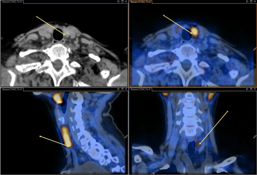
Diagnostics 2020, 10, 639                                                                         2 of 10

subtraction SPECT/CT images is more complicated than reviewing single isotope Tc-99m-sestamibi
SPECT/CT images. Thus, some nuclear medicine departments opt to use the more simple single isotope
SPECT/CT.
     This study aimed to evaluate the potential added information obtained from dual isotope
subtraction SPECT/CT, i.e., we intended to assess and compare two methods of dual isotope subtraction
scintigraphy, namely:

(1)   Dual isotope subtraction pinhole imaging (I-123 and Tc-99m-sestamibi) in combination with dual
      isotope subtraction SPECT/CT, henceforth termed “dual/dual”.
(2)   Dual isotope subtraction pinhole imaging in combination with single isotope SPECT/CT
      (Tc-99m-sestamibi), henceforth termed “dual/single”.

     The two methods were assessed in a retrospective, blinded, head-to-head comparison in a cohort
of consecutive PHPT patients who underwent post-imaging PTx.
     The null hypothesis was that the two methods did not differ regarding sensitivity, specificity,
or positive or negative predictive value.

2. Materials and Methods

2.1. Patients
      Patients were included retrospectively. All dual isotope subtraction scintigraphies performed at
the Department of Nuclear Medicine, Gentofte Hospital, Denmark from 1 January, 2017–31 December,
2017 were evaluated for inclusion. Exclusion criteria were incomplete scintigraphy (n = 4), post-imaging
surgery outside of the Capital Region of Denmark (due to unavailability of hospital files on surgical
notes) (n = 5), no post-imaging parathyroid surgery (n = 21), and previous thyroidectomy (n = 1)—the
latter rendering subtraction scintigraphy futile.
      Verifications of successful PTx were 1) a perioperative p-PTH decrease of ≥50%, 2) adenoma,
hyperplasia or carcinoma confirmed on postoperative pathology and 3) normalized p-PTH and Ca2+
levels approximately one month postoperatively.
      The study was conducted as a quality assurance study with permission from the hospital board at
Herlev and Gentofte Hospital, Denmark (workzone no. 18015429, date of approval: 5 July 2018).

2.2. Imaging Protocol
      Parathyroid scintigraphy was performed according to our standard protocol as follows:
Pinhole images were performed on Philips Skylight γ-camera (Philips Healthcare, Eindhoven,
The Netherlands) and SPECT/CT was performed on a Philips Precedence SPECT/CT scanner or a Siemens
Symbia Intevo SPECT/CT scanner (Siemens Healthcare, Erlangen, Germany). Patients received 12 MBq
(0.3 mCi) I-123 intravenously and, after 1.5 h, 600 MBq (160 mCi) Tc-99m-sestamibi. Dual isotope
pinhole images were obtained after 5 min (early) and again after 2 h (late) only if early images were
inconclusive. Dual isotope SPECT/CT of the neck and thorax was acquired shortly (15–30 min) after
the early pinhole images.
      Acquisition parameters: Energy windows were identical in planar and SPECT imaging (Tc-99m:
140 keV −7/+7; I-123: asymmetrical 159 keV −4/+10). The energy windows were chosen to minimize
Tc-99m spillover to the I-123-window [8]. Pinhole image parameters: 15 min; aperture size 3 mm.
SPECT imaging parameters: Dual-head, 180-degreee anterior orbit in a 90-degree angle; 128 angles,
14 s/angle; matrix 128 × 128. SPECT reconstruction used the vendor’s proprietary algorithm (Astonish®
or Flash3D® with attenuation and scatter correction). CT was done as a non-contrast enhanced lowdose
acquisition covering the neck and mediastinum and an additional diagnostic non-contrast enhanced
series covering only the thyroid and immediate surroundings.
      See Figures 1 and 2 for dual/dual and dual/single images.
Diagnostics 2020, 10, 639 3 of 10
Diagnostics 2020, 10, x FOR PEER REVIEW 3 of 10
Figure 1. Dual isotope planar pinhole subtraction scintigraphy and dual isotope subtraction
Figure 1. Dual isotope planar pinhole subtraction scintigraphy and dual isotope subtraction single-
single-photon emission computed tomography/computed tomography (SPECT/CT) images. (top image)
photon emission computed tomography/computed tomography (SPECT/CT) images. (top image)
Upper left: Tc-99m-sestamibi pinhole. Upper right: I-123 pinhole. Middle left and right: Varying
Upper left: Tc-99m-sestamibi pinhole. Upper right: I-123 pinhole. Middle left and right: Varying
degrees of subtraction. (bottom image) Dual isotope subtraction SPECT/CT with and without fusion.
degrees of subtraction. (bottom image) Dual isotope subtraction SPECT/CT with and without fusion.Diagnostics 2020, 10, 639 4 of 10
Diagnostics 2020, 10, x FOR PEER REVIEW 4 of 10
Figure Single isotope
2. Single
Figure 2. isotope SPECT/CT
SPECT/CT image
image (Tc-99m-sestamibi)
(Tc-99m-sestamibi) with
with and
and without fusion—same patient
without fusion—same patient
as in Figure 1.
as in Figure 1.
2.3. Image Analysis
2.3. Image Analysis
2.3.1. Prospective “Dual/Dual”
2.3.1. Prospective “Dual/Dual”
Scintigraphies were analyzed by a nuclear medicine specialist supervised and approved by one
Scintigraphies
particularly were in
experienced analyzed by a nuclear
parathyroid medicine
scintigraphy (MK).specialist supervised and approved by one
particularly
Pinholeexperienced in parathyroid
subtraction images scintigraphy
were analyzed (MK).
in Segami Oasis software (version 1.9, Columbia, MD,
Pinhole subtraction images were analyzed
USA) where variable degrees of subtraction were available in Segami Oasis software
through the(version
use of a1.9, Columbia,
slider MD,
until counts
USA) where variable degrees of subtraction were available through the
in the thyroid bed were equal to Tc-99m-sestamibi background counts on the neck. SPECT data use of a slider until counts in
the thyroid
were bed were
subtracted usingequal
Philips to Intellispace
Tc-99m-sestamibi
Portal background counts onThe
(Philips, Eindhoven, theNetherlands),
neck. SPECT data were
the initial
subtracted using Philips Intellispace Portal (Philips, Eindhoven, The Netherlands),
subtraction factor was determined by calculating the of ratio of I-123 and Tc-99m-sestamibi counts in the initial
subtraction
a factorofwas
thyroid region determined
interest (ROI) inbya calculating
representative the of ratio of
coronal I-123
slice butand Tc-99m-sestamibi
could later be modified counts in
at the
a thyroid region
reader’s discretion. of interest (ROI) in a representative coronal slice but could later be modified at the
reader’s
The discretion.
location of a putative hyperfunctioning parathyroid gland (HPG) was given in relation to the
The(left/right;
thyroid location ofupper/middle/lower
a putative hyperfunctioning parathyroid
third; ectopic). glandof(HPG)
Confidence was given
the findings wasingraded
relation
ontoa
the thyroid (left/right; upper/middle/lower
three-point scale (low (1), moderate (2), high (3)). third; ectopic). Confidence of the findings was graded on
a three-point scale (low
For the purpose (1),study,
of this moderate (2), high
all image (3)).
descriptions were reviewed and the number and location of
HPGsFor as the
wellpurpose of this study,
as the confidence scoreallwere
image descriptions
noted on a coded were
sheetreviewed and thewith
for comparison number and findings.
surgical location
of HPGs as well as the confidence score were noted on a coded sheet for comparison with surgical
findings.
2.3.2. Retrospective “Dual/Single”
2.3.2.All previously“Dual/Single”
Retrospective processed and recorded images/reconstructions were deleted, and images
subsequently anonymized to ensure re-analysis blinded to the original results.
All previously
Re-analyses wereprocessed
performedand recorded
by an images/reconstructions
experienced were deleted,
nuclear medicine specialist anda images
(MK) using similar
subsequently anonymized to ensure re-analysis blinded to the original results.
approach to the aforementioned, except only Tc-99m-sestamibi SPECT/CT was available while the
I-123Re-analyses
SPECT datasetwere performed
and, by an subtraction-SPECT/CT
consequently, experienced nuclear medicine
was not.specialist (MK) using a similar
approach to the aforementioned, except only Tc-99m-sestamibi SPECT/CT
Number and location of HPGs as well as a confidence score were recordedwas available while sheet
on a coded the I-
123 SPECT dataset and, consequently,
similar to the above-mentioned. subtraction-SPECT/CT was not.
Number and location of HPGs as well as a confidence score were recorded on a coded sheet
similar to the above-mentioned.
2.4. Surgery and Postoperative Follow-UpDiagnostics 2020, 10, 639 5 of 10
2.4. Surgery and Postoperative Follow-Up
Patients underwent surgery at Department of Otorhinolaryngology, Head and Neck Surgery in
The Capital Region of Denmark.
Surgery was guided by the scintigraphic findings and per-operative plasma-PTH-measurements
(where a decrease of >50% indicated successful removal of the HPG(s)).
Following surgery, histological evaluation was performed to confirm the diagnosis.
After approximately one month, postoperative plasma-PTH and Ca2+ were determined,
and normalization used to verify successful surgery.
2.5. Statistical Analyses
Location of each apparent HPG according to each imaging method was recorded along with
confidence score of each method. Location according to surgical notes and successful surgery confirmed
by perioperative PTH-decrease, postoperative pathology and postoperative PTH and Ca2+ served as
the reference standard.
The statistical analyses were conducted using ‘R’ version 3.6.0. Vienna, Austria. URL available
online: https://www.R-project.org/, accessed on 20 August 2020.Using McNemar’s Chi-square
test sensitivity, specificity, positive and negative predictive values (PPV and NPV), accuracy,
misclassification and Matthews Correlation Coefficient (MCC) were calculated for each modality.
Results were calculated per estimated parathyroid, rather than per patient, under the assumption of
four native parathyroids per patient. In order to compare the three classes of responder confidence
between the two methods of analysis, we created a 3 × 3 table and used the “McNemar–Bowker test”
in the statistical programme R.P values of 0.05 or less were considered statistically significant.
3. Results
Scintigraphies from 139 patients were evaluated for inclusion, and 106 patients were included
for reanalysis. Of these, eight patients had previously undergone parathyroid surgery, but still had
PHPT. The patients were predominantly female (84%) with a median age of 64 years (range 32–86).
Twenty-seven patients had previously undergone parathyroid scintigraphy. In eight of these,
scintigraphy had then been followed by parathyroid surgery. For 79 patients this was their primary
parathyroid scintigraphy. See Figure 3 for inclusion flow-chart. The eight patients who had undergone
previous parathyroid surgery had had nine parathyroid glands removed in total (ranging from zero to
each each). Thus, assuming four native parathyroid glands per patient, the 106 patients corresponded
to 415 parathyroid glands in total (i.e., (106 × 4) − 9 = 415).
The median time from scintigraphy to surgery was 101 days (range: 7–410 days). Median time
from surgery to the final postoperative blood-test was 46 days (range 11–276 days). See Table 1 for
baseline characteristics.
During surgery a total of 120 specimens were removed, pathology testing showed parathyroid
adenoma in 93 (90 patients), parathyroid hyperplasia in 18 (13 patients) and other tissue in 9 (normal
parathyroid tissue (5), normal thyroid tissue (1), lymph node (1), thymus (1) or cyst (1).
Preoperative Ca2+ and plasma-PTH were 1.47 mmol/L (range 1.25–1.85) and 12.15 ng/L (range
5.0–35.0), respectively. Of the 106 patients, 95 had normalized Ca2+ postoperatively. In five cases Ca2+
was continually slightly elevated, but patients were declared cured by their treating endocrinologist,
in three cases Ca2+ was continually elevated but decreased after reoperation, and in the final two
patients Ca2+ was continually elevated presumably due to no/insufficient removal of parathyroid
tissue but watchful waiting was chosen over re-operation. A single patient never turned up for
postoperative evaluation.
The average weight of the removed HPGs was 544 mg (range: 70–8000 mg).Diagnostics 2020, 10, 639 6 of 10
Diagnostics 2020, 10, x FOR PEER REVIEW 6 of 10
Figure 3.3.Inclusion
Figure Inclusionflowchart.
flowchart.A total of 106
A total of scintigraphies from between
106 scintigraphies January January
from between 1, 2017 and
1, December
2017 and
31, 2017 were
December eligible
31, 2017 wereforeligible
inclusion. Of these, 79
for inclusion. Ofwere
these,primary
79 werescintigraphies and in 27and
primary scintigraphies cases
in patients
27 cases
had undergone
patients previousprevious
had undergone parathyroid scintigraphy
parathyroid (27) and/or
scintigraphy parathyroid
(27) surgery (8).
and/or parathyroid surgery (8).
Table 1. Baseline patient characteristics.
The median time from scintigraphy to surgery was 101 days (range: 7–410 days). Median time
from surgery to the final postoperative blood-test
Number of Patients, n was 46 days (range106
11–276 days). See Table 1 for
baseline characteristics.
Gender, n (%) Female 89 (84%)
Male 17 (16%)
Table 1. Baseline patient characteristics.
Number of HGPs, n (%) None 5 (5%)
Number of Patients, n Single 91 (86%) 106
Gender, n (%) Female
Multiple 10 89 (9%) (84%)
Male 17 (16%)
Previous surgery, n (%)
Number of HGPs, n (%)
Yes None 8 5
(8%) (5%)
NoSingle 98 91 (93%) (86%)
Multiple Median 10 Range (9%)
Previous surgery, n (%) Yes 8 (8%)
Age 63.6 (32.0–86.4)
No 98 (93%)
Weight of removed parathyroid (mg) 544 (70–8000)
2+ (mmol/L)
Median Range
Preoperative Ca 1.47 (1.19–1.85)
Age 63.6 (32.0–86.4)
Preoperative PTH (ng/L)
Weight of removed parathyroid (mg)
15.5 544
(3.8–40.8) (70–8000)
Ca (mmol/L)
PreoperativeHPGs:
2+
Hyperfunctioning parathyroid glands 1.47 (1.19–1.85)
Preoperative PTH (ng/L)
PTH: Parathyroid hormone 15.5 (3.8–40.8)
HPGs: Hyperfunctioning parathyroid glands
PTH: Parathyroid hormone
During surgery a total of 120 specimens were removed, pathology testing showed parathyroid
adenoma in 93 (90 patients), parathyroid hyperplasia in 18 (13 patients) and other tissue in 9 (normal
parathyroid tissue (5), normal thyroid tissue (1), lymph node (1), thymus (1) or cyst (1).Diagnostics 2020, 10, 639 7 of 10
3.1. Analysis
The performance of the two methods differed significantly (p = 0.0006). When compared to the
reference standard dual/dual showed sensitivity, specificity, PPV, and NPV of 82% (95% CI 74%–88%),
93% (95% CI 90%–96%), 82% (95% CI 74%–88%), and 93% (95% CI 90%–96%), respectively. Dual/single
showed a lower sensitivity of 69% (95% CI 60%–77%) but comparable specificity, PPV, and NPV of
96% (95% CI 93%–98%), 87% (95% CI 78%–92%), and 89% (95% CI 85%–92%). Accuracy and MCC
were slightly higher using the dual/dual method while miss-classification ration (i.e., the inverse of
accuracy) was slightly lower. See Table 2 for details.
Table 2. Results—Per parathyroid gland. (106 patients, 415 parathyroid glands).
Dual/Dual Dual/Single
N (%) N (%)
True positives 92 (22) 77 (19)
True negatives 283 (68) 291 (70)
False positives 20 (5) 12 (3)
False negatives 20 (5) 35 (8)
N (95% CI) N (95% CI) Difference
Sensitivity 82.1 (74.0–88.1) 68.8 (59.7–76.6) 13.3
Specificity 93.4 (90.0–95.7) 96.0 (93.2–97.7) 2.6
PPV 82.1 (74.0–88.1) 86.5 (77.9–92.1) 4.4
NPV 93.4 (90.0–95.7) 89.3 (85.4–92.2) 4.1
Accuracy 90.4 (87.1–93.0) 88.7 (85.2–91.6) 1.7
Misclassification 9.6 (7.0–12.9) 11.3 (8.4–14.8) 1.7
MCC 76.1 70.6 5.5
The number of true positives, true negatives, false positives and false negatives as well as sensitivity,
specificity, positive and negative predictive values (PPV and NPV), Matthews Correlation Coefficient (MCC)
and miss-classification. Each modality is compared to the reference standard (i.e., surgery confirmed by pathology
and clinical follow up). p = 0.0006 (When comparing dual/dual to dual/single).
Subgroup analyses regarding patients with and without nodular thyroid, patients with or without
previous parathyroid surgery, and patients with a single HPG vs. multiglandular disease can be found
online in the Supplementary Materials (not discussed here).
Reader Confidence
Reader confidence according to type of analysis is displayed in Table 3 and graphically in
Figure 4. The level of reader confidence differed significantly (p = 0.007) with more readings with
“high confidence” with the dual/dual method.
Table 3. Scintigraphy “responder confidence” per parathyroid. N = number of hyperfunctioning
parathyroid glands found using each method.
N Low N (%) Moderate N (%) High N (%)
Dual/dual 111 29 (26%) 20 (18%) 62 (56%)
Dual/single 89 23 (26%) 25 (28%) 41 (46%)
Dual/dual: Dual isotope planar pinhole subtraction and SPECT/CT images (I-123 and Tc-99m-sestamibi).Dual/single:
Dual isotope planar pinhole subtraction (I-123 and Tc-99m-sestamibi) and single isotope SPECT/CT images
(Tc-99m-sestamibi).N Low N (%) Moderate N (%) High N (%)
Dual/dual 111 29 (26%) 20 (18%) 62 (56%)
Dual/single 89 23 (26%) 25 (28%) 41 (46%)
Dual/dual: Dual isotope planar pinhole subtraction and SPECT/CT images (I-123 and Tc-99m-
Diagnostics 2020, 10, 639
sestamibi).Dual/single: Dual isotope planar pinhole subtraction (I-123 and Tc-99m-sestamibi) and8 of 10
single isotope SPECT/CT images (Tc-99m-sestamibi).
Figure 4.
Figure Responder confidence
4. Responder confidence (low,
(low, moderate
moderate or
or high)
high) according
according to
to type
type of
of imaging
imaging analysis.
analysis.
4. Discussion
4. Discussion
In a clinical context, the ability to locate HPGs preoperatively (i.e., true positives) is far more
In a clinical context, the ability to locate HPGs preoperatively (i.e., true positives) is far more
important than the ability to dismiss healthy parathyroid glands (i.e., true negatives), we find that our
important than the ability to dismiss healthy parathyroid glands (i.e., true negatives), we find that
primary effect measure must be the sensitivities. In our study, the dual/dual method showed a higher
our primary effect measure must be the sensitivities. In our study, the dual/dual method showed a
sensitivity and thus was superior in the localization of HPGs and also yielded a higher level of reader
higher sensitivity and thus was superior in the localization of HPGs and also yielded a higher level
confidence. The dual/single method proved to have slightly higher specificity.
of reader confidence. The dual/single method proved to have slightly higher specificity.
We previously found sensitivity and specificity of 93% and 99% when using the dual/single
We previously found sensitivity and specificity of 93% and 99% when using the dual/single
method [4]. While the present study found a comparable specificity using the same method (96%),
method [4]. While the present study found a comparable specificity using the same method (96%),
the sensitivity was notably lower in this new cohort (69%). This is probably due to a more heterogeneous
the sensitivity was notably lower in this new cohort (69%). This is probably due to a more
study population in the current study. In the former study, only patients with no previous parathyroid
heterogeneous study population in the current study. In the former study, only patients with no
scintigraphy or PTx were prospectively included, while all patients studied in a defined timeframe
previous parathyroid scintigraphy or PTx were prospectively included, while all patients studied in
were evaluated in the present study. This included patients that had had previous off-site inconclusive
a defined timeframe were evaluated in the present study. This included patients that had had
imaging studies and patients with previous parathyroid/thyroid surgery thus potentially re-evaluating
previous off-site inconclusive imaging studies and patients with previous parathyroid/thyroid
patients with more elusive HPGs.
surgery thus potentially re-evaluating patients with more elusive HPGs.
Additionally, in recent years the patient population has changed due to more frequent incidentally
Additionally, in recent years the patient population has changed due to more frequent
diagnosed cases of primary hyperparathyroidism (most likely due to increased blood testing) which
incidentally diagnosed cases of primary hyperparathyroidism (most likely due to increased blood
would in turn cause the HPGs to be smaller and more difficult to locate [9,10]. This is in agreement
testing) which would in turn cause the HPGs to be smaller and more difficult to locate [9,10]. This is
with the fact that in our previous study the median HPG weight was 121 mg or 22% larger than in the
in agreement with the fact that in our previous study the median HPG weight was 121 mg or 22%
current study. Furthermore, differences may be contributed to the different set-ups—one a prospective
larger than in the current study. Furthermore, differences may be contributed to the different set-
study with designated readers, and one a retrospective study where the original analysis was used
ups—one a prospective study with designated readers, and one a retrospective study where the
for statistics.
original analysis was used for statistics.
Strengths of the study were in the large number of patients compared to previous publications
Strengths of the study were in the large number of patients compared to previous publications
(i.e., 50–96 patients [4–6]), and the consecutive inclusion of all patients referred for imaging. A weakness
(i.e., 50–96 patients [4–6]), and the consecutive inclusion of all patients referred for imaging. A
was that the dual/single analyses were performed retrospectively by a single, experienced reader
weakness was that the dual/single analyses were performed retrospectively by a single, experienced
(MK) while the dual/dual analyses were analyzed prospectively by different readers. However, the
reader (MK) while the dual/dual analyses were analyzed prospectively by different readers.
prospective dual/dual analyses were routinely supervised by the same experienced reader in order to
However, the prospective dual/dual analyses were routinely supervised by the same experienced
ensure consistency.
reader in order to ensure consistency.
Several previous publications have assessed imaging setups similar to ours. A combination of
subtraction pinhole and subtraction SPECT/CT (i.e., our dual/dual setup) has previously been found to
have sensitivity similar to ours or higher (81%–98%) and varying specificity (67%–99%) [11–13]. In these
studies the removed parathyroid glands were somewhat larger than in the present study (average size
700–1420 mg, compared to 544 mg in the present study) which, as mentioned, would likely increase
sensitivity. Furthermore, there is a difference in imaging criteria; i.e., Asseeva et al. discriminate
between “uniglandular disease”, “multiglandular disease” and “negative result” while we discriminate
by the location of HPGs [11]. Due to the limited sensitivity of the dual/single method, Krčálová et al.Diagnostics 2020, 10, 639 9 of 10
recommend adding subtraction SPECT/CT (i.e., dual/dual) or 18F-fluorocholin-positron emission
tomography / computed tomography (PET/CT) in order to increase sensitivity [14]. Others have
assessed subtraction pinhole alone and/or subtraction SPECT/CT alone, the former with reported
sensitivities and specificities of 75%–88% and 90% respectively, and the latter sensitivity of 86%–95%
and specificity 98%–100% [6,13]. In our experience very small HPG’s may be visible only on pinhole
images, therefore we do not recommend omitting planar pinhole imaging.
As mentioned, alternate protocols for preoperative imaging exist, such as 18 F-Choline and
11 C-Choline PET/CT. A recent review has shown that PET/CT has shown promise in the location of
HPGs with sensitivities > 90% [15]. However they conclude with a need for further testing in order to
confidently define the role of Choline PET/CT in preoperative diagnostics of PHPT [15].
5. Conclusions
Overall dual isotope subtraction SPECT/CT as an adjunct to dual isotope planar pinhole
subtraction scintigraphy was more sensitive than dual isotope planar pinhole subtraction scintigraphy
with Tc-99m sestamibi SPECT/CT. Moreover, dual isotope subtraction SPECT/CT resulted in higher
reader confidence.
Although slightly more complicated, we do recommend applying dual isotope subtraction
SPECT/CT in addition to planar pinhole subtraction imaging for preoperative parathyroid scintigraphy
in patients with primary hyperthyroidism. However, the preoperative imaging strategy must always
be adapted to local availability and expertise.
Supplementary Materials: The following are available online at http://www.mdpi.com/2075-4418/10/9/639/s1.
Author Contributions: Conceptualization, methodology, formal analysis, data curation and project administration
J.W.C. and M.K.; Software, investigation, supervision and writing—review and editing—M.K.; Writing—original
draft preparation—J.W.C.; All authors have read and agreed to the published version of the manuscript.
Funding: This research received no external funding.
Conflicts of Interest: The authors declare no conflict of interest.
References
1. Fraser, W.D. Hyperparathyroidism. Lancet 2009, 374, 145–158. [CrossRef]
2. Rejnmark, L. Danish Endocrine Society—Treatment Guideline for Primary Hyperparathyroidism.
Available online: http://www.endocrinology.dk/index.php/3-calcium-og-knoglemetaboliske-sygdomme/
nbv-endokrinologi-primaer-hyperparathyreoidisme-hypercalcaemi-calcium-creatinin-clearance-familiaer-
hypocalciurisk-hypercalcaemi-udredningsprogram-parathyreoidektomi-medicinsk-behandling-ovrig-
hyperparathyreoidisme (accessed on 23 June 2020).
3. Greenspan, B.S.; Dillehay, G.; Intenzo, C.; Lavely, W.C.; O’Doherty, M.; Palestro, C.J.; Scheve, W.; Stabin, M.G.;
Sylvestros, D.; Tulchinsky, M. SNM Practice Guideline for Parathyroid Scintigraphy 4.0. J. Nucl. Med. Technol.
2012, 40, 111–118. [CrossRef] [PubMed]
4. Krakauer, M.; Wieslander, B.; Myschetzky, P.S.; Lundstrøm, A.; Bacher, T.; Sørensen, C.H.; Trolle, W.;
Nygaard, B.; Bennedbæk, F.N. A Prospective Comparative Study of Parathyroid Dual-Phase Scintigraphy,
Dual-Isotope Subtraction Scintigraphy, 4D-CT, and Ultrasonography in Primary Hyperparathyroidism. Clin.
Nucl. Med. 2016, 41, 93–100. [CrossRef] [PubMed]
5. Rink, T.; Schroth, H.-J.; Holle, L.-H.; Garth, H. Limited sensitivity of parathyroid imaging with
(99m)Tc-sestamibi/(123)I subtraction in an endemic goiter area. J. Nucl. Med. 2002, 43, 1175–1180. [PubMed]
6. Hassler, S.; Ben-Sellem, D.; Hubele, F.; Constantinesco, A.; Goetz, C. Dual-Isotope 99mTc-MIBI/123I
Parathyroid Scintigraphy in Primary Hyperparathyroidism. Clin. Nucl. Med. 2014, 39, 32–36. [CrossRef]
[PubMed]
7. Karakas, E.; Schneider, R.; Rothmund, M.; Bartsch, D.K.; Schlosser, K. Initial Surgery for Benign Primary
Hyperparathyroidism: An Analysis of 1,300 Patients in a Teaching Hospital. World J. Surg. 2014, 38,
2011–2018. [CrossRef] [PubMed]Diagnostics 2020, 10, 639 10 of 10
8. Hindié, E.; Ugur, O.; Fuster, D.; O’Doherty, M.; Grassetto, G.; Ureña, P.; Kettle, A.; Gulec, S.A.; Pons, F.;
Rubello, D. 2009 EANM parathyroid guidelines. Eur. J. Nucl. Med. Mol. Imaging 2009, 36, 1201–1216.
[CrossRef] [PubMed]
9. Bilezikian, J.P.; Bandeira, L.; Khan, A.; Cusano, N.E. Hyperparathyroidism. Lancet 2018, 391, 168–178.
[CrossRef]
10. Yeh, M.W.; Ituarte, P.H.G.; Zhou, H.C.; Nishimoto, S.; Liu, I.-L.A.; Harari, A.; Haigh, P.I.; Adams, A.L. Incidence
and prevalence of primary hyperparathyroidism in a racially mixed population. J. Clin. Endocrinol. Metab.
2013, 98, 1122–1129. [CrossRef] [PubMed]
11. Asseeva, P.; Paladino, N.C.; Guerin, C.; Castinetti, F.; Vaillant-Lombard, J.; Abdullah, A.E.; Farman-Ara, B.;
Loundou, A.; Sebag, F.; Taïeb, D. Value of 123I/99mTc-sestamibi parathyroid scintigraphy with subtraction
SPECT/CT in primary hyperparathyroidism for directing minimally invasive parathyroidectomy. Am. J. Surg.
2019, 217, 108–113. [CrossRef] [PubMed]
12. Bhatt, P.R.; Klingensmith, W.C.; Bagrosky, B.M.; Walter, J.C.; McFann, K.K.; McIntyre, R.C., Jr.; Raeburn, C.D.;
Koo, P.J. Parathyroid Imaging with Simultaneous Acquisition of 99mTc-Sestamibi and 123I: The Relative
Merits of Pinhole Collimation and SPECT/CT. J. Nucl. Med. Technol. 2015, 43, 275–281. [CrossRef] [PubMed]
13. Tunninen, V.; Varjo, P.; Kauppinen, T.; Holm, A.; Eskola, H.; Seppänen, M. 99mTc-Sestamibi/123I Subtraction
SPECT/CT in Parathyroid Scintigraphy: Is Additional Pinhole Imaging Useful? Int. J. Mol. Imaging 2017,
2017, 1–8. [CrossRef] [PubMed]
14. Krčálová, E.; Horáček, J.; Nováková, E.; Cvejn, M.; Lazaráková, D.; Mikulecký, R.; Máslo, J.; Čepková, J.;
Tilšer, J.; Doležal, J. Dual Tracer 99mTc-Pertechnetate/99mTc-MIBI Dual-Time-Point SPECT/CT Parathyroid
Gland Assessment Regarding to Parathyroid Gland Size and Biochemical Parameters - Two Years Single
Imaging Centre Experience. Acta Medica (Hradec Kralove Czech Republic) 2019, 62, 1–5. [CrossRef] [PubMed]
15. Treglia, G.; Piccardo, A.; Imperiale, A.; Strobel, K.; Kaufmann, P.A.; Prior, J.O.; Giovanella, L. Diagnostic
performance of choline PET for detection of hyperfunctioning parathyroid glands in hyperparathyroidism:
A systematic review and meta-analysis. Eur. J. Nucl. Med. Mol. Imaging 2018, 46, 751–765. [CrossRef]
[PubMed]
© 2020 by the authors. Licensee MDPI, Basel, Switzerland. This article is an open access
article distributed under the terms and conditions of the Creative Commons Attribution
(CC BY) license (http://creativecommons.org/licenses/by/4.0/).You can also read